. 2024 Jun 5;4(5):100340.
doi: 10.1016/j.bpsgos.2024.100340. eCollection 2024 Sep.

Sex-Dependent Effects of Angiotensin Type 2 Receptor-Expressing Medial Prefrontal Cortex Interneurons in Fear Extinction Learning

Affiliations

Sex-Dependent Effects of Angiotensin Type 2 Receptor-Expressing Medial Prefrontal Cortex Interneurons in Fear Extinction Learning

Hannah C Smith et al. Biol Psychiatry Glob Open Sci. .

Abstract

Background: The renin-angiotensin system has been identified as a potential therapeutic target for posttraumatic stress disorder, although its mechanisms are not well understood. Brain angiotensin type 2 receptors (AT2Rs) are a subtype of angiotensin II receptors located in stress and anxiety-related regions, including the medial prefrontal cortex (mPFC), but their function and mechanism in the mPFC remain unexplored. Therefore, we used a combination of imaging, cre/lox, and behavioral methods to investigate mPFC-AT2R-expressing neurons in fear and stess related behavior.

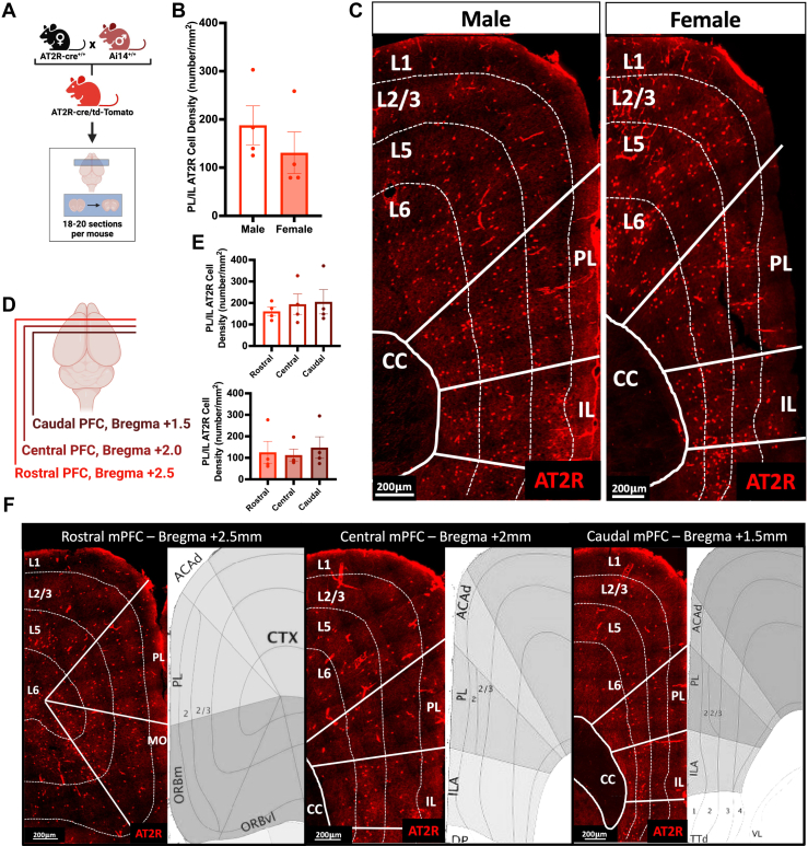
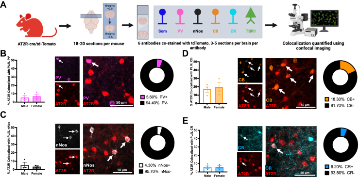
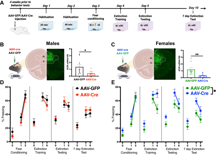
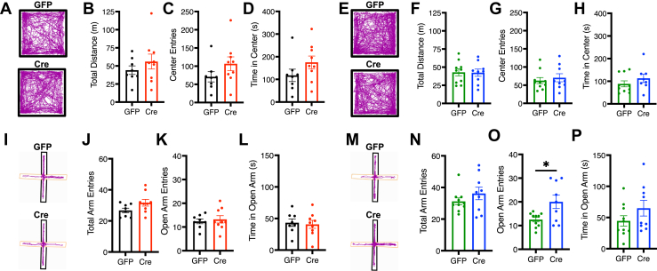
Methods: To characterize mPFC-AT2R-expressing neurons in the mPFC, AT2R-Cre/tdTomato male and female mice were used for immunohistochemistry. mPFC brain sections were stained with glutamatergic or interneuron markers, and density of AT2R+ cells and colocalization with each marker were quantified. To assess fear-related behaviors in AT2R-flox mice, we selectively deleted AT2R from mPFC neurons using a Cre-expressing adeno-associated virus. Mice then underwent Pavlovian auditory fear conditioning, elevated plus maze, and open field testing.

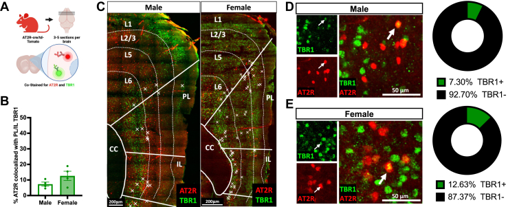
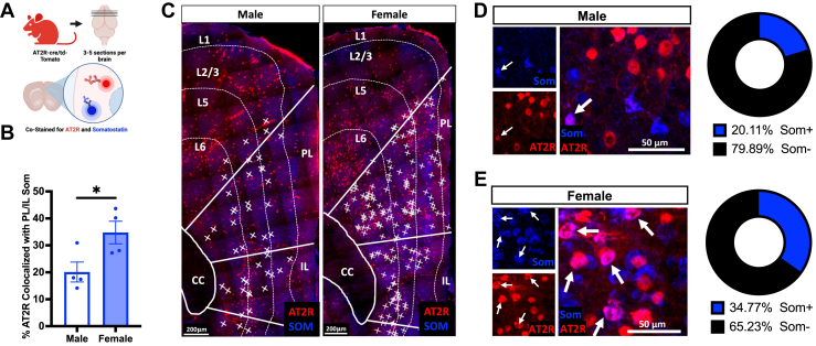
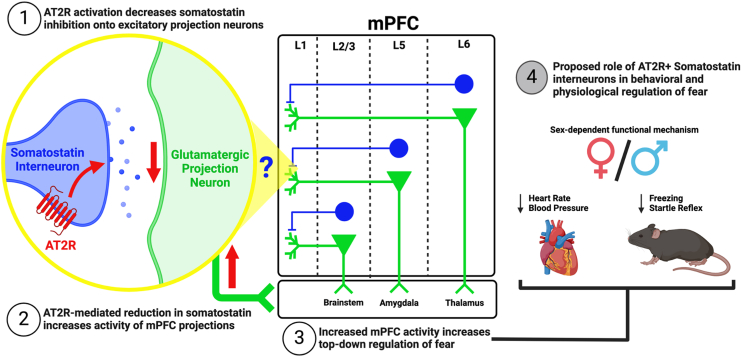
Results: Immunohistochemistry results revealed that AT2R was densely expressed throughout the mPFC and primarily expressed in somatostatin interneurons in a sex-dependent manner. Following fear conditioning, mPFC-AT2R Cre-lox deletion impaired extinction and increased exploratory behavior in female but not male mice, while locomotion was unaltered by mPFC-AT2R deletion in both sexes.

Conclusions: These results identify mPFC-AT2R+ neurons as a novel subgroup of somatostatin interneurons and reveal their role in regulating fear learning in a sex-dependent manner, potentially offering insights into novel therapeutic targets for posttraumatic stress disorder.

Keywords: AT2R; Angiotensin II; Fear extinction; Interneuron; PTSD; Prefrontal cortex.

Plain language summary

Posttraumatic stress disorder (PTSD) is a significant predictor of cardiovascular disease (CVD), although the underlying mechanisms are poorly understood. The brain renin-angiotensin system (RAS) is important for cardiovascular and emotional stress regulation and may better help understand the link between PTSD and CVD risk. Our research reveals that the brain angiotensin II type 2 receptor (AT2R) subtype is located on specific somatostatin (SOM+) interneurons in the medial prefrontal cortex (mPFC) and plays a role in fear memory extinction, particularly in females. These findings reveal a role for the mPFC-AT2R in fear-based learning and memory, offering potential insights into the mechanisms underlying the PTSD-CVD association and therapeutic strategies.


Figures

Figure 1
Figure 1
AT2R/tdTomato+ mPFC expression in male and female mice. (A) Generation of the AT2R-cre/tdTomato reporter mouse and experimental approach to test mPFC-AT2R distribution with immunohistochemistry. (B) AT2R+ cells (red) are equally expressed in the PL and IL regions of the mPFC of male (n = 4) and female (n = 4) mice (t6 = 0.96, p = .37). (C) Representative coronal sections showing laminar distribution of AT2R+ neurons in L2/3, L5, and L6 of the mPFC of male and female mice. (D) Approach for assessing distribution of AT2R+ neurons through the depth of the mPFC. (E) Rostral/caudal depth of the mPFC had no impact on the distribution of AT2R+ neurons in male (rostral mPFC: 160.9 ± 20.0 cells/mm2, central mPFC: 194.3 ± 43.4 cells/mm2, caudal mPFC: 205.4 ± 56.5 cells/mm2, F2,9 = 0.36, p = .71) or female (rostral mPFC: 125.7 ± 50.9 cells/mm2, central mPFC: 112.8 ± 28.0 cells/mm2, caudal mPFC: 147.8 ± 50.1 cells/mm2, F2,9 = 0.16, p = .85) brains. (F) Representative coronal sections showing laminar distribution of AT2R+ neurons throughout the rostral/caudal mPFC compared with reference images from the Allen Mouse Brain Atlas (mouse.brain-map.org) at the same slice positions as the confocal images. Scale bar = 200 μm. ACAd, dorsal anterior cingulate area; AT2R, angiotensin II type 2 receptor; CC, corpus callosum; CTX, cortex; IL, infralimbic cortex; ILA, infralimbic area; L, layer; MO, motor area; mPFC, medial PFC; ORBm, medial orbital area; ORBvl, ventrolateral orbital area; PFC, prefrontal cortex; PL, prelimbic cortex; TTd, dorsal taenia tecta; VL, ventrolateral area.
Figure 2
Figure 2
AT2R/tdTomato+ expression on excitatory mPFC neurons. (A) Experimental approach for costaining of AT2R-tdTomato+ cells (red) with TBR1 neurons (green) in the mPFC (n = 4/group). (B) Sex does not significantly affect AT2R colocalization with TBR1 in the PL and IL regions of the mPFC (male: 7.3% ± 1.3% colocalized, female: 12.6% ± 3.0% colocalized, t6 = 1.63, p = .15). (C) Representative coronal sections from male (left) and female (right) mPFC showing laminar distribution of AT2R/TBR1 colocalizations. Colocalization is indicated with X. (D, E) Representative coronal sections from the male (D) and female (E) mPFC. AT2R-tdTomato and TBR1 signals are colocalized (indicated with arrows). Right: percentage of AT2R+ neurons colocalized with TBR1 in the mPFC of male (D) and female (E) mice. Scale bar = 200 μm. AT2R, angiotensin II type 2 receptor; CC, corpus callosum; IL, infralimbic cortex; L, layer; mPFC, medial prefrontal cortex; PL, prelimbic cortex; TBR1, T-box brain transcription factor 1.
Figure 3
Figure 3
Characterization of AT2R/tdTomato+ in mPFC cell types. (A) Experimental approach to test mPFC AT2R+ cell type in the mPFC of male and female mice (n = 3/group). (B–E) Representative sections show that sex has no effect on AT2R (red) colocalization with (B) PV (pink) interneurons (male: 5.5% ± 2.5% colocalized, female: 6.9% ± 2.6% colocalized, t4 = 0.41, p = .70), (C) nNos (white) interneurons (male: 5.5% ± 3.3% colocalized, female: 3.5% ± 1.2% colocalized, t4 = 0.58, p = .59), (D) CB (orange) interneurons (male: 17.0% ± 4.0% colocalized, female: 19.6% ± 5.6% colocalized, t4 = 0.38, p = .72), or (E) CR (teal) interneurons (male: 6.1% ± 1.9% colocalized, female: 6.0% ± 1.7% colocalized, t4 = 0.03, p = .98). Colocalization indicated with arrows. Scale bar = 50 μm. AT2R, angiotensin II type 2 receptor; CB, calbindin; CR, calretinin; IL, infralimbic cortex; mPFC, medial prefrontal cortex; nNos, neuronal nitric oxide synthase; PL, prelimbic cortex; PV, parvalbumin; Som, somatostatin.
Figure 4
Figure 4
AT2R/tdTomato+ expression on mPFC SOM interneurons in female mice. (A) Experimental approach for costaining of AT2R-tdTomato+ cells (red) with SOM neurons (blue) in the mPFC (n = 4/group). (B) Females have significantly greater AT2R/SOM colocalizations in the PL and IL regions of the mPFC (males: 20.1% ± 3.7% colocalized, females: 34.8% ± 4.2% colocalized, t6 = 2.62, p = .03). (C) Representative coronal sections from male (left) and female (right) mPFC showing laminar distribution of AT2R/SOM colocalizations. Colocalizations are indicated with X. (D, E) Left: representative coronal sections from the male (D) and female (E) mPFC. AT2R-tdTomato and somatostatin signals are colocalized (indicated with arrows). Right: percentage of AT2R+ neurons colocalized with SOM in the mPFC of male (D) and female (E) mice. Scale bar = 200 μm. ∗p < .05. AT2R, angiotensin II type 2 receptor; CC, corpus callosum; IL, infralimbic cortex; L, layer; mPFC, medial prefrontal cortex; PL, prelimbic cortex; SOM/Som, somatostatin.
Figure 5
Figure 5
AT2R cre/lox mPFC deletion delays extinction learning in females but not males. (A) Experimental protocol for the auditory cue-dependent fear extinction test. (B) Left: strategy for injection of AAV-cre (red) or AAV-GFP (black) into the mPFC of male AT2R-flox mice and representative injection image in the mPFC. Right: injection of AAV-cre into the mPFC successfully decreased expression of AT2R in males (GFP: 1.35 ± 0.38 fold change, Cre: 0.40 ± 0.11 fold change, t17 = 2.54, p = .02). (C) Left: strategy for injection of AAV-cre (blue) or AAV-GFP (green) into the mPFC of female AT2R-flox mice and representative injection image in the mPFC. Right: injection of AAV-cre into the mPFC decreased expression of AT2R in females (GFP: 1.20 ± 0.28 fold change, Cre: 0.32 ± 0.07 fold change, t18 = 3.28, p < .01). (D) AT2R deletion from the male mPFC does not impact fear acquisition or extinction learning across 3 testing days in males (F1,17 = 0.09, p = .76). (E) In females, AT2R deletion from the mPFC significantly inhibits extinction learning (F1,20 = 5.35, p = .03). n = 9–12/group. Blocks represent groupings of 5 conditioned stimuli exposures; for example, block 1 represents conditioned stimuli 1–5, while block 6 represents conditioned stimuli 25–30. ∗p < .05, ∗∗p < .01. AAV, adeno-associated virus; AT2R, angiotensin II type 2 receptor; GFP, green fluorescent protein; IL, infralimbic cortex; mPFC, medial prefrontal cortex; mRNA, messenger RNA; PL, prelimbic cortex.
Figure 6
Figure 6
Effects of AT2R cre/lox medial prefrontal cortex deletion on behavioral assays of locomotion and exploration in males and females. (A, E) Representative locomotor traces of GFP- and Cre-injected males (AAV-GFP: black; AAV-Cre: red) and females (AAV-GFP: green; AAV-Cre: blue) in the open field test. (A–H) AT2R deletion has no impact on locomotion [(B)t15 = 0.97, p = .35; (F)t17 = 0.07, p = .95, total distance traveled] or generalized anxiety [(C)t15 = 1.49, p = .16; (G)t17 = 0.57, p = .58, center entries; (D)t15 = 1.47, p = .16; (H)t17 = 1.11, p = .29, time in center] in males or females. (I, M) Representative locomotor traces of GFP- and Cre-injected males and females in the elevated plus maze test. (I–P) AT2R deletion has no impact on exploratory behavior [(J) total arm entries, t15 = 1.84, p = .09] or generalized anxiety in males [(K) open arm entries, t15 = 0.45, p = .66; (L) time in open arm, t15 = 0.29, p = .78]. Medial prefrontal cortex AT2R deletion increases some aspects of exploratory behavior in females [(N) total arm entries, t17 = 1.14, p = .27; (O) open arm entries, t17 = 2.64, p = .02; (P) open arm time, t17 = 1.38, p = .19]. n = 8–10/group. ∗p < .05. AT2R, angiotensin II type 2 receptor; GFP, green fluorescent protein.
Figure 7
Figure 7
Proposed mechanism for mPFC-AT2R+ interneurons role in conditioned fear. (1) Activation of mPFC-AT2R on somatostatin interneurons of the mPFC decreases the activity of these interneurons. (2) This decreases inhibition of glutamatergic projection neurons and increases their firing to downstream regions. (3) Because of preferential somatostatin inhibition on brainstem (L2/3), amygdala (L5), and thalamic (L6) projecting excitatory neurons, mPFC top-down regulation is increased to these regions [see (26)]. (4) Potential role of mPFC+AT2R interneurons in the physiology of conditioned fear. AT2R, angiotensin II type 2 receptor; L, layer; mPFC, medial prefrontal cortex.

Similar articles

Cited by

References

    1. Lancaster C.L., Teeters J.B., Gros D.F., Back S.E. Posttraumatic stress disorder: Overview of evidence-based assessment and treatment. J Clin Med. 2016;5:105. - PMC - PubMed
    1. Edmondson D., von Känel R. Post-traumatic stress disorder and cardiovascular disease. Lancet Psychiatry. 2017;4:320–329. - PMC - PubMed
    1. Brudey C., Park J., Wiaderkiewicz J., Kobayashi I., Mellman T.A., Marvar P.J. Autonomic and inflammatory consequences of posttraumatic stress disorder and the link to cardiovascular disease. Am J Physiol Regul Integr Comp Physiol. 2015;309:R315–R321. - PMC - PubMed
    1. Kimerling R., Allen M.C., Duncan L.E. Chromosomes to social contexts: Sex and gender differences in PTSD. Curr Psychiatry Rep. 2018;20:114. - PubMed
    1. Seligowski A.V., Duffy L.A., Merker J.B., Michopoulos V., Gillespie C.F., Marvar P.J., et al. The renin-angiotensin system in PTSD: A replication and extension. Neuropsychopharmacology. 2021;46:750–755. - PMC - PubMed

LinkOut - more resources