Review
. 2024 Jul-Aug;16(4):e1983.
doi: 10.1002/wnan.1983.

Theranostic nanoparticles for detection and treatment of pancreatic cancer

Affiliations
Review

Theranostic nanoparticles for detection and treatment of pancreatic cancer

Happy Agarwal et al. Wiley Interdiscip Rev Nanomed Nanobiotechnol. 2024 Jul-Aug.

Abstract

Pancreatic ductal adenocarcinoma (PDAC) is one of the most recalcitrant cancers due to its late diagnosis, poor therapeutic response, and highly heterogeneous microenvironment. Nanotechnology has the potential to overcome some of the challenges to improve diagnostics and tumor-specific drug delivery but they have not been plausibly viable in clinical settings. The review focuses on active targeting strategies to enhance pancreatic tumor-specific uptake for nanoparticles. Additionally, this review highlights using actively targeted liposomes, micelles, gold nanoparticles, silica nanoparticles, and iron oxide nanoparticles to improve pancreatic tumor targeting. Active targeting of nanoparticles toward either differentially expressed receptors or PDAC tumor microenvironment (TME) using peptides, antibodies, small molecules, polysaccharides, and hormones has been presented. We focus on microenvironment-based hallmarks of PDAC and the potential for actively targeted nanoparticles to overcome the challenges presented in PDAC. It describes the use of nanoparticles as contrast agents for improved diagnosis and the delivery of chemotherapeutic agents that target various aspects within the TME of PDAC. Additionally, we review emerging nano-contrast agents detected using imaging-based technologies and the role of nanoparticles in energy-based treatments of PDAC. This article is categorized under: Implantable Materials and Surgical Technologies > Nanoscale Tools and Techniques in Surgery Therapeutic Approaches and Drug Discovery > Nanomedicine for Oncologic Disease Diagnostic Tools > In Vivo Nanodiagnostics and Imaging.

Keywords: active targeting; drug delivery; imaging; nanoparticle; pancreatic cancer.


Conflict of interest statement

Conflict of Interest

The authors declare no conflicts of interest.

Figures

Figure 1.
Figure 1.
Liposome assembly and characterization. (A) Schematic of liposome self-assembly with active functionalization and drug encapsulation (B) DLS measurement of naked liposomes demonstrating average diameter of 113 nm (C) SEM analysis of liposomes.
Figure 2.
Figure 2.
Tumor-specific accumulation of intravenously injected syndecan-1 conjugated targeted liposomes versus non-specific accumulation of untargeted liposomes as evidenced by ex vivo fluorescence imaging. Signal intensities reflect the accumulation of dye-encapsulated liposomes. Image adapted and printed with permission from reference (Yin et al., 2015) licensed under Creative Commons Attribution 4.0 International License (http://creativecommons.org/licenses/by/4.0/).
Figure 3.
Figure 3.
EGFR targeted micelles deliver miRNA and Gemcitabine to pancreatic tumors (A) Bioluminescence Images (BLI) images show representative mice from each treatment group (unmodified, IgG-, C225-micelles with GEM and miR-205, and C225-micelles with GEM and scrambled miRNA) (B) BLI photon intensity measurements. The data is presented as the mean ± SEM (standard error of the mean), observed differences in treatment responses are statistically significant by **p < 0.01 and *p < 0.05 compared to the reference group (C225-micelles containing GEM and miR-205 treated group) (C) Schematic Representation of Micelle Self-Assembly with Covalently Attached Drugs, Peptide Linkers, and Copolymers, reprinted with permission from reference (Mondal et al., 2017).
Figure 4.
Figure 4.
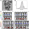
Targeted gold nanocages identify pancreatic tumors. (A) TEM images (B) Statistical size of gold nanocages (C) Fluorescence image of mice with BxPC-3 xenograft, administered with Sur-AuNc-Gd-Cy7 nanoprobe via intravenous injection at different time intervals showing a positive targeting capability of the survivin-bounded gold nanoprobe. Images are adapted and reprinted with permission from reference (Song et al., 2023), Creative Commons Attribution 4.0 (CC BY) license (https://creativecommons.org/licenses/by/4.0/).
Figure 5.
Figure 5.
In vivo visualization of UPA-targeted mesoporous silica nanoparticles for delivery of contrast agents to pancreatic cancer. (A) Tomographic optoacoustic images of pancreatic nanoparticle accumulation. Organs identified: yellow arrow = pancreas tumor, green arrow = liver, red arrow = spleen, white arrow = kidney. (B) TEM image of mesoporous silica nanoparticles (C) MSOT of tissue phantoms with untargeted and UPA-targeted MSN and their quantification data. Image adapted from (Gurka et al., 2016).
Figure 6.
Figure 6.
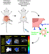
Schematic illustration of Plectin-1 targeted SPIONs based tumor imaging. (A) Schematic of SPIONs assembly (B) Intratumoral injection and tumor interaction (C) Multimodality images demonstrating differential uptake of SPIONs in mice treated with the targeted nanoparticles as compared to control. Image adapted from (Zhang et al., 2016).

References

    1. Abdolahinia ED, Nadri S, Rahbarghazi R, Barar J, Aghanejad A, & Omidi Y (2019). Enhanced penetration and cytotoxicity of metformin and collagenase conjugated gold nanoparticles in breast cancer spheroids. Life sciences, 231, 116545. - PubMed
    1. Adamska A, Domenichini A, & Falasca M (2017). Pancreatic ductal adenocarcinoma: current and evolving therapies. International journal of molecular sciences, 18(7), 1338. - PMC - PubMed
    1. Ahmed H, Gomte SS, Prathyusha E, Prabakaran A, Agrawal M, & Alexander A (2022). Biomedical applications of mesoporous silica nanoparticles as a drug delivery carrier. Journal of Drug Delivery Science and Technology, 76, 103729.
    1. Ait-Oudhia S, Straubinger RM, & Mager DE (2012). Meta-analysis of nanoparticulate paclitaxel delivery system pharmacokinetics and model prediction of associated neutropenia. Pharmaceutical research, 29, 2833–2844. - PubMed
    1. Alakhov V, Klinski E, Li S, Pietrzynski G, Venne A, Batrakova E, Bronitch T, & Kabanov A (1999). Block copolymer-based formulation of doxorubicin. From cell screen to clinical trials. Colloids and surfaces B: Biointerfaces, 16(1–4), 113–134.

Publication types

MeSH terms

LinkOut - more resources