. 2024 Dec;39(1):2388207.
doi: 10.1080/14756366.2024.2388207. Epub 2024 Aug 14.

Crystal structure of dihydroneopterin aldolase from Mycobacterium tuberculosis associated with 8-mercaptoguanine, and development of novel S8-functionalized analogues as inhibitors: Synthesis, enzyme inhibition, in vitro toxicity and antitubercular activity

Affiliations

Crystal structure of dihydroneopterin aldolase from Mycobacterium tuberculosis associated with 8-mercaptoguanine, and development of novel S8-functionalized analogues as inhibitors: Synthesis, enzyme inhibition, in vitro toxicity and antitubercular activity

Alexia de Matos Czeczot et al. J Enzyme Inhib Med Chem. 2024 Dec.

Abstract

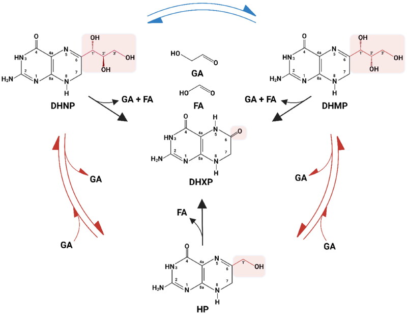
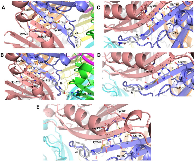
The crystallographic structure of the FolB enzyme from Mycobacterium tuberculosis (MtFolB), complexed with its inhibitor 8-mercaptoguanine (8-MG), was elucidated at a resolution of 1.95 Å. A novel series of S8-functionalized 8-MG derivatives were synthesised and evaluated as in vitro inhibitors of dihydroneopterin aldolase (DHNA, EC 4.1.2.25) activity of MtFolB. These compounds exhibited IC50 values in the submicromolar range. Evaluation of the activity for five compounds indicated their inhibition mode and inhibition constants. Molecular docking analyses were performed to determine the enzyme-inhibitor intermolecular interactions and ligand conformations upon complex formation. The inhibitory activities of all compounds against the M. tuberculosis H37Rv strain were evaluated. Compound 3e exhibited a minimum inhibitory concentration in the micromolar range. Finally, Compound 3e showed no apparent toxicity in both HepG2 and Vero cells. The findings presented herein will advance the quest for novel, specific inhibitors targeting MtFolB, an attractive molecular target for TB drug development.

Keywords: 8-mercaptoguanine; Dihydroneopterin aldolase inhibitors; Mycobacterium tuberculosis; SAR.


Conflict of interest statement

No potential conflict of interest was reported by the author(s).

Figures

Figure 1.
Figure 1.
Aldolase, epimerase, and oxygenase reactions catalysed by MtFolB. 7,8-dihydroneopterin (DHNP) and 7,8-dihydromonapterin (DHMP) are interconverted by the epimerase activity of MtFolB (blue arrows). Both DHNP and DHMP are reversibly converted to 6-hydroxymethyl-7,8-dihydropterin (HP) and glycolaldehyde (GA) by MtFolB aldolase activity (red arrows). Both DHNP and DHMP are also irreversibly converted to 7,8-dihydroxanthopterin (DHXP), GA and formic acid (FA), while HP are irreversibly converted to DHXP and FA by the MtFolB oxygenase activity (black arrows).
Scheme 1.
Scheme 1.
Reagents and reactants. Conditions: i = NaOH (0.5 M), EtOH, 25 °C, 2–6 h.
Figure 2.
Figure 2.
Three-dimensional structure of octameric MtFolB complexed with 8-MG and their 2D and 3D interactions representation. (A) The 3D crystallographic structure of MtFolB complexed with 8-MG revels that MtFolB forms an octamer upon interaction with 8-MG. MtFolB is represented as cartoon, with each chain is represented in a distinct colour: A (green), B (cyan), C (magenta), D (yellow), E (salmon), F (gray), G (slate blue), and H (orange). 8-MG is present between each monomer and is represented in stick model. (B) 2D interaction diagrams between 8-MG and MtFolB residues, with hydrogen bonds (black) and π–π stacking interactions (green) shown in dashed lines. (C) An enlarged view of 3D interaction with 8-MG. The main residues that interact by polar contacts with 8-MG are shown as sticks. 3D figures were generated using PyMOL v2.5 and 2D figures using PoseView.
Figure 3.
Figure 3.
Determination of half-maximum inhibition concentrations (IC50 values) of compounds 3a-n against MtFolB. IC50 values were determined performing initial velocity curves of enzymatic activity using 0.3 µM of MtFolB, 1.5 µM of DHNP and various concentrations of inhibitors. The IC50 value of each compound provided here were obtained using Equation (1) and plotted using Systat Software SigmaPlot 14.
Figure 4.
Figure 4.
Determination of the inhibition mechanism for compounds 3a-e on aldolase activity of MtFolB. (A) Compound 3a (0 – 0.8 µM). The Lineweaver − Burk plot displays a pattern of intersection at the y-axis, which indicates competitive inhibition. (B) Compound 3b (0 – 0.6 µM). The Lineweaver − Burk plot displays a pattern of intersection at the y-axis, which indicates competitive inhibition. (C) Compound 3c (0 – 0.9 µM). The Lineweaver − Burk plot displays a pattern of intersection to the left of the y-axis towards DHNP, which is diagnostic of non-competitive inhibition. (D) Compound 3d (0 – 2.5 µM). The Lineweaver − Burk plot displays a pattern of intersection at the y-axis, which indicates competitive inhibition. (E) Compound 3e (0 – 7 µM). The Lineweaver − Burk plot displays a pattern of intersection to the left of the y-axis towards DHNP, which is diagnostic of non-competitive inhibition.
Figure 5.
Figure 5.
3D interactions between compounds 3a-e and MtFolB obtained by molecular docking. An enlarged view of 3D interaction between compounds 3a (A), 3b (B), 3c (C), 3d (D) and 3e (E) with binding pocket of MtFolB. MtFolB is represented as cartoon, with each chain represented in a distinct colour: A (green), E (salmon), and G (slate blue); the main MtFolB residues that interact by polar contacts with the compounds are visualised as sticks, with carbon atoms coloured according to their respective MtFolB chain, nitrogen atoms in blue, and oxygen atoms in red. Compounds 3a-e are represented in stick model, with each atom represented in a distinct colour: carbon (gray), oxygen (red), nitrogen (blue), and sulphur (yellow). The distances of hydrogen bonds are provided in Angstrom (Å). Figures were obtained using PyMOL v2.5.

References

    1. World Health Organization . Global tuberculosis report. 2023.
    1. The End TB Strategy . World Health Organization. 2015.
    1. World Health Organization . Global tuberculosis report. 2022. 27:326–336.
    1. Yang L, Hu X, Chai X, Ye Q, Pang J, Li D, Hou T.. Opportunities for overcoming tuberculosis: Emerging targets and their inhibitors. Drug Discov Today. 2022;27(1):326–336. - PubMed
    1. Falcão VC, Villela AD, Rodrigues-Junior VS, Pissinate K, Eichler P, Pinto AF, Basso LA, Santos DS, Bizarro CV.. Validation of Mycobacterium tuberculosis dihydroneopterin aldolase as a molecular target for anti-tuberculosis drug development. Biochem Biophys Res Commun. 2017;485(4):814–819. - PubMed

MeSH terms

LinkOut - more resources