. 2024 Sep 11;15(9):e0192224.
doi: 10.1128/mbio.01922-24. Epub 2024 Aug 14.

CD81 fusion alters SARS-CoV-2 Spike trafficking

Affiliations

CD81 fusion alters SARS-CoV-2 Spike trafficking

Allaura S Cone et al. mBio. .

Abstract

The severe acute respiratory syndrome coronavirus 2 (SARS-CoV-2) pandemic caused the biggest public health crises in recent history. Many expect future coronavirus introductions into the human population. Hence, it is essential to understand the basic biology of these viruses. In natural infection, the SARS-CoV-2 Spike (S) glycoprotein is co-expressed with all other viral proteins, which modify cellular compartments to maximize virion assembly. By comparison, most of S is degraded when the protein is expressed in isolation, as in current molecular vaccines. To probe the maturation pathway of S, we redirected its maturation by fusing S to the tetraspanin protein CD81. CD81 is a defining constituent of extracellular vesicles (EVs) or exosomes. EVs are generated in large numbers by all cells, extruded into blood and lymph, and transfer cargo between cells and systemically (estimated 1012 EVs per mL plasma). EVs, like platelets, can be transfused between unrelated donors. When fusing the proline-stabilized form of strain Delta S into the flexible, large extracellular loop of CD81 rather than being degraded in the lysosome, S was extruded into EVs. CD81-S fusion containing EVs were produced in large numbers and could be isolated to high purity. Purified CD81::S EVs bound ACE2, and S displayed on individual EV was observed by cryogenic electron microscopy (EM). The CD81::S-fusion EVs were non-toxic and elicited an anti-S trimer and anti-RBD antibody response in mice. This report shows a design path to maximize viral glycoprotein assembly and release without relying on the co-expression of potentially pathogenic nonstructural viral proteins.

Importance: The severe acute respiratory syndrome coronavirus 2 pandemic caused the biggest public health crises in recent history. To understand the maturation pathway of S, we fused S to the tetraspanin protein CD81. The resulting molecule is secreted in extracellular vesicles and induces antibodies in mice. This may be a general design path for viral glycoprotein vaccines.

Keywords: CD63; CD81; SARS-CoV-2; Spike; TSPAN; coronavirus; dSTORM; exosome; extracellular vesicles; tetraspanins.


Conflict of interest statement

D.P.D., A.S.C., R.P.M., and Y.Z. declare competing interests with respect to the possible commercialization (WO 2024/ 073397) of some of the information presented. These are managed by the University of North Carolina. The authors have no other relevant affiliations or financial involvement with any organization or entity with a financial interest in or conflict with the subject matter or materials discussed in the paper apart from those disclosed.

Figures

Fig 1
Fig 1
Map of constructs that lists the names, a diagram, the features, and if the S protein has an intact furin cleavage insert. Additionally, we also list if the construct was tested in cell lysate and if we have seen the construct present on EVs using dSTORM. Blue is for CD81, red is for D614GS, blue is for wild-type S, green is for GFP, and open box is for the His tag.
Fig 2
Fig 2
Expression of recombinant recombinant CD81::S constructs. (A–D) Pictographs of constructs are used in this research. (A) A stabilized spike-his construct with the FCI removed as indicated by the yellow triangle within the turquoise outline and a bacterial trimerization domain: SWT[2P]-FCI::His. (B) Wild-type Spike strain WA1 with the D614G SNV containing the FCI is in red with GFP (in green) on the C-terminus: D614GSWA1::GFP. (C) CD81 molecule (in blue) with GFP on the C-terminus: CD81::GFP. (D) Non-stabilized Spike strain WA1 with the D614G SNV containing the FCI (in red) cloned into the large extracellular loop of CD81 with GFP on the C-terminus: CD81::GFP::D614GSWA1. (E) EVs size and concentration after purification from cells stably expressing CD81::GFP or CD81::GFP::D614GSWA1. EVs were then diluted in water and analyzed using the ZetaView. The size and concentration of the particles were measured with three reads per experiment and three separate experiments. (F–H) Western blot analysis of whole cell lysates. (F) Cell lysates from U-2 OS cells stably express the constructs. (G) HEK293T cells were transfected with CD81::GFP, D614GSWA1::GFP, or CD81::GFP::D614GSWA1. Then, the cells were harvested, lysed, and probed against Spike and the loading control vinculin. (H) HEK293T cells were transfected with CD81::GFP D614GSWA1::GFP, and the constructs were described in Table S1. The lysates were then run on an SDS-PAGE gel and probed with the indicated antibodies. NTC stands for no transfection control. (I–J) Western blot analysis of EV lysate. (I) Analysis of EV protein enrichment. Cell lysate was used as a control. EVs were harvested from U-2 OS cells stably expressing CD81::GFP or CD81::GFP::D614GSWA1. These EVs are enriched in proteins important for biogenesis, including CD63 and Syntenin-1, and do not have the cellular protein actin. (J) EVs purified from these cell lines were lysed and run on an SDS-PAGE gel. Two anti-S antibodies (mab N and mab P) were used to show S expression in the lysate. All experiments were conducted in at ≥3 biological replicates; * indicates a reprobed blot (hence the upper band in lane 2 representing CD81-GFP).
Fig 3
Fig 3
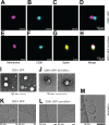
CD81::S fusion protein has altered localization. Cells were seeded on coverslips and transfected with CD81::GFP, D614GSWA1::GFP, or CD81::GFP::D614GSWA1, in addition to a non-transfected control. Twenty-four hours post-transfection, the cells were fixed with methanol and stained with a CD81 antibody. (A–D) Cells with no DNA added. (E–H) Cells transfected with CD81::GFP. The GFP signal is diffused through the cell and on the plasma membrane. (I–L) Cells transfected with D614GSWA1::GFP. The GFP signal is located around the nucleus. (M–P) Cells transfected with CD81::GFP::D614GSWA1. The GFP signal is diffused through the cell. The CD81 antibody does not bind to this construct since the Spike covers the binding area. All experiments were conducted in at ≥3 biological replicates. The yellow arrow highlights different vesicle localization ofD614GSWA1::GFP as compared to CD81::GFP::D614GSWA1. Scale bar = 10 µm.
Fig 4
Fig 4
Single particle analysis using dSTORM, TEM, and CryoEM. EVs harvested from cells expressing CD81::GFP or CD81::GFP::D614GSWA1 were analyzed using different techniques. First, EVs were seeded onto Ibidi 8-well glass bottom chamber slides. The EVs were stained with a CD81-Alexa488 antibody and then a S-Alexa594 antibody. After staining, the EVs were washed and placed in an oxygen scavenging buffer (B3; ONI) and imaged using the Nanoimager from Oxford Nanoimaging. Example images are taken from EVs the backbone only CD81::GFP EVs (A–D) or the CD81::GFP::D614GSWA1 EVs (E–H). Next, EVs were stained using uranyl acetate seeded onto a carbon-coated copper grid and imaged using TEM. (I) CD81::GFP EVs and (J) CD81::GFP::D614GSWA1 EVs images taken using TEM. Finally, EV samples were absorbed onto a copper grid, then snap-frozen in ethane/propane to be imaged using cryo-EM. (K) CD81::GFP EVs and (L) CD81::GFP::D614GSWA1 images taken using cryo-EM. All experiments were conducted in at ≥3 biological replicates.
Fig 5
Fig 5
Stabilization of S increases the expression levels on EV. (A) CD81-Delta Spike-GFP with hexa-proline mutations and the FCI removed, CD81::GFP::D614GSdelta[4P]-FCI, the primary construct used in the rest of the paper. (B) CD81::GFP::D614GSWA1-FCI with the FCI removed. (C) CD81::GFP::D614GSdelta[4P] with hexa-proline mutations but with the FCI intact. (D) Example images of cryo-EM images. EV samples were absorbed onto a copper grid, then snap-frozen in ethane/propane to be imaged using cryo-EM. (E–I) First, EVs were seeded onto Ibidi 8-well glass bottom chamber slides. The EVs were stained with a CD81-488 antibody and then a Spike-594 antibody. After staining, the EVs were washed and placed in an oxygen scavenging buffer (B3; ONI) and imaged using the Nanoimager from ONI. Representative dSTORM images taken from EVs, the backbone only CD81::GFP EVs (E) or the stabilized CD81::GFP::D614GSdelta[4P]-FCI EVs (F). During image acquisition, at least 2,000 EVs taken from three separate frames were analyzed using ONI CODI software. The Excel reports were analyzed in R to create pie charts to determine the percent of EVs carrying S (G) or the percent of EVs positive for CD81, S, or both. (H and I) Similar to G, the amount of EVs positive for CD81 or S. All experiments were conducted in at ≥3 biological replicates.
Fig 6
Fig 6
CD81::S EV bind to ACE2. EVs from (A) CD81::GFP, (B) CD81::GFP::D614GSWA1-FCI, or (C) CD81::GFP::D614GSdelta[4P]-FCI were seeded onto Ibidi 8-well glass bottom chamber slides. The EVs were then incubated with purified ACE2 (Sino Biological, Cat# 10108-H08H-B) conjugated to Alexa fluor 594. After staining, the EVs were washed and placed in an oxygen scavenging buffer (B3; ONI) and imaged using the Nanoimager from Oxford Nanoimaging. During image acquisition, at least 2,000 EVs taken from three separate frames were analyzed using ONI CODI software. The Excel reports were analyzed in R to create pie charts to determine the percent of EVs that bound to ACE2 or had CD81-GFP signal (D–F). Similar to panels D and F, the amount of EVs positive for CD81 or Spike is graphed in panel R (G and H). Vero cells overexpressing human ACE2 were transfected with (I–K) no DNA, (L–N) D614GSpikeWA1::GFP, (O–Q) CD81::GFP, (R–T) CD81::GFP::D614GSWA1, or (U–W) CD81::GFP::D614GSWA1-FCI. Previous research showed that co-expression of S and ACE2 induces syncytia formation. Scale bar = 10 µm.
Fig 7
Fig 7
Stabilized CD81::GFP::D614GSdelta[4P]-FCI EV induce S-Trimer and RBD antibodies. (A) Illustration of the treatment plan and boost regimen. The numbers on top indicate days of injection or (day 35) collection. The group size was n = 10. (B) Western blot assay to test sera for the presence of anti-S antibodies. The target SWT[2P]-FCI::His was collected from conditioned cell media and purified using a His column. Protein was run on an SDS-PAGE gel. Serum from mice injected with PBS or EV-CD81::GFP::D614GSdelta[4P]-FCI was incubated with the membranes at 1:100 dilution and detected with anti-murine total IgG-conjugated to horseradish peroxidase. Weak bands on the left and numbers indicate molecular weight markers in kDa. (C) Results of an ELISA assay testing for the presence of S-trimer-specific antibodies. This uses a commercial ELISA (Acro Biosystems RAS-T023). Shown is a box and whisker plot of the range, median, first and third quartile overlayed with individual data points for either the S negative group (blue) or the S positive group (brown). The amount of anti-S-specific IgG is shown on the vertical axis in ng/mL on a log 10 scale. Significance is indicated by the number of stars with ****: P ≤ 0.0001 by one-way analysis of variance with multiple comparisons. (D) Western blot assay to test sera for the presence of RBD antibodies. Purified RBD protein (ACRO) was run on an SDS-PAGE gel. Serum from mice injected with PBS or EV-S was incubated with the membranes at 1:100 dilution and detected with anti-murine total IgG-conjugated to horseradish peroxidase. Weak bands on the left and numbers indicate molecular weight markers in kDa.

References

    1. Corbett KS, Edwards DK, Leist SR, Abiona OM, Boyoglu-Barnum S, Gillespie RA, Himansu S, Schäfer A, Ziwawo CT, DiPiazza AT, et al. 2020. SARS-CoV-2 mRNA vaccine design enabled by prototype pathogen preparedness. Nature 586:567–571. doi: 10.1038/s41586-020-2622-0 - DOI - PMC - PubMed
    1. Jackson LA, Anderson EJ, Rouphael NG, Roberts PC, Makhene M, Coler RN, McCullough MP, Chappell JD, Denison MR, Stevens LJ, et al. 2020. An mRNA vaccine against SARS-CoV-2 - preliminary report. N Engl J Med 383:1920–1931. doi: 10.1056/NEJMoa2022483 - DOI - PMC - PubMed
    1. Barouch DH, Stephenson KE, Sadoff J, Yu J, Chang A, Gebre M, McMahan K, Liu J, Chandrashekar A, Patel S, Le Gars M, de Groot AM, Heerwegh D, Struyf F, Douoguih M, van Hoof J, Schuitemaker H. 2021. Durable humoral and cellular immune responses 8 months after Ad26.COV2.S vaccination. N Engl J Med 385:951–953. doi: 10.1056/NEJMc2108829 - DOI - PMC - PubMed
    1. Sadoff J, Gray G, Vandebosch A, Cárdenas V, Shukarev G, Grinsztejn B, Goepfert PA, Truyers C, Fennema H, Spiessens B, et al. 2021. Safety and efficacy of single-dose Ad26.COV2.S vaccine against Covid-19. N Engl J Med 384:2187–2201. doi: 10.1056/NEJMoa2101544 - DOI - PMC - PubMed
    1. Dunkle LM, Kotloff KL, Gay CL, Áñez G, Adelglass JM, Barrat Hernández AQ, Harper WL, Duncanson DM, McArthur MA, Florescu DF, et al. 2022. Efficacy and safety of NVX-CoV2373 in adults in the United States and Mexico. N Engl J Med 386:531–543. doi: 10.1056/NEJMoa2116185 - DOI - PMC - PubMed

LinkOut - more resources