. 2024 Sep 2;221(9):e20240391.
doi: 10.1084/jem.20240391. Epub 2024 Aug 14.

Transcription of HIV-1 at sites of intact latent provirus integration

Affiliations

Transcription of HIV-1 at sites of intact latent provirus integration

Ana Rafaela Teixeira et al. J Exp Med. .

Erratum in

  • Correction: Transcription of HIV-1 at sites of intact latent provirus integration.
    Teixeira AR, Bittar C, Silva Santos GS, Oliveira TY, Huang AS, Linden N, Ferreira IATM, Murdza T, Muecksch F, Jones RB, Caskey M, Jankovic M, Nussenzweig MC. Teixeira AR, et al. J Exp Med. 2025 Jun 2;222(6):e2024039105162025c. doi: 10.1084/jem.2024039105162025c. Epub 2025 May 23. J Exp Med. 2025. PMID: 40408268 Free PMC article. No abstract available.

Abstract

HIV-1 antiretroviral therapy is highly effective but fails to eliminate a reservoir of latent proviruses, leading to a requirement for life-long treatment. How the site of integration of authentic intact latent proviruses might impact their own or neighboring gene expression or reservoir dynamics is poorly understood. Here, we report on proviral and neighboring gene transcription at sites of intact latent HIV-1 integration in cultured T cells obtained directly from people living with HIV, as well as engineered primary T cells and cell lines. Proviral gene expression was correlated to the level of endogenous gene expression under resting but not activated conditions. Notably, latent proviral promoters were 100-10,000× less active than in productively infected cells and had little or no measurable impact on neighboring gene expression under resting or activated conditions. Thus, the site of integration has a dominant effect on the transcriptional activity of intact HIV-1 proviruses in the latent reservoir, thereby influencing cytopathic effects and proviral immune evasion.


Conflict of interest statement

Disclosures: R.B. Jones reports personal fees from ViiV Healthcare outside the submitted work. M.C Nussenzweig reports personal fees from Gilead and personal fees from Frontier Biosciences outside the submitted work; in addition, M.C. Nussenzweig has a patent to 3BNC117 issued (Gilead) and a patent to 10-1074 issued (Gilead). No other disclosures were reported.

Figures

Figure 1.
Figure 1.
Selected integration sites and production of reporter T cell clones. (A) Table of selected integration sites. (B) Schematic representation of the methods used to produce reporter T cell lines (see Materials and methods for details). Created with https://BioRender.com.
Figure 2.
Figure 2.
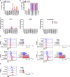
Jurkat reporter lines. (A) Histograms show GFP fluorescence (x axis) per normalized counts (y axis) for each integration site studied (iStrains, red) and control (blue), in both resting (upper panel) and PMA/ionomycin-activated (lower panel) conditions. (B) Graphs show relative LTR (left panel), eGFP (middle panel), and nLuciferase (right panel) expression assessed by qPCR under resting (blue) and PMA/ionomycin-activated (red) conditions. Bars represent the mean relative expression from two independent assays (biological replicates) ± SD. (C) LTR transcripts per cell (y axis), determined by qPCR, for each integration-positive clone and control, under resting (blue) and activated (red) conditions. CD4+ T cells infected with HIV-1YU2 served as positive control (black bar). Bars represent the mean of two independent assays (biological replicates) ± SD. (D) Relative expression determined by qPCR for host genes neighboring their respective reporter proviruses (iStrains, full bars) and control (striped bars), under resting (left panel, blue), and PMA/ionomycin-activated (right panel, red) conditions. Bars represent the mean relative expression of two independent assays (biological replicates) ± SD. Expression of the host gene was averaged across multiple clones to the same reporter integration site. (E) Correlation between the relative expression of LTR (y axis) for each Jurkat clone and their respective host gene (x axis), averaged across multiple clones to the same reporter integration site, under resting (left panel, blue) and PMA/ionomycin-activated (right panel, red) conditions. Pearson’s correlation coefficients, r, and two-tailed P values were computed for each condition.
Figure 3.
Figure 3.
Primary CD4 + T cell reporter lines. (A) Histograms show GFP fluorescence (x axis) per normalized counts (y axis) for each integration site studied (iStrains, red) and control (blue) in both resting (upper panel) and CD3/CD28-activated (lower panel) conditions. (B) Graphs show relative LTR (left panel), eGFP (middle panel), and nLuciferase (right panel) expression assessed by qPCR under resting (blue) and CD3/CD28-activated (red) conditions. Bars represent the mean relative expression from two independent assays (biological replicates) ± SD. (C) LTR transcripts per cell (y axis), determined by qPCR, for each integration-positive clone and control, under resting (blue) and CD3/CD28-activated (red) conditions. CD4+ T cells infected with HIV-1YU2 served as positive control (black bar). Bars represent the mean of two independent assays (biological replicates) ± SD. (D) Relative expression determined by qPCR for host genes neighboring their respective reporter proviruses (iStrains, full bars) and control (striped bars) under resting (left panel, blue) and CD3/CD28-activated (right panel, red) conditions. Bars represent the mean relative expression of two independent assays (biological replicates) ± SD. (E) Correlation between the relative expression of LTR (y axis) for each primary T cell clone and their respective host gene (x axis), averaged across multiple clones to the same reporter integration site, under resting (left panel, blue) and activated (right panel, red) conditions. Pearson’s correlation coefficients, r, and two-tailed P values were computed for each condition.
Figure S1.
Figure S1.
GFP expression in reporter cell lines. (A and B) Percentage of GFP+ cells as measured by flow cytometry for each integration-positive clone and control, under resting (blue) and PMA/ionomycin- and CD3/CD28-activated (red) conditions, respectively for Jurkat (A) and primary CD4+ T (B) cells. Bars represent the mean of two independent experiments (biological replicates) ± SD. (C) Graphs showing relative LTR (left panel), eGFP (middle panel), and nLuciferase (right panel) expression assessed by qPCR in Jurkat cell clones under resting (blue) and CD3/CD28-activated (red) conditions. Bars represent the mean relative expression from two independent assays (biological replicates) ± SD. (D and E) Representative histograms (left panels) show GFP fluorescence (x axis) per normalized counts (y axis) for Jurkat clones iZNF460, iKDM2A, iKCNA3a, and iATP2B4a (D), and primary CD4+ T cell clone iKDM2A (E), cultured for 24 h under resting conditions or in the presence of different LRAs or cell activation stimuli (PMA/ionomycin for Jurkat cells, anti-CD3/CD28 monoclonal antibodies for primary cells). Graphs (right panels) show the percentage of GFP+ cells measured by flow cytometry for the same conditions. Bars represent the mean of two independent experiments (biological replicates) ± SD.
Figure S2.
Figure S2.
Expression of host genes carrying reporter proviruses in Jurkat clones. Relative expression of host genes PYKFYVE and Fiz1 in Jurkat clones (full bars) and control (striped bars), under resting (blue) and activated (red) conditions. Bars represent the mean relative expression ± SD for each gene for three technical replicates, for two independent assays (biological replicates, I and II).
Figure 4.
Figure 4.
Chromatin accessibility. (A and B) Chromatin accessibility in Jurkat (A) and primary CD4+ T cell clones (B) as measured by ATAC-seq for the reporter construct at each integration site and control under resting conditions. (C) Graph shows ATAC-seq for ATP2B4 encompassing the reporter integration site in control and clones that carry the reporter in KDM2A, ATP2B4. Blue shading indicates the site of reporter integration. Graphs were generated by averaging the normalized reads from three technical replicates for each clone.
Figure S3.
Figure S3.
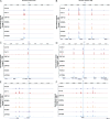
Chromatin accessibility around reporter construct integration site in Jurkat cell clones. (A–G) Chromatin accessibility measured by ATAC-seq in a 200,000 kb window of the genome around each of the integration sites for all Jurkat clones: chr13 (A), chr18 (ZNF407, B), chr12 (ZNF140, C), chr11 (KDM2A, D), chr19 (ZNF460, E), chr1 (KCNA3, F), and chr1 (ATP2B4, G). Graphs were generated by averaging the normalized reads from three technical replicates for each clone.
Figure S4.
Figure S4.
Chromatin accessibility around reporter construct integration site in primary CD4 + T cell clones. (A–F) Chromatin accessibility measured by ATAC-seq in a 200,000 kb window of the genome around each of the integration sites for all primary CD4+ T cell clones: chr13 (A), chr12 (ZNF140, B), chr11 (KDM2A, C), chr19 (ZNF460, D), chr1 (KCNA3, E), and chr1 (ATP2B4, F). Graphs were generated by averaging the normalized reads from three technical replicates for each clone.
Figure 5.
Figure 5.
HIV-infected cells from PLWH. (A) Schematic representation of the methods used to grow out HIV-1–infected cells from ART-suppressed individuals (Weymar et al., 2022). Created with https://BioRender.com. (B and C) UMAP of 10X single-cell gene expression data showing the position of the cells expressing the latent clones’ specific TCR (red dots) (B) and the fraction of latent cells in each cluster (C), for participants 603 (upper panels) and 5104 (lower panels) from ex vivo cells (left panels, data from Weymar et al. [2022]), and cultured cells under resting (middle panels) and activated (right panels) conditions. (D) Histograms show HIV-1 Gag p24 expression in HIV+ cells (red) and non-infected cells (blue) of cultures derived from 603 to 5104, under resting (upper panel) and activated (lower panel) conditions. (E) Relative expression of LTR (purple bars), gag (red bars), and env (blue bars) by qPCR for 603 and 5104 HIV+ cells under resting and activated conditions. Bars represent the mean relative expression of two independent assays (biological replicates) ± SD. (F) LTR transcripts per cell determined by qPCR, in cells from 603 to 5104 and in Jurkat and primary T cells reporter lines with proviruses integrated into ATP2B4, under resting (blue) and activated (red) conditions and HIV-1YU2 controls. Bars represent the mean of two independent experiments (biological replicates) ± SD. (G) Relative expression determined by 10X Genomics single-cell mRNA sequencing of host gene neighboring HIV-1 proviral integration (ZNF486) in HIV-infected (HIV+) and non-infected (HIV) cells from the same cell population under resting (blue bars) and activated (red bars) conditions. Bars represent the mean of the respective population from one assay ± standard deviation representing population variance.
Figure S5.
Figure S5.
HIV-1 RNA splice variants. Sashimi plot showing scRNA-seq reads of 10X single-cell gene-expression data mapping to the HIV genome (bottom), in infected cells of participant 5104 under resting conditions. Splice junctions are represented as arcs connecting exons and the histogram represents the read coverage at each junction. The number of reads mapping to each junction is indicated by the numbers associated with each arc.

Update of

References

    1. Amemiya, H.M., Kundaje A., and Boyle A.P.. 2019. The ENCODE blacklist: Identification of problematic regions of the genome. Sci. Rep. 9:9354. 10.1038/s41598-019-45839-z - DOI - PMC - PubMed
    1. Antar, A.A.R., Jenike K.M., Jang S., Rigau D.N., Reeves D.B., Hoh R., Krone M.R., Keruly J.C., Moore R.D., Schiffer J.T., et al. . 2020. Longitudinal study reveals HIV-1-infected CD4+ T cell dynamics during long-term antiretroviral therapy. J. Clin. Invest. 130:3543–3559. 10.1172/JCI135953 - DOI - PMC - PubMed
    1. Bachmann, N., von Siebenthal C., Vongrad V., Turk T., Neumann K., Beerenwinkel N., Bogojeska J., Fellay J., Roth V., Kok Y.L., et al. . 2019. Determinants of HIV-1 reservoir size and long-term dynamics during suppressive ART. Nat. Commun. 10:3193. 10.1038/s41467-019-10884-9 - DOI - PMC - PubMed
    1. Battivelli, E., Dahabieh M.S., Abdel-Mohsen M., Svensson J.P., Tojal Da Silva I., Cohn L.B., Gramatica A., Deeks S., Greene W.C., Pillai S.K., and Verdin E.. 2018. Distinct chromatin functional states correlate with HIV latency reactivation in infected primary CD4(+) T cells. Elife. 7:e34655. 10.7554/eLife.34655 - DOI - PMC - PubMed
    1. Bui, J.K., Sobolewski M.D., Keele B.F., Spindler J., Musick A., Wiegand A., Luke B.T., Shao W., Hughes S.H., Coffin J.M., et al. . 2017. Proviruses with identical sequences comprise a large fraction of the replication-competent HIV reservoir. PLoS Pathog. 13:e1006283. 10.1371/journal.ppat.1006283 - DOI - PMC - PubMed