. 2024 Aug 20;121(34):e2319724121.
doi: 10.1073/pnas.2319724121. Epub 2024 Aug 14.

Zfp697 is an RNA-binding protein that regulates skeletal muscle inflammation and remodeling

Affiliations

Zfp697 is an RNA-binding protein that regulates skeletal muscle inflammation and remodeling

Jorge C Correia et al. Proc Natl Acad Sci U S A. .

Abstract

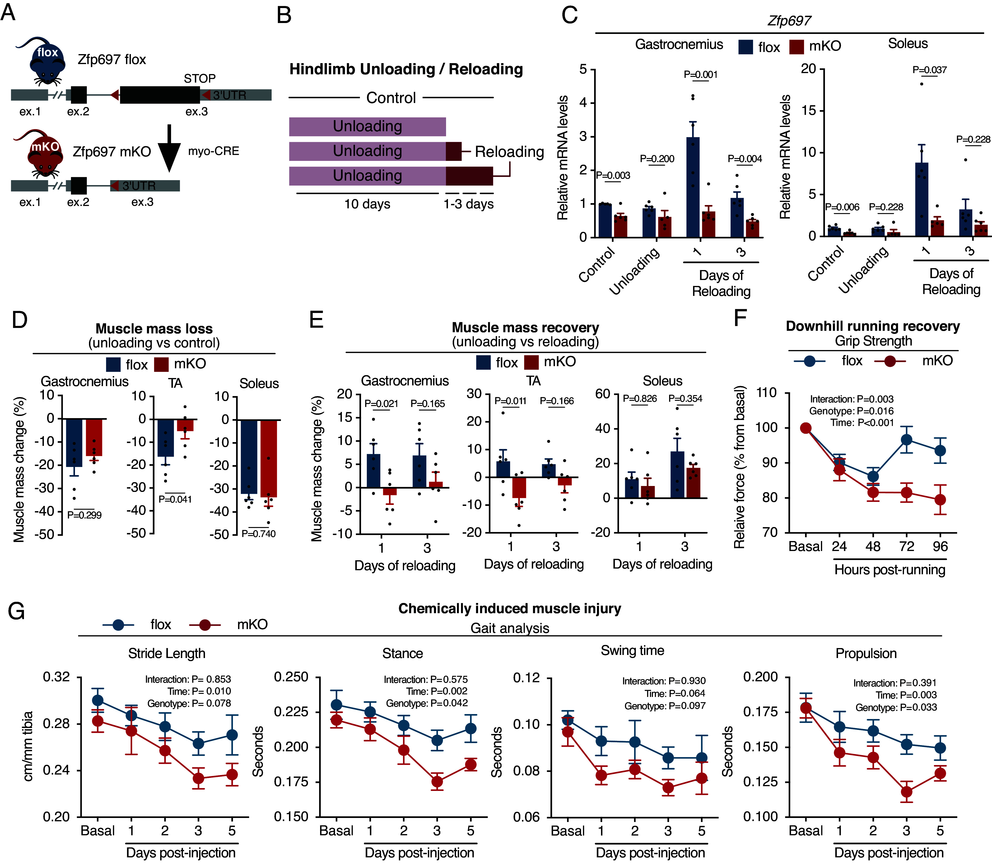
Skeletal muscle atrophy is a morbidity and mortality risk factor that happens with disuse, chronic disease, and aging. The tissue remodeling that happens during recovery from atrophy or injury involves changes in different cell types such as muscle fibers, and satellite and immune cells. Here, we show that the previously uncharacterized gene and protein Zfp697 is a damage-induced regulator of muscle remodeling. Zfp697/ZNF697 expression is transiently elevated during recovery from muscle atrophy or injury in mice and humans. Sustained Zfp697 expression in mouse muscle leads to a gene expression signature of chemokine secretion, immune cell recruitment, and extracellular matrix remodeling. Notably, although Zfp697 is expressed in several cell types in skeletal muscle, myofiber-specific Zfp697 genetic ablation in mice is sufficient to hinder the inflammatory and regenerative response to muscle injury, compromising functional recovery. We show that Zfp697 is an essential mediator of the interferon gamma response in muscle cells and that it functions primarily as an RNA-interacting protein, with a very high number of miRNA targets. This work identifies Zfp697 as an integrator of cell-cell communication necessary for tissue remodeling and regeneration.

Keywords: RNA-binding protein; Zfp697; inflammation; muscle atrophy; skeletal muscle.


Conflict of interest statement

Competing interests statement:G.W.Y. is an Scientific Advisory Board (SAB) member of Jumpcode Genomics and a co-founder, member of the Board of Directors, on the SAB, equity holder, and paid consultant for Locanabio and Eclipse BioInnovations. G.W.Y. is a distinguished visiting professor at the National University of Singapore. G.W.Y.’s interests have been reviewed and approved by the University of California, San Diego in accordance with its conflict-of-interest policies.

Figures

Fig. 1.
Fig. 1.
Zfp697 expression increases during muscle recovery from atrophy. (A) Overview of the mouse hindlimb unloading and reloading protocol. (B) Change in mouse gastrocnemius mass (normalized by tibia length) during hindlimb unloading and reloading (n = 6 to 18). One-way ANOVA with Tukey’s multiple comparisons test. (C) RNA-seq of mouse gastrocnemius muscle after 10 d of hindlimb unloading, 10 d of unloading followed by 1 d of reloading, and controls (n = 2 to 4 per condition). Heatmap highlights genes differentially expressed between unloading compared with control mice, and reloading compared with unloading. (D) GSEA for hallmark pathways in mouse gastrocnemius after hindlimb unloading and reloading. FDR, false discovery rate. (E and F) MA plots of gene expression levels in mouse gastrocnemius after 10 d of unloading compared with control, and 1 d of reloading compared with 10 d of unloading. Red dots indicate genes with significantly increased expression, while blue dots indicate genes with significantly decreased expression (Padj < 0.05). Known and putative transcriptional factors are highlighted. TF, transcription factor; Padj, adjusted P-value. (G) Schematic representation of the Zfp697 protein and its predicted glutamine-rich and zinc finger structural domains. The numbers shown refer to amino acid positions in the mouse Zfp697 protein. (H) Zfp697 gene expression in mouse gastrocnemius during hindlimb unloading and reloading (n = 6 to 8). One-way ANOVA with Tukey’s multiple comparisons test. Data represent mean values and error bars represent SEM.
Fig. 2.
Fig. 2.
Zfp697/ZNF697 expression is transiently elevated during recovery from injury. (A) Zfp697 gene expression in mouse gastrocnemius (gastroc) 16 h after clenbuterol injection (i.p. 2 mg/kg, n = 9 to 11). Two-tailed Student’s t test. (B) Zfp697 gene expression in mouse quadriceps following a single bout of treadmill running at 0, 3, 6, 12, and 24 h, compared with the rest control (n = 4 to 6). One-way ANOVA with Dunnett’s multiple comparisons test. (C) Zfp697 gene expression in mouse gastroc. after muscle injury induced by i.m. BaCl2 injection in the gastroc. of one leg (Right) and control solution in the same site of the contralateral leg (Left). Muscles were harvested 6, 24, and 72 h postinjection (n = 6 per time point). Two-way ANOVA with Šídák’s multiple comparisons test. (D) ZNF697 gene expression in muscle biopsies taken before and 24 h after a single bout of high-intensity interval training in recreationally active subjects (Control) and elite athletes (Elite) (n = 6 to 7). Two-way ANOVA with Šídák’s multiple comparisons test. (E) ZNF697 gene expression in human skeletal muscle after exercise (data from the MetaMEx database). (F) Zfp697 gene expression in skeletal muscles of mice with muscular dystrophy with myositis (mdm) compared with wild-type (WT) controls (dataset GSE210263). Two-tailed Student’s t test. (G) Zfp697 gene expression in mouse gastroc. during cancer cachexia progression (dataset GSE114820). One-way ANOVA with Dunnett’s multiple comparisons test. (H) Zfp697 gene expression in mouse gastroc. during aging (dataset GSE145480). One-way ANOVA with Dunnett’s multiple comparisons test. (I) Zfp697 gene expression in nuclei isolated from soleus or tibialis anterior (TA) of 5-mo-old mice (dataset GSE147127). (J) Zfp697 gene expression in mononuclear cells isolated from regenerating muscle (dataset GSE143437).
Fig. 3.
Fig. 3.
Zfp697 is necessary for IFNg signal transduction in myotubes. (A) Zfp697 gene expression in mouse primary myotubes with Zfp697 gain- or loss-of-function. For gain-of-function experiments, fully differentiated myotubes were transduced with adenovirus expressing GFP alone (Ad-GFP) or together with Zfp697 (Ad-Zfp697; n = 5 independent experiments). For loss- of-function, cells were transduced with adenovirus expressing Zfp697-specific (Ad-shZfp697) or scrambled control shRNAs (Ad- shControl; n = 3 independent experiments). Two-tailed Student’s paired t test vs. respective control. (B) Gene expression analysis of chemokines in mouse primary myotubes with Zfp697 gain- or loss-of-function. Two-tailed Student’s paired t test vs. respective control. (C) GSEA for hallmark pathways in mouse primary myotubes overexpressing Zfp697. FDR, false discovery rate. (D) MA plot showing mean gene expression vs. log2 fold-change in mouse primary myotubes overexpressing Zfp697. Differentially expressed genes are shown in blue (Padj < 0.05). IFNγ response genes are highlighted in red. Padj, adjusted P-value. (E and F) Gene expression analysis of Zfp697, chemokines, and IFNγ response genes in mouse primary myotubes after Zfp697 loss-of-function and treated with IFNγ or control (20 ng/mL for 8 h; n = 3 independent experiments). Two-way ANOVA with Tukey’s multiple comparisons test. Data represent mean values and error bars represent SEM.
Fig. 4.
Fig. 4.
Skeletal muscle-specific Zfp697 knockout blunts recovery from injury. (A) Schematic representation of the strategy adopted to generate skeletal-muscle specific Zfp697 knockout mice (Zfp697-mKO) and flox controls. Red arrows indicate loxP sites location. (B) Experimental approach for hindlimb unloading and reloading of Zfp697-mKO and flox littermates. (C) Zfp697 gene expression in the gastrocnemius (Left panel) and soleus (Right panel) of Zfp697-mKO and flox littermates following hindlimb unloading and reloading. Two-tailed Student’s t test compared with time-matched flox controls. (D) Muscle mass change after 10 d of hindlimb unloading comparing Zfp697-mKO and flox littermates with preunloading genotype-matched controls (n = 6). Two-tailed Student’s t test. (E) Muscle mass change after 1 or 3 d of hindlimb reloading comparing Zfp697-mKO and flox littermates with genotype-matched 10 d of unloaded mice (n = 6). Two-way ANOVA with Šídák’s multiple comparisons test. (F) Change in body-weight-normalized grip strength following a single bout of strenuous downhill running in Zfp697-mKO mice and flox littermates (n = 10). Repeated measures two-way ANOVA with Šídák’s multiple comparisons test. (G) Gait analysis of Zfp697-mKO mice and floxed littermates (flox, n = 5; mKO, n = 4) at baseline and following chemically induced muscle injury. Cardiotoxin was intramuscularly injected in the gastrocnemius and TA muscles of one leg (Right) and control solution in the same site of contralateral leg (Left). Repeated measures two-way ANOVA. Data represent mean values and error bars represent SEM.
Fig. 5.
Fig. 5.
Zfp697 mKOs fail to activate gene expression necessary for regeneration. (A) Principal component analysis for RNA-seq performed in the gastrocnemius muscle of control Zfp697 flox and mKO, after 10 d of hindlimb unloading and 3 d of reloading (n = 3 per condition and genotype). (B) Heatmap of differentially expressed genes for the interaction effect between conditions and genotypes. (C) GSEA for hallmark pathways accounting for the interaction effect between conditions (control, unloading, and reloading) and genotypes (mKO and flox). FDR, false discovery rate. (D) Heatmap of genes belonging to inflammatory and interferon-gamma response pathways. (E) Fraction of muscle-resident cell populations identified by digital cytometry (CIBERSORTx) applied to bulk muscle RNA-seq data from control, hindlimb unloaded, and reloaded Zfp697 flox and mKO mice (n = 3 per condition and genotype). Two-way ANOVA with Šídák’s multiple comparisons test. *P < 0.05 between indicated cell types. (F) Immunostaining for proliferating cells using KI67 in gastrocnemius muscles of Zfp697 flox and mKO and respective quantification (n = 4 per condition and genotype). Two-way ANOVA with Šídák’s multiple comparisons test. P-values represent comparison with time-matched flox controls. (Scale bar, 500 μm.)

Update of

References

    1. Chow L. S., et al. , Exerkines in health, resilience and disease. Nat. Rev. Endocrinol. 18, 273–289 (2022). - PMC - PubMed
    1. Furrer R., Hawley J. A., Handschin C., The molecular athlete: Exercise physiology from mechanisms to medals. Physiol. Rev. 103, 1693–1787 (2023), 10.1152/physrev.00017.2022. - DOI - PMC - PubMed
    1. Hindi S. M., Millay D. P., All for one and one for all: Regenerating skeletal muscle. Cold Spring Harb. Perspect. Biol. 14, a040824 (2022). - PMC - PubMed
    1. Joe A. W. B., et al. , Muscle injury activates resident fibro/adipogenic progenitors that facilitate myogenesis. Nat. Cell Biol. 12, 153–163 (2010). - PMC - PubMed
    1. Bernard C., Zavoriti A., Pucelle Q., Chazaud B., Gondin J., Role of macrophages during skeletal muscle regeneration and hypertrophy-Implications for immunomodulatory strategies. Physiol. Rep. 10, e15480 (2022). - PMC - PubMed

LinkOut - more resources