. 2024 Nov 1;134(21):e181839.
doi: 10.1172/JCI181839.

TET3-overexpressing macrophages promote endometriosis

Affiliations

TET3-overexpressing macrophages promote endometriosis

Haining Lv et al. J Clin Invest. .

Abstract

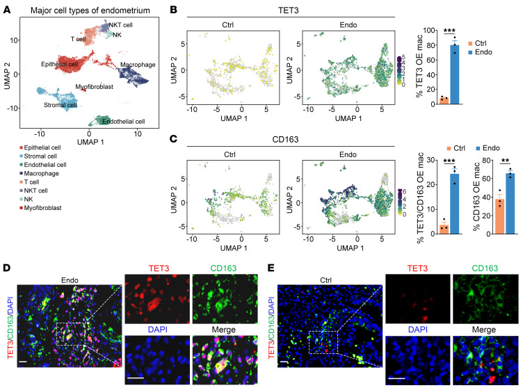
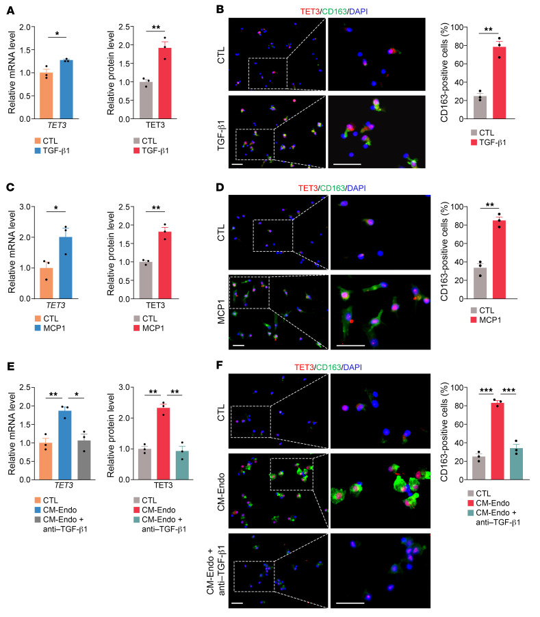
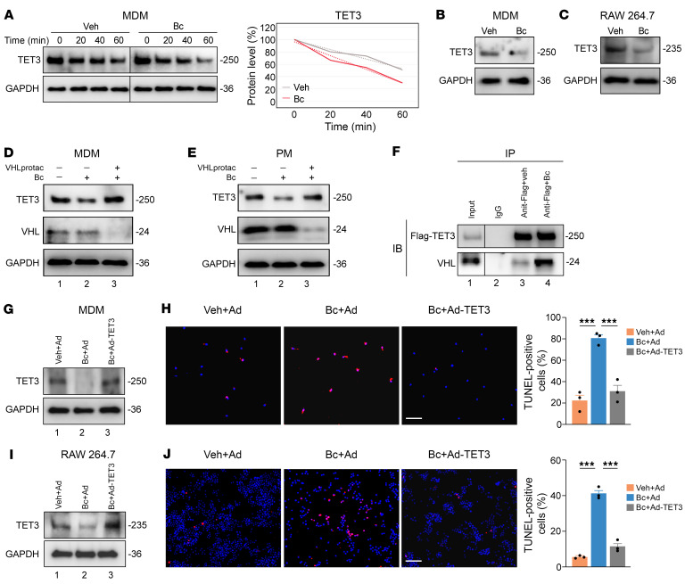
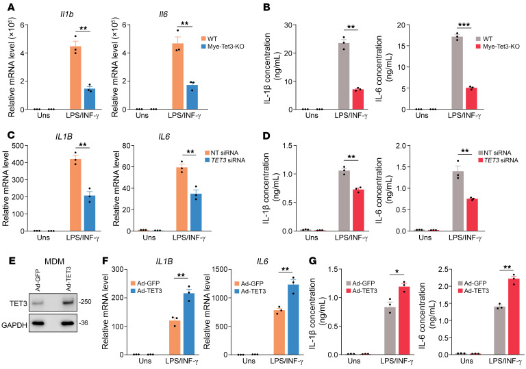
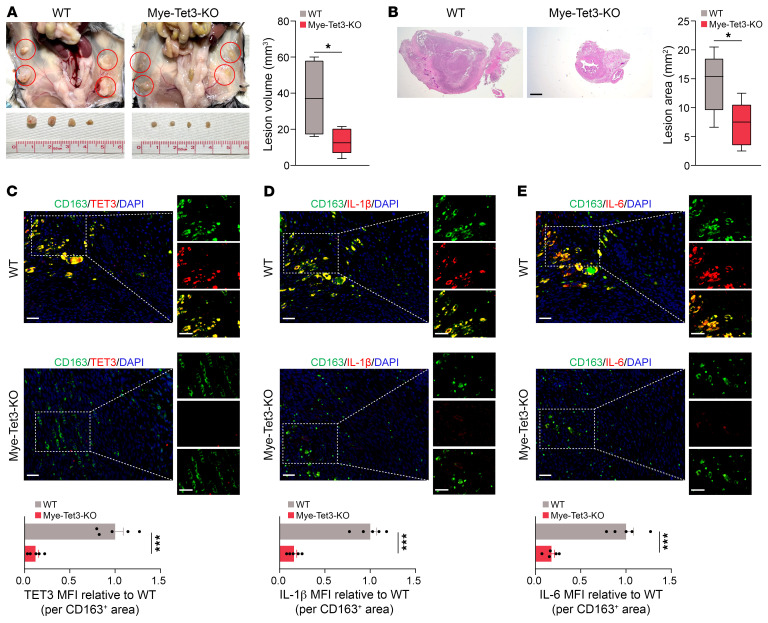
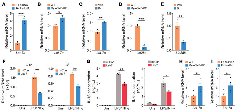
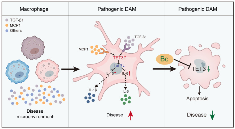
Endometriosis is a debilitating, chronic inflammatory disease affecting approximately 10% of reproductive-age women worldwide with no cure. While macrophages have been intrinsically linked to the pathophysiology of endometriosis, targeting them therapeutically has been extremely challenging due to their high heterogeneity and because these disease-associated macrophages (DAMs) can be either pathogenic or protective. Here, we report identification of pathogenic macrophages characterized by TET3 overexpression in human endometriosis lesions. We show that factors from the disease microenvironment upregulated TET3 expression, transforming macrophages into pathogenic DAMs. TET3 overexpression stimulated proinflammatory cytokine production via a feedback mechanism involving inhibition of let-7 miRNA expression. Remarkably, these cells relied on TET3 overexpression for survival and hence were vulnerable to TET3 knockdown. We demonstrated that Bobcat339, a synthetic cytosine derivative, triggered TET3 degradation in both human and mouse macrophages. This degradation was dependent on a von Hippel-Lindau (VHL) E3 ubiquitin ligase whose expression was also upregulated in TET3-overexpressing macrophages. Furthermore, depleting TET3-overexpressing macrophages either through myeloid-specific Tet3 ablation or using Bobcat339 strongly inhibited endometriosis progression in mice. Our results defined TET3-overexpressing macrophages as key pathogenic contributors to and attractive therapeutic targets for endometriosis. Our findings may also be applicable to other chronic inflammatory diseases where DAMs have important roles.

Keywords: Inflammation; Macrophages; Obstetrics/gynecology; Pain; Reproductive biology.


Figures

Figure 1
Figure 1. TET3 OE macrophages are abundant in human peritoneal endometriosis lesions.
(A) UMAP plot displaying total cells from normal endometrium (Ctrl, n = 3) and endometriosis lesions (Endo, n = 3). (B) UMAP showing macrophage TET3 expression in Ctrl (n = 3) and Endo (n = 3), with bar graph displaying percentages of TET3 OE macrophages on the right. (C) UMAP showing macrophage CD163 expression in Ctrl (n = 3) and Endo (n = 3). Bar graphs on the right show percentages of CD163 OE and TET3/CD163 double-OE macrophages. Data are represented as mean ± SEM. **P < 0.01; ***P < 0.001, 2-tailed Student’s t test. (D and E) Representative immunofluorescence staining of TET3 (red), CD163 (green), and nuclei (blue) from human endometriosis tissue (n = 7) and normal endometrial tissue (n = 5). The panels on the right are zoomed-in images from the left. Scale bars: 40 μm.
Figure 2
Figure 2. Macrophage TET3 expression is upregulated by inflammatory mediators in human MDMs.
(A) qRT-PCR of TET3 mRNA (left) and IHC quantification of TET3 protein (right) from MDMs treated with control media (CTL) or TGF-β1 at a final concentration of 10 ng/mL for 48 hours. MFI of TET3 in B was used to quantify TET3 protein expression. (B) Representative photomicrographs and corresponding statistical analysis of immunostaining of TET3 and CD163 in MDMs treated as in A. (C) qRT-PCR of TET3 mRNA (left) and IHC quantification of TET3 protein (right) from MDMs treated with CTL or MCP1 at a final concentration of 200 ng/mL for 24 hours. MFI of TET3 (red) in D was used to quantify TET3 protein expression. (D) Representative photomicrographs and corresponding statistical analysis of immunostaining of TET3 (red) and CD163 (green) in MDMs treated as in C. (E) qRT-PCR of TET3 mRNA (left) and IHC quantification of TET3 protein (right) from MDMs treated with CTL, CM-Endo, or CM-Endo plus TGF-β1 antibody at a final concentration of 10 ng/mL for 72 hours. MFI of TET3 in F was used to quantify TET3 protein expression. (F) Representative photomicrographs and corresponding statistical analysis of immunostaining of TET3 and CD163 in MDMs treated as in E. For quantification of immunostaining, n = 3 randomly selected areas per group were used. All data are represented as mean ± SEM. *P < 0.05; **P < 0.01; ***P < 0.001, 2-tailed Student’s t test. Scale bars: 40 μm.
Figure 3
Figure 3. TET3 knockdown leads to apoptosis of TET3 OE macrophages.
(A) qRT-PCR of TET3 and TET2 mRNAs isolated from MDMs treated with CM-Endo and transfected with nontargeting (NT) siRNA or TET3 siRNA for 24 hours. n = 3 per group in technical replicates. (B) Representative immunoblots for TET3 and TET2 from MDMs treated as in A. Protein sizes in kDa are marked on the right. Proteins were isolated after 48 hours of transfection. (C) Representative photomicrographs and corresponding statistical analysis of TUNEL+ (red) MDMs treated as in A. TUNEL assays were performed after 48 hours of transfection. n = 3 randomly selected areas per group. (D) qRT-PCR of Tet3 and Tet2 mRNAs isolated from RAW 264.7 cells transfected with NT siRNA or Tet3 siRNA for 24 hours. n = 3 per group in technical replicates. (E) Representative immunoblots for TET3 and TET2 from RAW 264.7 cells treated as in D. Proteins were isolated after 48 hours of transfection. (F) Representative photomicrographs and corresponding statistical analysis of TUNEL+ RAW 264.7 cells treated as in D. TUNEL assays were performed after 48 hours of transfection. n = 3 randomly selected areas per group. *P < 0.05; **P < 0.01; ***P < 0.001, 2-tailed Student’s t test. Scale bars: 40 μm.
Figure 4
Figure 4. Bc induces apoptosis of TET3 OE macrophages.
(A) Human MDMs were incubated with Veh or Bc at a final concentration of 10 μM for 2 hours, followed by time-course analysis of TET3 in the presence of CHX at a final concentration of 50 μg/mL. Cells were harvested at 0, 20, 40, and 60 minutes after addition of CHX. Human MDMs in CM-Endo (B) and RAW 264.7 cells (C) were incubated with Veh or Bc at a final concentration of 10 μM for 24 hours. Proteins were extracted and analyzed. (D) Immunoblots for TET3 and VHL in MDMs pretreated with or without VHLprotac at the concentration of 5 μM for 18 hours, followed by exposure to Bc at 10 μM for 8 hours. (E) PMs from WT mice primed with 30 ng/mL of TGF-β1 were conditioned by VHLprotac 5 μM for 18 hours, followed by exposure to Bc at 10 μM for 8 hours and Western blot analysis. (F) co-IP of Flag-TET3 and endogenous VHL in H1299 cells transfected with Ad-TET3, with or without the presence of Bc at 50 μM for 2 hours. MDMs (G and H) (primed with 10 ng/mL of TGF-β1) and RAW 264.7 cells (I and J) were incubated with Veh plus GFP-expressing adenovirus (Veh+Ad), Bc at 10 μM plus Ad (Bc+Ad), or Bc at 10 μM plus TET3-expressing adenovirus (Bc+Ad-TET3) for 48 hours, followed by immunoblotting and TUNEL assays. Representative immunoblots, photomicrographs and corresponding statistical analysis are shown. For TUNEL assay, n = 3 randomly selected areas per group. All data are represented as mean ± SEM. ***P < 0.001, 1-way ANOVA with Tukey’s post test (H and J). Scale bars: 40 μm. The dashed dividing lines (A and F) indicate splicing of noncontiguous lanes from the same blots.
Figure 5
Figure 5. TET3 knockdown reduces macrophage expression of IL-1β and IL-6.
(A) qRT-PCR of Il1b and Il6 mRNAs of cultured PMs isolated from Mye-Tet3–KO mice or WT controls and treated with 10 ng/mL LPS plus 20 ng/mL IFN-γ. RNAs were isolated after 6 hours of LPS/IFN-γ stimulation. Uns, unstimulated. n = 3 mice per genotype. (B) ELISA analysis (after 6 hours of LPS/IFN-γ stimulation) of IL-1β and IL-6 of cultured PMs treated as in A. n = 3 mice per genotype. (C) Human MDMs primed with 10 ng/mL of TGF-β1 were transfected with NT siRNA or TET3 siRNA. After 48 hours of transfection, cells were stimulated with 10 ng/mL LPS plus 20 ng/mL IFN-γ for 8 hours, followed by RNA extraction and qRT-PCR of IL-1B and IL-6 mRNAs. n = 3 biological replicates. (D) ELISA analysis (after 8 hours of LPS/IFN-γ stimulation) of IL-1β and IL-6 of MDMs following treatment as in C. (E) Human MDMs were infected with Ad-GFP or Ad-TET3. The next day, proteins were extracted, followed by Western blot analysis. The Ad-TET3–infected cells showed approximately 5-fold TET3 overexpression as compared with Ad-GFP–infected cells. (F) Human MDMs were infected with Ad-GFP or Ad-TET3. The next day, cells were stimulated with 10 ng/mL LPS plus 20 ng/mL IFN-γ for 8 hours, followed by RNA extraction and qRT-PCR of IL-1B and IL-6 mRNAs. n = 3 biological replicates. (G) ELISA analysis (after 8 hours of LPS/IFN-γ stimulation) of IL-1β and IL-6 of MDMs following treatment as in F. All data are represented as mean ± SEM. *P < 0.05; **P < 0.01; ***P < 0.001, 2-tailed Student’s t test.
Figure 6
Figure 6. Myeloid-specific TET3-KO decreases endometriosis burden.
(A) Representative photographs and corresponding statistical analysis of endometriosis lesions (marked by red circles). n = 5 mice per genotype. (B) Representative photomicrographs and corresponding statistical analysis of endometriosis lesions stained with H&E. n = 5 mice per genotype. Scale bar: 500 μm. (C) Immunostaining of TET3 (red) in CD163+ macrophages (green) and quantification of macrophage TET3 MFI in endometriosis lesions. n = 5 mice per genotype. (D) Immunostaining of IL-1β (red) and CD163+ macrophages (green) and quantification of macrophage IL-1β MFI in endometriosis lesions. n = 5 mice per genotype. (E) Immunostaining of IL-6 (red) and CD163+ macrophages (green) and quantification of macrophage IL-6 MFI in endometriosis lesions. n = 5 mice per genotype. *P < 0.05; ***P < 0.001. Scale bars: 40 μm (CE).
Figure 7
Figure 7. Bc recapitulates the therapeutic effects of myeloid-specific TET3-KO.
(A) Experimental design. (B) Representative photographs and corresponding statistical analysis of endometriosis lesions (marked by red circles). n = 5 mice per group. (C) Representative photomicrographs and corresponding statistical analysis of endometriosis lesions stained with H&E. n = 5 mice per group. Scale bar: 500 μm. (D) Immunostaining of TET3 (red) in CD163+ macrophages (green) and quantification of macrophage TET3 MFI in endometriosis lesions. n = 5 mice per group treated as indicated. (E) Immunostaining of IL-1β (red) and CD163+ macrophages (green) and quantification of macrophage IL-1β MFI in endometriosis lesions. n = 5 mice per group treated as indicated. (F) Immunostaining of IL-6 (red) and CD163+ macrophage (green) and quantification of macrophage IL-6 MFI in endometriosis lesions. n = 5 mice per group treated as indicated. All data are represented as mean ± SEM. *P < 0.05; **P < 0.01; ***P < 0.001, 2-tailed Student’s t test. Scale bar: 40 μm (DF).
Figure 8
Figure 8. TET3 enhances IL-1β and IL-6 expression by decreasing let-7 miRNA levels.
(A) qRT-PCR of let-7a in RAW 264.7 cells transfected with NT siRNA or Tet3 siRNA. RNA was isolated at 24 hours after transfection. n = 3 technical replicates. (B) qRT-PCR of let-7a in PMs isolated from WT and Mye-Tet3–KO mice. n = 3 mice per genotype. (C) PMs were isolated from WT mice and treated with TGF-β1 at a final concentration of 30 ng/mL. After 48 hours, Veh or Bc was added at a final concentration of 10 μM and incubation carried out for 48 hours. RNAs were extracted and analyzed by qRT-PCR. n = 3 mice per group. (D) qRT-PCR of Lin28b mRNA in PMs isolated from WT and Mye-Tet3–KO mice. n = 3 mice per genotype. (E) qRT-PCR of Lin28b mRNA in PMs isolated from WT mice and treated as in C. n = 3 mice per group. (F) qRT-PCR of Il-1b and Il-6 mRNAs of PMs isolated from WT mice and transfected with control miRNA (miCon) or let-7a mimic and stimulated with 10 ng/mL LPS plus 20 ng/mL IFN-γ. RNAs were isolated after 6 hours of LPS/IFN-γ stimulation. n = 3 mice per group. (G) ELISA results of IL-1β (after 6 hours of LPS/IFN-γ stimulation) and IL-6 (after 10 hours of LPS/IFN-γ stimulation) of PMs treated as in F. n = 3 mice per group. (H) Relative let-7a miRNA levels in endometriosis lesions from WT and Mye-tet3–KO mice. n = 4 animals per genotype. (I) Relative let-7a miRNA levels in endometriosis lesions from Veh- or Bc-treated mice. n = 4 animals per group. All data are represented as mean ± SEM. *P < 0.05; **P < 0.01; ***P < 0.001, 2-tailed Student’s t test.
Figure 9
Figure 9. A proposed model.
Macrophages exist as a heterogeneous population. Factors from the disease microenvironment induce TET3 overexpression in a subset of them. TET3 overexpression leads to genome-wide gene expression changes, which transform these macrophages into pathogenic DAMs. Though these macrophages are likely not identical in terms of other molecular features/surface markers, they share the feature of being proinflammatory and addicted to TET3 overexpression for survival. TET3 stimulates IL-1β and IL-6 production by inhibiting let-7 miRNA expression. Bc induces TET3 degradation, thereby eradicating TET3-overexpressing DAMs and inhibiting disease progression.

Comment in

  • Targeting TET3 in macrophages provides a concept strategy for the treatment of endometriosis

References

    1. Taylor HS, et al. Endometriosis is a chronic systemic disease: clinical challenges and novel innovations. Lancet. 2021;397(10276):839–852. doi: 10.1016/S0140-6736(21)00389-5. - DOI - PubMed
    1. Bulun SE, et al. Endometriosis and adenomyosis: shared pathophysiology. Fertil Steril. 2023;119(5):746–750. doi: 10.1016/j.fertnstert.2023.03.006. - DOI - PubMed
    1. Giudice LC, et al. Time for global health policy and research leaders to prioritize endometriosis. Nat Commun. 2023;14(1):8028. doi: 10.1038/s41467-023-43913-9. - DOI - PMC - PubMed
    1. Bulun SE, et al. Epithelial mutations in endometriosis: link to ovarian cancer. Endocrinology. 2019;160(3):626–638. doi: 10.1210/en.2018-00794. - DOI - PMC - PubMed
    1. Patzkowsky K. Rethinking endometriosis and pelvic pain. J Clin Invest. 2021;131(20):e154876. doi: 10.1172/JCI154876. - DOI - PMC - PubMed