

Optimized Protocol for Intestinal Swiss Rolls and Immunofluorescent Staining of Paraffin Embedded Tissue
- PMID: 39141543
- PMCID: PMC12212290
- DOI: 10.3791/66977
Optimized Protocol for Intestinal Swiss Rolls and Immunofluorescent Staining of Paraffin Embedded Tissue
Abstract
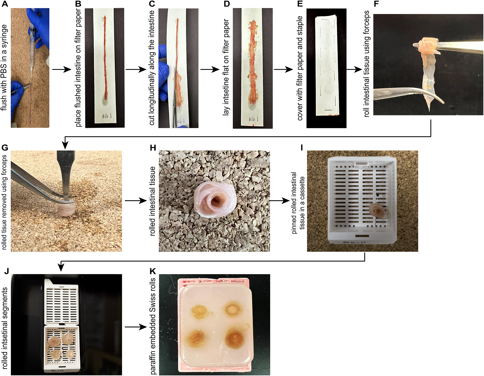
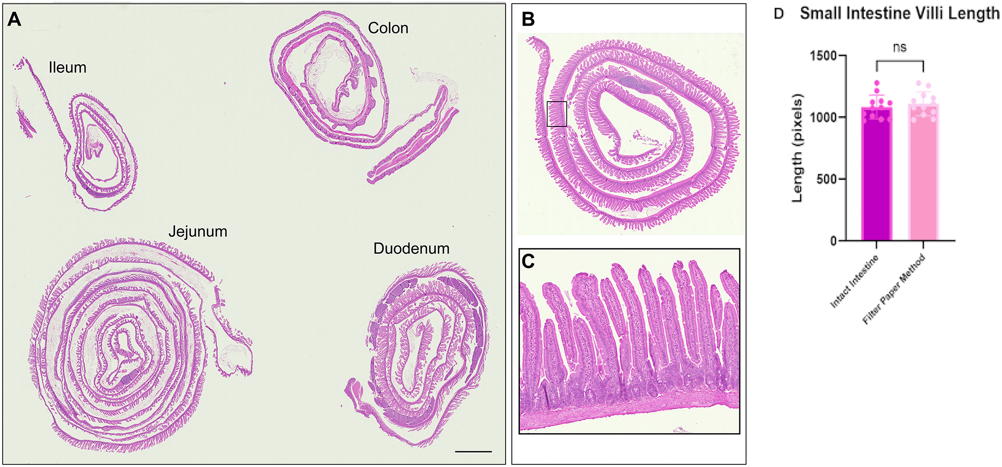
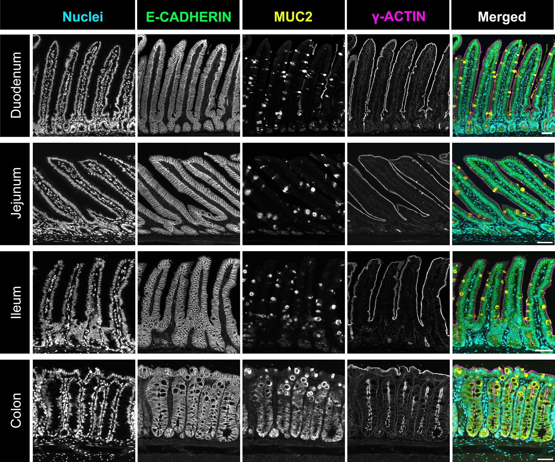
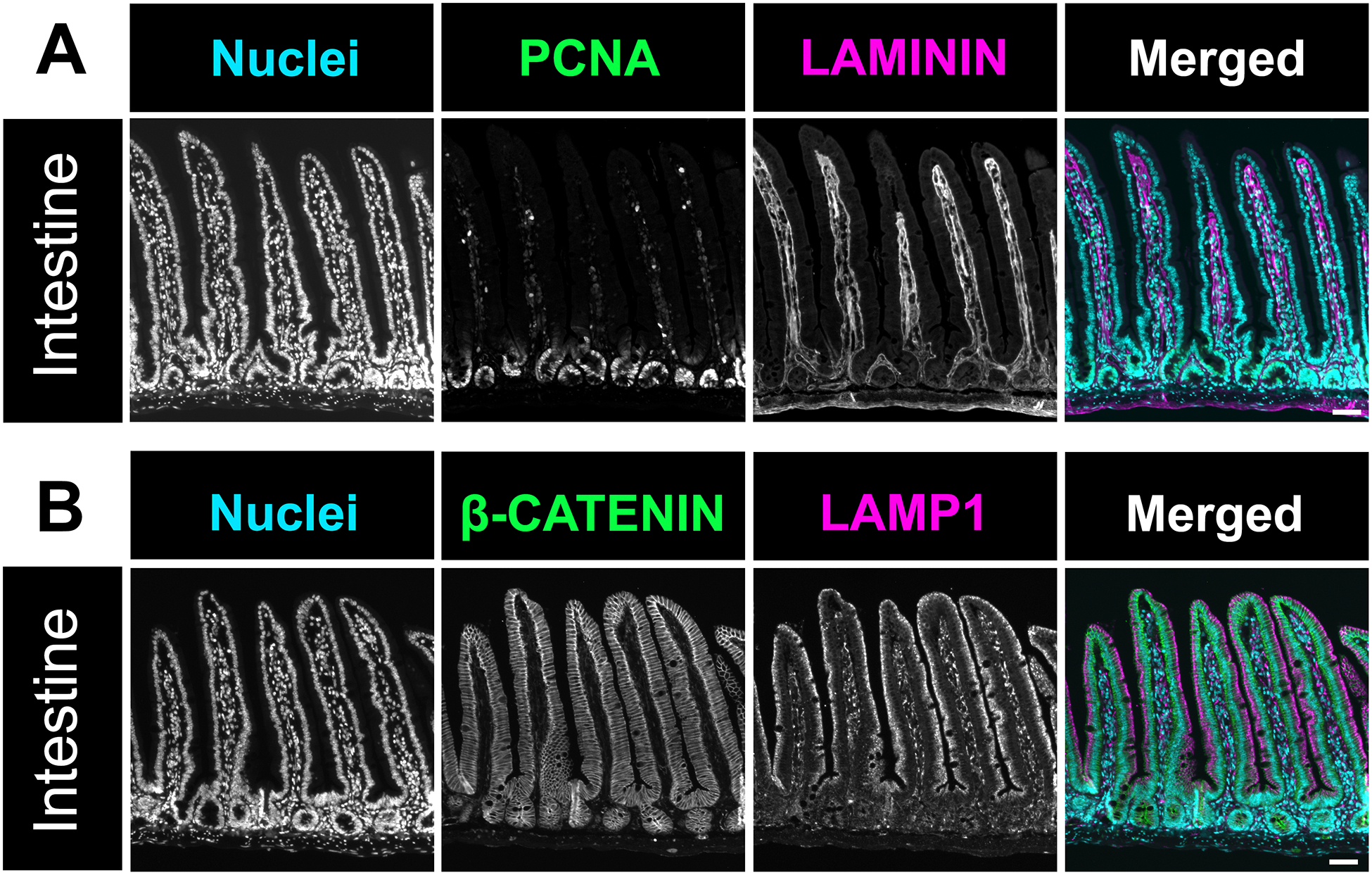
The intestine is a complex organ composed of the small and the large intestines. The small intestine can be further divided into duodenum, jejunum, and ileum. Each anatomical region of the intestine has a unique function that is reflected by differences in cellular structure. Investigating changes in the intestine requires an in-depth analysis of different tissue regions and cellular alterations. To study the intestine and visualize large pieces of tissue, researchers commonly use a technique known as intestinal Swiss rolls. In this technique, the intestine is divided into each anatomical region and fixed in a flat orientation. Then, the tissue is carefully rolled and processed for paraffin embedding. Proper tissue fixation and orientation is an often-overlooked laboratory technique but is critically important for downstream analysis. Additionally, improper Swiss rolling of intestinal tissue can damage the fragile intestinal epithelium, leading to poor tissue quality for immunostaining. Ensuring well-fixed and properly oriented tissue with intact cellular structures is a crucial step that ensures optimal visualization of intestinal cells. We present a cost-effective and simple method for making Swiss rolls to include all sections of the intestine in a single paraffin-embedded block. We also describe optimized immunofluorescence staining of intestinal tissue to study various aspects of the intestinal epithelium. The following protocol provides researchers with a comprehensive guide to obtaining high-quality immunofluorescence images through intestinal tissue fixation, Swiss-roll technique, and immunostaining. Employing these refined approaches preserves the intricate morphology of the intestinal epithelium and fosters a deeper understanding of intestinal physiology and pathobiology.
Figures
References
-
- Louvard D, Kedinger M, Hauri HP The differentiating intestinal epithelial cell: Establishment and maintenance of functions through interactions between cellular structures. Annu Rev Cell Biol. 8, 157–195 (1992). - PubMed
-
- Rieger J, Pelckmann LM, Drewes B Animal models of allergic disease: Methods and protocols. Springer US, New York, NY: (2021).
-
- Webster JD, Miller MA, Dusold D, Ramos-Vara J Effects of prolonged formalin fixation on the immunohistochemical detection of infectious agents in formalin-fixed, paraffin-embedded tissues. Vet Pathol. 47 (3), 529–535 (2010). - PubMed
-
- Hayashi Y, Koike M, Matsutani M, Hoshino T Effects of fixation time and enzymatic digestion on immunohistochemical demonstration of bromodeoxyuridine in formalin-fixed, paraffin-embedded tissue. J Histochem Cytochemis. 36 (5), 511–514 (1988). - PubMed
-
- Werner M, Chott A, Fabiano A, Battifora H Effect of formalin tissue fixation and processing on immunohistochemistry. Am J Surg Pathol. 24 (7), 1016–1019 (2000). - PubMed
Publication types
MeSH terms
Grants and funding
LinkOut - more resources
Full Text Sources
