. 2025 Jul:73:593-605.
doi: 10.1016/j.jare.2024.08.017. Epub 2024 Aug 12.

Targeting NGF but not VEGFR1 or BDNF signaling reduces endometriosis-associated pain in mice

Affiliations

Targeting NGF but not VEGFR1 or BDNF signaling reduces endometriosis-associated pain in mice

Tiago H Zaninelli et al. J Adv Res. 2025 Jul.

Abstract

Introduction: Endometriosis is a chronic inflammatory disease that affects ∼10 % of women. A significant fraction of patients experience limited or no efficacy with current therapies. Tissue adjacent to endometriosis lesions often exhibits increased neurite and vascular density, suggesting that disease pathology involves neurotrophic activity and angiogenesis.

Objectives: We aim to evaluate the potential for key tyrosine-kinase-receptor-coupled neurotrophic molecules to contribute to endometriosis-associated pain in mice.

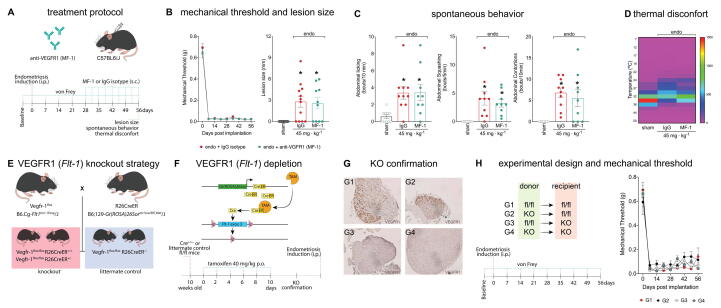
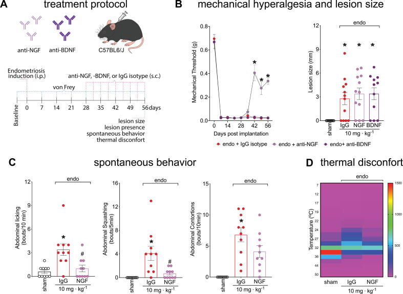
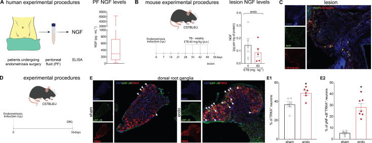
Methods: Peritoneal fluid was collected from endometriosis patients undergoing surgery and the levels of NGF and VEGFR1 regulators (VEGFA, VEGFB, PLGF, and sVEGFR1) were quantified by ELISA. VEGFR1 regulator concentrations were used to calculate VEGFR1 occupancy. We used genetic depletion, neutralizing antibodies, and pharmacological approaches to specifically block neurotrophic ligands (NGF or BDNF) or receptors (VEGFR1, TRKs) in a murine model of endometriosis-associated pain. Endometriosis-associated pain was measured using von Frey filaments, quantification of spontaneous abdominal pain-related behavior, and thermal discomfort. Disease parameters were evaluated by lesion size and prevalence. To evaluate potential toxicity, we measured the effect of entrectinib dose and schedule on body weight, liver and kidney function, and bone structure (via micro-CT).

Results: We found that entrectinib (pan-Trk inhibitor) or anti-NGF treatments reduced evoked pain, spontaneous pain, and thermal discomfort. In contrast, even though calculated receptor occupancy revealed that VEGFR1 agonist levels are sufficient to support signaling, blocking VEGFR1 via antibody or tamoxifen-induced knockout did not reduce pain or lesion size in mice. Targeting BDNF-TrkB with an anti-BDNF antibody also proved ineffective. Notably, changing dosing schedule to once weekly eliminated entrectinib-induced bone-loss without decreasing efficacy against pain.

Conclusions: This suggests NGF-TrkA signaling, but not BDNF-TrkB or VEGF-VEGFR1, mediates endometriosis-associated pain. Moreover, entrectinib blocks endometriosis-associated pain and reduces lesion sizes. Our results also indicated that entrectinib-like molecules are promising candidates for endometriosis treatment.

Keywords: BDNF; Neurotrophins; VEGF; VEGFR1; Visceral pain.


Conflict of interest statement

Declaration of competing interest The authors declare that they have no known competing financial interests or personal relationships that could have appeared to influence the work reported in this paper.

Figures

None
Graphical abstract
Fig. 1
Fig. 1
Levels of VEGFR1 ligands are increased in the peritoneal fluid of endometriosis patients and in mouse lesions. (A) experimental procedures for peritoneal fluid collection from endometriosis patients. (B) The levels of VEGFA, VEGFB, PlGF, and sVEGFR1 in peritoneal fluid samples determined by Ella®. Data are presented in box and whisker charts representing the levels of each mediator in pg/mL. (C) Calculated VEGFR1 occupancy as determined by previously published dissociation constant (Kd) and half maximal effective concentrations (EC50) , , . Note that in published data, occupancy is approximately proportional to signalling . (D) Experimental procedures and timepoints for tissue collection in our mouse model. (E) 56 days after endometriosis induction, mouse lesions were collected for determination of VEGFA levels by ELISA. Results are presented as mean ± SEM of VEGFA levels, n = 10 (sham – uterine horn tissue), and n = 9 (endo) mice per group (*P<0.05 vs. sham by two-tailed Student’s t-test). (F) Profile of VEGFR1 expression in DRG nociceptors determined by immunohistochemistry 56 days after endometriosis induction.
Fig. 2
Fig. 2
VEGFR1 neutralization or ablation do not reduce endometriosis-associated pain or thermal discomfort in mice. (A) Scheme of the treatment protocol with anti-VEGFR1 (MF-1) antibody and IgG control. (B) Mechanical hyperalgesia was determined using von Frey filaments before (zero) and after (7, 14, 21, 28, 35, 42, and 56 days) endometriosis induction. Results are presented as mean ± SEM of mechanical threshold, n = 10 mice per group. (C) Spontaneous behaviours measurements. For abdominal licking, the total number of times that mice directly groomed the abdominal region (without going to any other body region before or after the behaviours) was quantified for 10 min. For abdominal squashing, the number of times the mice pressed the lower abdominal region against the floor was quantified for 5 min. Sham mice did not display abdominal squashing. Abdominal contortions were quantified for 10 min by counting the number of contractions of the abdominal muscle together with the stretching of hind limbs. Sham mice did not display abdominal contortions. Results are expressed as mean ± SEM of abdominal licking, squashing, and contortions bouts per minute, n = 9–12. (*P<0.05 vs. sham by ANOVA followed by Tukey’s post-test). (D) Thermal discomfort heatmap. Heatmap shows mean time spent in each temperature zone for IgG control- or MF-1-treated mice. Data are presented as mean ± SEM of the amplitude of permanence in seconds in each thermal zone during 60 min, n = 9–12. (E) Breeding scheme for generation of VEGFR1 tamoxifen-induced cre-dependent KO strategy. (F) Scheme of tamoxifen-induced cre-dependent VEGFR1 KO and tamoxifen treatment protocol. (G) DRG neuron representative images for VEGFR1 KO confirmation determined by IHC analysis. G1-G2 from littermate controls and G3-G4 from KO mice n = 10. (H) Endometriosis induction protocol scheme with receptor and donor combinations. Mechanical response before (zero) and after (7, 14, 21, 28, 35, 42, and 56 days) endometriosis induction using von Frey filaments. Results are presented as mean ± SEM of mechanical threshold, n = 10 mice per group.
Fig. 3
Fig. 3
Entrectinib, a pan-Trk inhibitor, reduces endometriosis-associated pain and thermal discomfort. (A) Scheme of the treatment protocol with entrectinib. T1: 15 mg/kg every other day; T2: 20 mg/kg three times a week; and T3: 60 mg/kg weekly. In all treatment schedules, the maximum weekly dose is 60 mg/kg. (B left) Mechanical response to von Frey filaments before (zero) and after (7, 14, 21, 28, 35, 42, and 56 days) endometriosis induction. Results are presented as mean ± SEM of mechanical threshold, n = 10 mice per group (*P<0.05 vs. vehicle-treated group by 2-way ANOVA). (B right) Lesion size. Results are presented as mean ± SEM of lesion size in mm, n = 10–19 mice per group (*P<0.05 vs. sham, # P<0.05 vs. vehicle-treated group by ANOVA followed by Tukey’s post-test). (C) Thermal discomfort heatmap. Data are presented as mean ± SEM of the amplitude of permanence in seconds in each thermal zone during 60 min. (D) Spontaneous behaviour measurements. For abdominal licking, the total number of times that mice directly groomed the abdominal region (without going to any other body region before or after the behaviour) was quantified for 10 min. For abdominal squashing, the number of times the mice pressed the lower abdominal region against the floor was quantified for 5 min. Sham mice did not display abdominal squashing. Abdominal contortions were quantified for 10 min by counting the number of contractions of the abdominal muscle together with stretching of hind limbs. Sham mice did not display abdominal contortions. Results are expressed as mean ± SEM of abdominal licking, squashing, and contortions bouts per minute, n = 7–10. (*P<0.05 vs. sham, # P<0.05 vs. vehicle-treated group by ANOVA followed by Tukey’s post-test).
Fig. 4
Fig. 4
NGF, but not BDNF neutralization, reduces endometriosis-associated pain and thermal discomfort in mice. (A) Scheme of the treatment protocol with IgG control, anti-NGF, or anti-BDNF antibodies. (B) Mechanical hyperalgesia was measured using von Frey filaments before (zero) and after (7, 14, 21, 28, 35, 42, and 56 days) endometriosis induction. Results are presented as mean ± SEM of mechanical threshold, n = 10–12 mice per group (*P<0.05 vs. IgG treated group by 2-way ANOVA (left) or ANOVA followed by Tukey’s post-test (right)). (C) Spontaneous behaviour measurements. For abdominal licking, the total number of times that mice directly groomed the abdominal region (without going to any other body region before or after the behaviour) was quantified for 10 min. For abdominal squashing, the number of times the mice pressed the lower abdominal region against the floor was quantified for 5 min. Sham mice did not display abdominal squashing. Abdominal contortions were quantified for 10 min by counting the number of contractions of the abdominal muscle together with stretching of hind limbs. Sham mice did not display abdominal contortions. Results are expressed as mean ± SEM of abdominal licking, squashing, and contortions bouts per minute, n = 10 mice per group. (*P<0.05 vs. sham, # P<0.05 vs. IgG control by ANOVA followed by Tukey’s post-test). (D) Thermal discomfort heatmap. Heatmap shows mean time spent in each temperature zone for IgG control-, anti-NGF-, or anti-BDNF-treated mice. Data are presented as mean ± SEM of the amplitude of permanence in seconds in each thermal zone during 60 min, n = 10 mice per group.
Fig. 5
Fig. 5
NGF-TrkA signaling is activated during endometriosis. (A) Scheme of NGF measurements in the peritoneal fluid of endometriosis patients. Results are presented in box and whisker charts in pg/mL. n = 44. (B) Mouse experimental procedures and NGF levels in endometriotic lesions. Results are presented as mean ± SEM of NGF in picograms per milligram of tissue. n = 5 per group. (C) representative image from endometriotic lesions stained for NGF and beta-III tubulin. (D) Scheme of experimental methods for DRG immunohistochemistry. DRGs were dissected at 56 dpi. (E) Representative images of DRG neurons stained for TrkA (red) and p-NF-kB (green) and imaged with confocal microscopy, and the quantification of (E1) TrkA+ and (E2) pNF-κB+TrkA+ neurons of sham and endometriosis lesion-bearing mice. Results are presented as mean ± SEM of the percentage of positive neurons. n = 6 or 7 mice per group. Two-tailed Student’s T-test (*P<0.05 vs. sham). (For interpretation of the references to color in this figure legend, the reader is referred to the web version of this article.)
Fig. 6
Fig. 6
Weekly treatment with entrectinib does not induce weight change, liver or kidney toxicity, or bone loss in mice. (A) Scheme of experimental protocol for determination of entrectinib safety using the different treatment schedules. (B) Mouse weight was determined weekly. Results are presented as mean ± SEM in grams, n = 7–10 mice per group. (C) Kidney function was determined by measuring urea plasma levels. Results are presented as mean ± SEM of urea levels, n = 7–10 mice per group. (D) Liver function was determined by measuring ALT and AST plasma levels. Blood and femur were collected 56 dpi (after 4 weeks of treatment). Results are presented as mean ± SEM of AST and ALT levels, n = 7–10 mice per group. (E) Micro computed tomography of bone (femur) was used to determine bone loss. Results are presented as mean ± SEM, n = 5 mice per group. (*P<0.05 vs. vehicle by ANOVA followed by Tukey’s post-test.).

References

    1. Zondervan K.T., Becker C.M., Missmer S.A. Endometriosis. N Engl J Med. 2020;382:1244–1256. doi: 10.1056/NEJMra1810764. - DOI - PubMed
    1. Parasar P., Ozcan P., Terry K.L. Endometriosis: Epidemiology, Diagnosis and Clinical Management. Curr Obstet Gynecol Rep. 2017;6:34–41. doi: 10.1007/S13669-017-0187-1/TABLES/2. - DOI - PMC - PubMed
    1. Laux-Biehlmann A., d’Hooghe T., Zollner T.M. Menstruation pulls the trigger for inflammation and pain in endometriosis. Trends Pharmacol Sci. 2015;36:270–276. doi: 10.1016/j.tips.2015.03.004. - DOI - PubMed
    1. McKinnon B.D., Bertschi D., Bersinger N.A., Mueller M.D. Inflammation and nerve fiber interaction in endometriotic pain. Trends Endocrinol Metab. 2015;26:1–10. doi: 10.1016/j.tem.2014.10.003. - DOI - PubMed
    1. Bulun S.E., Yildiz S., Adli M., Chakravarti D., Parker J.B., Milad M., et al. Endometriosis and adenomyosis: shared pathophysiology. Fertil Steril. 2023;119:746–750. doi: 10.1016/J.FERTNSTERT.2023.03.006. - DOI - PubMed

MeSH terms

Substances

LinkOut - more resources