Review
. 2024 Aug 14;33(173):240064.
doi: 10.1183/16000617.0064-2024. Print 2024 Jul.

Pathogenesis-driven treatment of primary pulmonary alveolar proteinosis

Affiliations
Review

Pathogenesis-driven treatment of primary pulmonary alveolar proteinosis

Sara Lettieri et al. Eur Respir Rev. .

Abstract

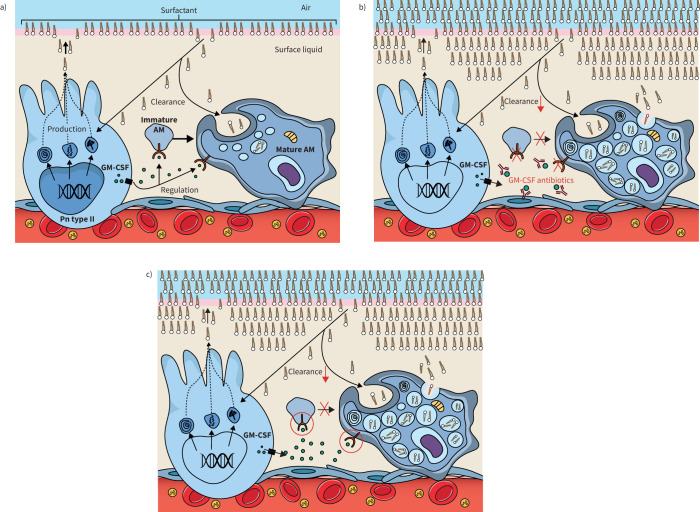
Pulmonary alveolar proteinosis (PAP) is a syndrome that results from the accumulation of lipoproteinaceous material in the alveolar space. According to the underlying pathogenetic mechanisms, three different forms have been identified, namely primary, secondary and congenital. Primary PAP is caused by disruption of granulocyte-macrophage colony-stimulating factor (GM-CSF) signalling due to the presence of neutralising autoantibodies (autoimmune PAP) or GM-CSF receptor genetic defects (hereditary PAP), which results in dysfunctional alveolar macrophages with reduced phagocytic clearance of particles, cholesterol and surfactant. The serum level of GM-CSF autoantibody is the only disease-specific biomarker of autoimmune PAP, although it does not correlate with disease severity. In PAP patients with normal serum GM-CSF autoantibody levels, elevated serum GM-CSF levels is highly suspicious for hereditary PAP. Several biomarkers have been correlated with disease severity, although they are not specific for PAP. These include lactate dehydrogenase, cytokeratin 19 fragment 21.1, carcinoembryonic antigen, neuron-specific enolase, surfactant proteins, Krebs von Lungen 6, chitinase-3-like protein 1 and monocyte chemotactic proteins. Finally, increased awareness of the disease mechanisms has led to the development of pathogenesis-based treatments, such as GM-CSF augmentation and cholesterol-targeting therapies.


Conflict of interest statement

Conflict of interest: F. Bonella reports consultancy fees from Boehringer Ingelheim, Sanofi, BMS and CSL-Behring, payment or honoraria for lectures, presentations, manuscript writing or educational events from Boehringer Ingelheim and Sanofi, support for attending meetings from Boehringer Ingelheim, Astra Zeneca and Atyr, and participation on a data safety monitoring board or advisory board with Boehringer Ingelheim, Sanofi and BMS. I. Campo reports consultancy fees from Partner Therapeutics, and participation on a data safety monitoring board or advisory board with Savara. All other authors have nothing to disclose.

Figures

FIGURE 1
FIGURE 1
Typical high-resolution computed tomography appearance of pulmonary alveolar proteinosis, characterised by diffuse and geographically distributed ground glass opacifications with thickening of the septa (crazy paving aspect).
FIGURE 2
FIGURE 2
Periodic acid–Schiff stain. The optical microscopy evaluation of bronchoalveolar lavage fluid reveals foamy macrophages (solid arrow), cell debris and diffuse periodic acid–Schiff-positive material (dashed arrow). Magnification: ×100.
FIGURE 3
FIGURE 3
a) During normal homeostasis, pulmonary surfactant is synthesised, packaged and secreted by type II pneumocytes, then migrates to the air–liquid interface. Granulocyte−macrophage colony-stimulating factor (GM-CSF) secreted from type II pneumocytes binds to GM-CSF receptors on immature alveolar macrophages (AMs) promoting maturation, as well as regulating phagocytosis, immune and other nonimmune functions in mature AMs. Clearance of surfactant occurs via uptake and recycling in type II pneumocytes (Pn) and via phagocytosis and catabolism in AMs. b) In autoimmune pulmonary alveolar proteinosis (PAP), anti-GM-CSF antibodies disrupt maturation and activation of AMs, leading to intracellular build-up of lipid, impaired clearance and accumulation of surfactant in the alveolus. c) Conversely, in hereditary PAP, mutations in the α and β chains of the GM-CSF receptor results in conformational changes and reduced receptor function or cell surface expression, again leading to impaired AM function and accumulation of surfactant in the alveolus.

Similar articles

Cited by

References

    1. McCarthy C, Avetisyan R, Carey BC, et al. Prevalence and healthcare burden of pulmonary alveolar proteinosis. Orphanet J Rare Dis 2018; 13: 129. doi: 10.1186/s13023-018-0846-y - DOI - PMC - PubMed
    1. Inoue Y, Trapnell BC, Tazawa R, et al. Characteristics of a large cohort of patients with autoimmune pulmonary alveolar proteinosis in Japan. Am J Respir Crit Care Med 2008; 177: 752–762. doi: 10.1164/rccm.200708-1271OC - DOI - PMC - PubMed
    1. Seymour JF, Presneill JJ. Pulmonary alveolar proteinosis: progress in the first 44 years. Am J Respir Crit Care Med 2002; 166: 215–235. doi: 10.1164/rccm.2109105 - DOI - PubMed
    1. Rosen SH, Castleman B, Liebow AA. Pulmonary alveolar proteinosis. N Engl J Med 1958; 258: 1123–1142. doi: 10.1056/NEJM195806052582301 - DOI - PubMed
    1. McCarthy C, Carey BC, Trapnell BC. Autoimmune pulmonary alveolar proteinosis. Am J Respir Crit Care Med 2022; 205: 1016–1035. doi: 10.1164/rccm.202112-2742SO - DOI - PMC - PubMed

MeSH terms

Substances

LinkOut - more resources