Recreating chronic respiratory infections in vitro using physiologically relevant models
- PMID: 39142711
- PMCID: PMC11322828
- DOI: 10.1183/16000617.0062-2024
Recreating chronic respiratory infections in vitro using physiologically relevant models
Abstract
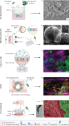
Despite the need for effective treatments against chronic respiratory infections (often caused by pathogenic biofilms), only a few new antimicrobials have been introduced to the market in recent decades. Although different factors impede the successful advancement of antimicrobial candidates from the bench to the clinic, a major driver is the use of poorly predictive model systems in preclinical research. To bridge this translational gap, significant efforts have been made to develop physiologically relevant models capable of recapitulating the key aspects of the airway microenvironment that are known to influence infection dynamics and antimicrobial activity in vivo In this review, we provide an overview of state-of-the-art cell culture platforms and ex vivo models that have been used to model chronic (biofilm-associated) airway infections, including air-liquid interfaces, three-dimensional cultures obtained with rotating-wall vessel bioreactors, lung-on-a-chips and ex vivo pig lungs. Our focus is on highlighting the advantages of these infection models over standard (abiotic) biofilm methods by describing studies that have benefited from these platforms to investigate chronic bacterial infections and explore novel antibiofilm strategies. Furthermore, we discuss the challenges that still need to be overcome to ensure the widespread application of in vivo-like infection models in antimicrobial drug development, suggesting possible directions for future research. Bearing in mind that no single model is able to faithfully capture the full complexity of the (infected) airways, we emphasise the importance of informed model selection in order to generate clinically relevant experimental data.
Copyright ©The authors 2024.
Conflict of interest statement
Conflict of interest: The authors declare no competing interests.
Figures

Similar articles
-
Bacteria in the respiratory tract-how to treat? Or do not treat?Int J Infect Dis. 2016 Oct;51:113-122. doi: 10.1016/j.ijid.2016.09.005. Int J Infect Dis. 2016. PMID: 27776777 Review.
-
Sweetening Inhaled Antibiotic Treatment for Eradication of Chronic Respiratory Biofilm Infection.Pharm Res. 2018 Feb 7;35(3):50. doi: 10.1007/s11095-018-2350-4. Pharm Res. 2018. PMID: 29417313
-
Antibiofilm Activities of Borneol-Citral-Loaded Pickering Emulsions against Pseudomonas aeruginosa and Staphylococcus aureus in Physiologically Relevant Chronic Infection Models.Microbiol Spectr. 2022 Oct 26;10(5):e0169622. doi: 10.1128/spectrum.01696-22. Epub 2022 Oct 4. Microbiol Spectr. 2022. PMID: 36194139 Free PMC article.
-
Insights into host-pathogen interactions from state-of-the-art animal models of respiratory Pseudomonas aeruginosa infections.FEBS Lett. 2016 Nov;590(21):3941-3959. doi: 10.1002/1873-3468.12454. Epub 2016 Nov 1. FEBS Lett. 2016. PMID: 27730639 Review.
-
Staphylococcus aureus Biofilm Growth on Cystic Fibrosis Airway Epithelial Cells Is Enhanced during Respiratory Syncytial Virus Coinfection.mSphere. 2018 Aug 15;3(4):e00341-18. doi: 10.1128/mSphere.00341-18. mSphere. 2018. PMID: 30111629 Free PMC article.
Cited by
-
Aggregatibacter is inversely associated with inflammatory mediators in sputa of patients with chronic airway diseases and reduces inflammation in vitro.Respir Res. 2024 Oct 12;25(1):368. doi: 10.1186/s12931-024-02983-z. Respir Res. 2024. PMID: 39395980 Free PMC article.
-
Corticosteroids modulate biofilm formation and virulence of Pseudomonas aeruginosa.Biofilm. 2025 May 29;9:100289. doi: 10.1016/j.bioflm.2025.100289. eCollection 2025 Jun. Biofilm. 2025. PMID: 40539031 Free PMC article.
References
Publication types
MeSH terms
Substances
LinkOut - more resources
Full Text Sources
Medical
Miscellaneous
