. 2024 Aug 14;15(1):7000.
doi: 10.1038/s41467-024-51318-5.

Lamin A/C deficiency-mediated ROS elevation contributes to pathogenic phenotypes of dilated cardiomyopathy in iPSC model

Affiliations

Lamin A/C deficiency-mediated ROS elevation contributes to pathogenic phenotypes of dilated cardiomyopathy in iPSC model

Hangyuan Qiu et al. Nat Commun. .

Abstract

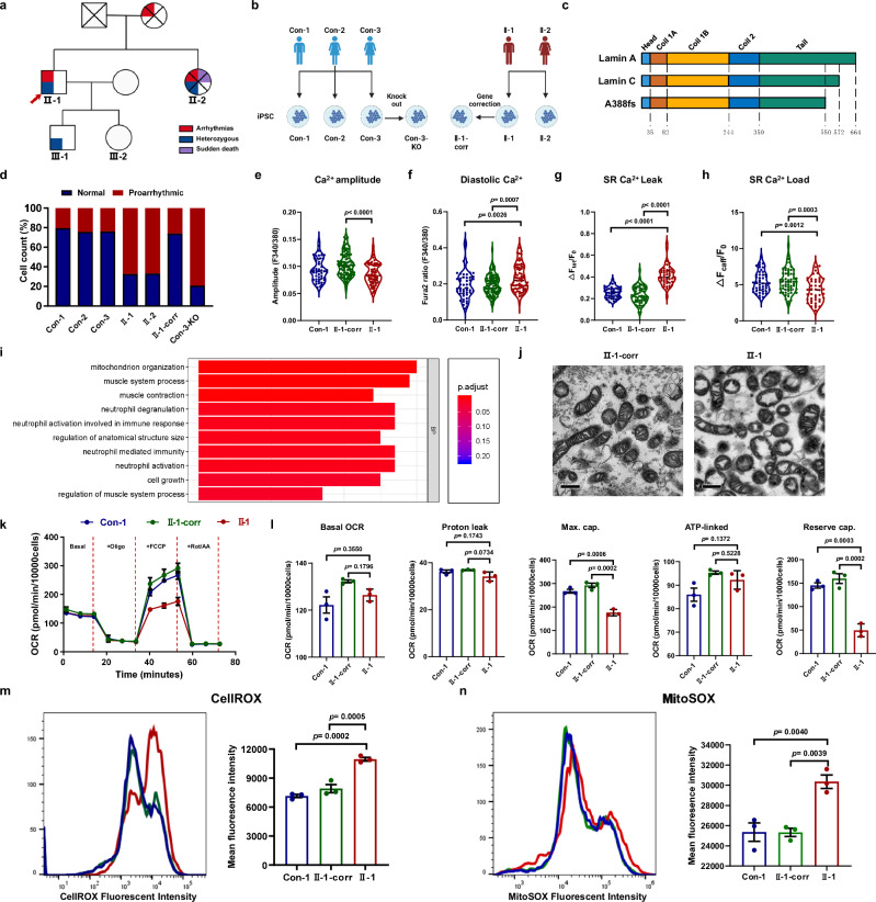
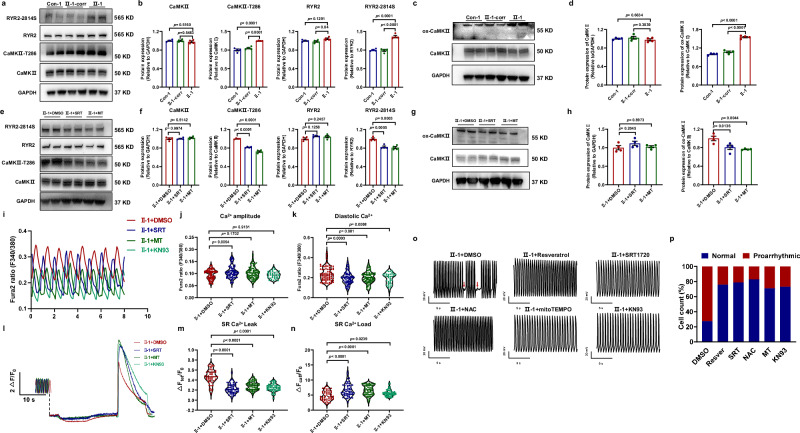
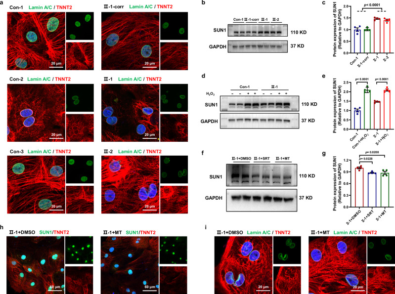
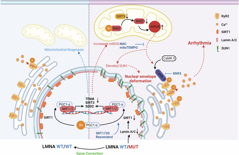
Mutations in the nuclear envelope (NE) protein lamin A/C (encoded by LMNA), cause a severe form of dilated cardiomyopathy (DCM) with early-onset life-threatening arrhythmias. However, molecular mechanisms underlying increased arrhythmogenesis in LMNA-related DCM (LMNA-DCM) remain largely unknown. Here we show that a frameshift mutation in LMNA causes abnormal Ca2+ handling, arrhythmias and disformed NE in LMNA-DCM patient-specific iPSC-derived cardiomyocytes (iPSC-CMs). Mechanistically, lamin A interacts with sirtuin 1 (SIRT1) where mutant lamin A/C accelerates degradation of SIRT1, leading to mitochondrial dysfunction and oxidative stress. Elevated reactive oxygen species (ROS) then activates the Ca2+/calmodulin-dependent protein kinase II (CaMKII)-ryanodine receptor 2 (RYR2) pathway and aggravates the accumulation of SUN1 in mutant iPSC-CMs, contributing to arrhythmias and NE deformation, respectively. Taken together, the lamin A/C deficiency-mediated ROS disorder is revealed as central to LMNA-DCM development. Manipulation of impaired SIRT1 activity and excessive oxidative stress is a potential future therapeutic strategy for LMNA-DCM.


Conflict of interest statement

The authors declare no competing interests.

Figures

Fig. 1
Fig. 1. Lamin A/C haploinsufficiency causes arrhythmias, abnormal Ca2+ handling, mitochondrial dysfunction, and ROS elevation.
a Pedigree of the family carrying a novel heterozygous mutation in LMNA. Individuals who were dead are marked with a fork. The proband is indicated by the red arrow. b Schematic diagram of the iPSC lines used in the study. c. Predicted effect of the LMNA A388fs mutation on the two splicing products lamin A and C. d Bar graph to compare the occurrence of arrhythmias among control (Con−1, Con-2, Con-3) iPSC-CMs, A388fs mutant (II-1, II-2) iPSC-CMs, gene-corrected (GC) (II−1-corr) iPSC-CMs, and LMNA KO (Con-3-KO) iPSC-CMs. e, f Violin graphs to compare the Ca2+ transient amplitude and diastolic [Ca2+]i among Con−1, II-1-corr, and II−1 iPSC-CMs. n = 49 (Con−1), 71 (II-1), 94 (II-1-corr) cells. g, h Violin graphs to compare the RYR2-mediated SR Ca2+ leak and SR Ca2+ load among Con-1, II-1-corr, and II-1 iPSC-CMs. n = 44 (II-1), 46 (Con-1), 60 (II-1-corr) cells. i The GO analysis identified a significant change in mitochondrial organization. j Representative graphs of mitochondria in II-1-corr and II-1 iPSC-CMs by transmission electron microscopy. Scale bar, 0.5 μm. k Analysis of oxygen consumption in Con-1, II-1-corr, and II-1 iPSC-CMs. n = 3 independently biological repeats. l Bar graphs to compare key parameters of OCR among Con-1, II-1-corr, and II-1 iPSC-CMs, including basal OCR, proton leak OCR, maximal capacity (max. cap.), ATP-linked OCR, and reserved capacity (reserved cap.). n = 3 independently biological repeats. m, n Cellular and Mitochondrial ROS levels in Con-1, II-1-corr, and II-1 iPSC-CMs measured by flow cytometry of cells stained with CellROX or MitoSOX. n = 3 independently biological repeats. Data are presented as mean ± SEM in kn. p values were calculated by Dunnett’s multiple comparisons test in eh, ln and two-sided unpaired t-test in i. Figure 1b created with BioRender.com was released under a Creative Commons Attribution-NonCommercial-NoDerivs 4.0 International license. Source data are provided as a Source Data file.
Fig. 2
Fig. 2. Accelerated SIRT1 degradation contributes to the mitochondrial dysfunction and excessive ROS level in A388fs iPSC-CMs.
a. Representative graphs of staining of SIRT1 (red) and lamin A/C (green) in Con-1 and mutant (II-1, II-2) iPSC-CMs. DAPI indicates nuclear staining (blue). Scale bar, 60 μm. b Bar graph to compare the SIRT1 intensity among different groups in a. n = 21 (Con-1), 38 (II-1), 41 (II-2) cells. c Western blot analysis revealed that lamin A was detected in anti-SIRT1 immunoprecipitates, and SIRT1 was detected in anti-lamin A/C immunoprecipitates. n = 4 independently biological repeats. d, e Western blot analysis of protein expression of SIRT1 in control (Con-1, Con-2, Con-3), II-1-corr, and mutant (II-1, II-2) iPSC-CMs. n = 3 independently biological repeats. f Bar graph to compare the mRNA expression of SIRT1 between II-1-corr and II-1 iPSC-CMs. n = 5 independently biological repeats. g Western blot analysis of SIRT1 expression in Con-3, Con-3-KO, II−1-corr, and II-1 iPSC-CMs after CHX treatment. h Line graph to compare the degradation rate of SIRT1 expression among different groups in g. n = 3 (Con-3, II-1-corr, II-1), 4 (Con-3-KO) independently biological repeats. i Bar graphs to compare the mRNA expression of TFAM, SIRT3, and SOD2 between II-1-corr and II-1 iPSC-CMs. n = 3 (TFAM-Con-3), 4 (others), and 8 (TFAM-II-1) independently biological repeats. j Analysis of oxygen consumption in II-1-corr and II-1 iPSC-CMs with or without treatment of SRT1720 (SRT). n = 3 independently biological repeats. k Bar graphs to compare key parameters of OCR among different groups in j. n = 3 independently biological repeats. l Mitochondrial ROS levels in Con-1, II-1-corr, and II-1 iPSC-CMs treated with DMSO, and II-1 iPSC-CMs treated with SRT. n = 3 independently biological repeats. m Cellular ROS levels in Con-1, II-1-corr, and II-1 iPSC-CMs treated with DMSO, II-1 iPSC-CMs treated with MT or SRT. n = 3 independently biological repeats. Data are presented as mean ± SEM in b, e, f, hm. p values were calculated by Dunnett’s multiple comparisons test in b, i, l, m, Nested t-test in e, and two-sided unpaired t-test in f, h, k. Source data are provided as a Source Data file.
Fig. 3
Fig. 3. Activated ROS-CAMKII-RYR2 pathway links down-regulated SIRT1 and arrhythmic phenotype in A388fs iPSC-CMs.
a, b Western blot analysis of total CaMKII, p-CaMKII (CaMKII-T286), total RYR2, and p-RYR2 (RYR2−2814S) expression in Con−1, II-1-corr, and II-1 iPSC-CMs. n = 4 independently biological repeats. c, d Western blot analysis of protein expression of total CaMKII and oxidized CaMKII (M281-M282 oxidation; ox-CaMKII) in Con-1, II-1-corr, and II-1 iPSC-CMs. n = 4 independently biological repeats. e, f Western blot analysis of total CaMKII, p-CaMKII, total RYR2, and p-RYR2 expression in II-1 iPSC-CMs treated with DMSO, SRT, or MT, respectively. n = 4 independently biological repeats. g, h Western blot analysis of protein expression of total and oxidized CaMKII in II-1 iPSC-CMs treated with DMSO, SRT or mitoTEMPO (MT). n = 4 independently biological repeats. i Representative Ca2+ transient traces recorded from II-1 iPSC-CMs treated with DMSO, SRT, MT or KN93, respectively. j, k Violin graphs to compare the Ca2+ transient amplitude and diastolic [Ca2+]i among different groups in i. n = 37 (II-1 + KN93), 74 (II-1 + SRT), 76 (II-1 + MT), 80 (II-1 + DMSO) cells. l Representative traces of cytosolic Ca2+ fluorescence in II-1 iPSC-CMs treated with DMSO, SRT, MT, or KN93 in NT solution and exposed to 0 Na+, 0 Ca2+ solution containing tetracaine (Tet) and caffeine (Caff). mn Violin graphs to compare the RYR2-mediated SR Ca2+ leak and SR Ca2+ load among different groups in l. n = 49 (II-1 + DMSO), 54 (II-1 + KN93), 55 (II-1 + SRT), and 58 (II-1 + MT) cells. o Representative electrophysiological measurements of spontaneous action potentials in II-1 iPSC-CMs treated with DMSO, resveratrol (resver), SRT, NAC, MT, or KN93, respectively. p Bar graph to compare the occurrence of arrhythmias among different groups in o. Data are presented as mean ± SEM in b, d, f, h. p values were calculated by Dunnett’s multiple comparisons test in b, d, f, h, j, k, m, n. Source data are provided as a Source Data file.
Fig. 4
Fig. 4. ROS-mediated protein accumulation of SUN1 confers the abnormal NE structure in A388fs iPSC-CMs.
a. Representative graphs of staining of lamin A/C (green) and cardiac-specific marker TNNT2 (red) in control (Con-1, Con-2, and Con-3), II-1-corr, and mutant (II−1, II-2) iPSC-CMs. DAPI indicates nuclear staining (blue). Scale bar, 20 μm. The experiment was repeated four times with similar results. b, c Western blot analysis of SUN1 expression in Con−1, II-1-corr, and mutant (II−1, II-2) iPSC-CMs. n = 4 independently biological repeats. d, e Western blot analysis of SUN1 expression in Con−1 and II-1 iPSC-CMs with or without the treatment of H2O2. n = 4 independently biological repeats. f, g Western blot analysis of SUN1 expression in II−1 iPSC-CMs treated with DMSO, SRT or MT. n = 4 independently biological repeats. h Representative graphs of staining of SUN1 (green) and cardiac-specific marker TNNT2 (red) in II−1 iPSC-CMs treated with DMSO or MT. DAPI indicates nuclear staining (blue). Scale bar, 60 μm. The experiment was repeated three times with similar results. i Representative graphs of staining of lamin A/C (green) and cardiac-specific marker TNNT2 (red) in II−1 iPSC-CMs treated with DMSO or MT. DAPI indicates nuclear staining (blue). Scale bar, 20 μm. The experiment was repeated three times with similar results. Data are presented as mean ± SEM in c, e, g. p values were calculated by Nested t-test in c, two-sided unpaired t-test in e, and Dunnett’s multiple comparisons test in g. Source data are provided as a Source Data file.
Fig. 5
Fig. 5. Genetic inhibition of SIRT1 in isogenic control iPSC-CMs activates ROS-CAMKII-RYR2 pathway and recapitulates pathogenic phenotypes of A388fs iPSC-CMs.
a, b Western blot analysis of SIRT1, total CaMKII, ox-CaMKII, p-CaMKII, total RYR2, p-RYR2, and SUN1 expression in II-1-corr iPSC-CMs treated with scrambled siRNA (NC), SIRT1 siRNA (KD), or SIRT1 siRNA and SRT (KD + SRT), respectively. n = 4 independently biological repeats. c Cellular ROS levels in II−1-corr iPSC-CMs treated with scrambled siRNA, SIRT1 siRNA, or SIRT1 siRNA and SRT, respectively. n = 3 independently biological repeats. d Representative Ca2+ transient traces recorded from II−1-corr iPSC-CMs treated with scrambled siRNA, SIRT1 siRNA, or SIRT1 siRNA and SRT, respectively. e, f Bar graphs to compare the Ca2+ transient amplitude and diastolic [Ca2+]i among different groups in d. n = 19 (KD), 21 (NC, KD + SRT) cells. g Representative traces of cytosolic Ca2+ fluorescence in II−1-corr iPSC-CMs treated with scrambled siRNA, SIRT1 siRNA, or SIRT1 siRNA and SRT, respectively in NT solution and exposed to 0 Na+, 0 Ca2+ solution containing Tet and Caff. h, i Bar graphs to compare the RYR2-mediated SR Ca2+ leak and SR Ca2+ load among different groups in g. n = 24 (KD), 26 (KD + SRT), 30 (NC) cells. j Representative electrophysiological measurements of spontaneous action potentials in II-1-corr iPSC-CMs treated with scrambled siRNA, SIRT1 siRNA, or SIRT1 siRNA and SRT, respectively. k Bar graph to compare the occurrence of arrhythmias among different groups in j. l Representative graphs of staining of lamin A/C (green) and cardiac-specific marker TNNT2 (red) in II−1-corr iPSC-CMs treated with scrambled siRNA, SIRT1 siRNA, or SIRT1 siRNA and SRT, respectively. DAPI indicates nuclear staining (blue). Scale bar, 20 μm. m Bar graph to compare the occurrence of abnormal NE structure among different groups in l. n = 6 views from 3 independent experiments. Data are presented as mean ± SEM in b, c, e, f, h, i, m. p values were calculated by Dunnett’s multiple comparisons test in b, c, e, f, h, i, m. Source data are provided as a Source Data file.
Fig. 6
Fig. 6. Overexpression of SIRT1 rescues the pathogenic phenotypes in A388fs iPSC-CMs through suppressing the ROS-CAMKII-RYR2 pathway.
a, b Western blot analysis of SIRT1, total CaMKII, ox-CaMKII, CaMKII-T286, total RYR2, p-RYR2, and SUN1 expression in II−1 iPSC-CMs overexpressed vector only (negative control, NC), or II-1 iPSC-CMs with SIRT1 overexpression (SIRT1 OE). n = 3 (total CaMKII, ox-CaMKII), 6 (SIRT1, CaMKII-T286, total RYR2, p-RYR2, SUN1) independently biological repeats. c Cellular ROS levels in NC and SIRT1 OE. n = 3 independently biological repeats. d Representative Ca2+ transient traces recorded from NC and SIRT1 OE. e Bar graphs to compare the Ca2+ transient amplitude and diastolic [Ca2+]i between the two groups in d. n = 20 (II−1-OE), 22 (II−1-NC) cells. f Representative traces of cytosolic Ca2+ fluorescence in NC and SIRT1 OE in NT solution and exposed to 0 Na+, 0 Ca2+ solution containing Tet and Caff. g Bar graphs to compare the RYR2-mediated SR Ca2+ leak and SR Ca2+ load between the two groups in f. n = 25 (II-1-OE), 28(II−1-NC) cells. h Representative electrophysiological measurements of spontaneous action potentials in NC and SIRT1 OE. i Bar graph to compare the occurrence of arrhythmias between the two groups in h. j Representative graphs of staining of lamin A/C (green) and cardiac-specific marker TNNT2 (red) in NC and SIRT1 OE. DAPI indicates nuclear staining (blue). Scale bar, 20 μm. k Bar graph to compare the occurrence of abnormal NE structure between the two groups in j. n = 6 views from 3 independent experiments. Data are presented as mean ± SEM in b, c, e, g, k. p values were calculated by two-sided unpaired t-test in b, c, e, g, k. Source data are provided as a Source Data file.
Fig. 7
Fig. 7. Proposed work model.
The novel mutation A388fs in the LMNA gene leads to lamin A/C haploinsufficiency. This causes abnormal Ca2+ handling, arrhythmias, and a disformed nuclear envelope (NE) in mutant iPSC-CMs. The reduced expression of lamin A/C in A388fs iPSC-CMs leads to accelerated degradation of the lamin A-binding protein sirtuin 1 (SIRT1), resulting in mitochondrial dysfunction and oxidative stress. On one hand, elevated ROS activates Ca2+/calmodulin-dependent protein kinase II (CaMKII). This then promotes ryanodine receptor 2 (RYR2)-mediated sarcoplasmic reticulum (SR) Ca2+ leak and subsequently causes elevated diastolic [Ca2+]i, abnormal Ca2+ handling, and arrhythmias in A388fs iPSC-CMs. Rescue of these phenotypes can be achieved via the application of the CaMKII inhibitor KN93. On the other hand, excessive ROS production accelerates the accumulation of the nuclear protein SUN1, which then contributes to NE deformation in A388fs iPSC-CMs. In this case, both arrhythmias and nuclear defects can be rescued via the application of SIRT1 activators (either SRT1720 or resveratrol) or ROS scavengers (either NAC or mitoTEMPO). Figure 7 created with BioRender.com was released under a Creative Commons Attribution-NonCommercial-NoDerivs 4.0 International license.

References

    1. Schultheiss, H.-P. et al. Dilated cardiomyopathy. Nat. Rev. Dis. Prim.5, 32 (2019). 10.1038/s41572-019-0084-1 - DOI - PMC - PubMed
    1. Richardson, P. Report of the 1995 World Health Organization/International Society and Federation of Cardiology Task Force on the definition and classification of cardiomyopathies. Circulation93, 841–842 (1996). 10.1161/01.CIR.93.5.841 - DOI - PubMed
    1. Hershberger, R. E., Hedges, D. J. & Morales, A. Dilated cardiomyopathy: the complexity of a diverse genetic architecture. Nat. Rev. Cardiol.10, 531–547 (2013). 10.1038/nrcardio.2013.105 - DOI - PubMed
    1. Rosenbaum, A. N., Agre, K. E. & Pereira, N. L. Genetics of dilated cardiomyopathy: practical implications for heart failure management. Nat. Rev. Cardiol.17, 286–297 (2020). 10.1038/s41569-019-0284-0 - DOI - PubMed
    1. Capell, B. C. & Collins, F. S. Human laminopathies: nuclei gone genetically awry. Nat. Rev. Genet7, 940–952 (2006). 10.1038/nrg1906 - DOI - PubMed

MeSH terms

LinkOut - more resources