. 2024 Aug 14;14(1):18874.
doi: 10.1038/s41598-024-69864-9.

Effect of magnification error and axial length on circumpapillary capillary density and retinal nerve fiber layer thickness

Affiliations

Effect of magnification error and axial length on circumpapillary capillary density and retinal nerve fiber layer thickness

Kaho Akiyama et al. Sci Rep. .

Abstract

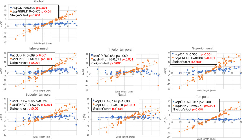
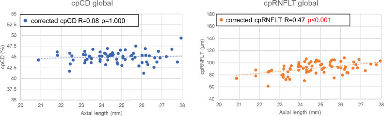
This study aimed to evaluate the effect of magnification error and axial length (AL) on circumpapillary capillary density (cpCD) and circumpapillary retinal nerve fiber layer thickness (cpRNFLT) in healthy eyes. Seventy-two healthy eyes of 72 subjects with AL 24.7 ± 1.5 mm (range: 20.9-28.0 mm) were enrolled in this retrospective cross-sectional study and underwent optical coherence tomography angiography scanning. Magnification corrected measurement areas were obtained using AL upon which corrected cpCD, cpRNFLT values were determined. Relationships between AL and the percentage difference between corrected and uncorrected values (ΔcpCD, ΔcpRNFLT) as well as the effect of AL on magnification corrected cpCD, cpRNFLT were evaluated. ΔcpCD significantly increased with AL in the global, inferior nasal and superior nasal sectors (all p < 0.001). ΔcpRNFLT significantly increased with AL in global and all sectors (all p < 0.001) and the correlations were significantly stronger than that of ΔcpCD-AL in all sectors (all p < 0.001). Corrected cpCD did not associate with AL while corrected cpRNFLT demonstrated a significant positive association with AL in the global (p = 0.005) and temporal sector (p < 0.001). Magnification error led to a significant underestimation of cpCD in eyes with longer AL although its underestimation and the effect of AL was smaller in comparison to that of cpRNFLT.

Keywords: High myopia; Magnification correction; Optical coherence tomography angiography; Superficial retinal vessel density.


Conflict of interest statement

Dr Iwase has received financial support (research instruments) by Carl Zeiss, Meditec. Dr Saito and Dr Aihara received honorarium by Carl Zeiss, Meditec. Others declare no potential conflict of interests.

Figures

Figure 1
Figure 1
Scatter plots of global and Garway-Heath sectoral Δcircumpapillary capillary density (cpCD), Δcircumpapillary retinal nerve fiber layer thickness (cpRNFLT) and axial length (AL). R and p values are the results of Pearson’s correlation. ΔcpRNFLT demonstrated significantly stronger correlation with AL in comparison with ΔcpCD in global and all sectors (all p < 0.001).
Figure 2
Figure 2
Scatter plots of uncorrected and corrected global circumpapillary vessel density (cpCD), circumpapillary retinal nerve fiber layer thickness (cpRNFLT) and axial length (AL). R and p values are the results of Pearson’s correlation.
Figure 3
Figure 3
Illustration of magnification uncorrected and corrected measurement area of circumpapillary capillary density (cpCD) and measurement circle of circumpapillary retinal nerve fiber layer thickness (cpRNFLT) in an eye with 27.76 mm axial length. Global and sectoral cpCD and cpRNFLT values are presented. IN: Inferior nasal; IT: Inferior temporal; SN: Superior nasal; ST: Superior temporal; N: nasal; T: temporal. (a) The red area represents the magnification uncorrected measurement area for cpCD. Global and sectoral uncorrected cpCD are shown in the red square. (b) The yellow area represents the magnification corrected measurement area for cpCD. Global and sectoral corrected cpCD are shown in the yellow square. (c) The red circle represents the magnification uncorrected measurement circle for cpRNFLT. Global and sectoral uncorrected cpRNFLT are shown in the red square. (d) The yellow circle represents the magnification corrected measurement circle for cpRNFLT. Global and sectoral corrected cpRNFLT are shown in the yellow square.

References

    1. Kang, S. H., Hong, S. W., Im, S. K., Lee, S. H. & Ahn, M. D. Effect of myopia on the thickness of the retinal nerve fiber layer measured by Cirrus HD optical coherence tomography. Invest. Ophthalmol. Vis. Sci.51, 4075–4083 (2010). 10.1167/iovs.09-4737 - DOI - PubMed
    1. Ueda, K. et al. Effects of axial length and age on circumpapillary retinal nerve fiber layer and inner macular parameters measured by 3 types of SD-OCT instruments. J. Glaucoma25, 383–389 (2016). 10.1097/IJG.0000000000000216 - DOI - PubMed
    1. Garway-Heath, D. F. et al. Measurement of optic disc size: Equivalence of methods to correct for ocular magnification. Br. J. Ophthalmol.82, 643–649 (1998). 10.1136/bjo.82.6.643 - DOI - PMC - PubMed
    1. Hirasawa, H. et al. Peripapillary retinal nerve fiber layer thickness determined by spectral-domain optical coherence tomography in ophthalmologically normal eyes. Arch. Ophthalmol.128, 1420–1426 (2010). 10.1001/archophthalmol.2010.244 - DOI - PubMed
    1. Budenz, D. L. et al. Determinants of normal retinal nerve fiber layer thickness measured by Stratus OCT. Ophthalmology.114, 1046–1052 (2007). 10.1016/j.ophtha.2006.08.046 - DOI - PMC - PubMed

LinkOut - more resources