Observational Study
. 2024 Nov;27(4):797-808.
doi: 10.1007/s10456-024-09939-3. Epub 2024 Aug 14.

Maternal serum PlGF associates with 3D power doppler ultrasound markers of utero-placental vascular development in the first trimester: the rotterdam periconception cohort

Affiliations
Observational Study

Maternal serum PlGF associates with 3D power doppler ultrasound markers of utero-placental vascular development in the first trimester: the rotterdam periconception cohort

Eline S de Vos et al. Angiogenesis. 2024 Nov.

Abstract

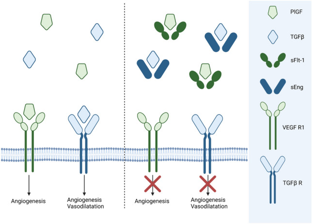
Objective (s): Circulating angiogenic factors are used for prediction of placenta-related complications, but their associations with first-trimester placental development is unknown. This study investigates associations between maternal angiogenic factors and utero-placental vascular volume (uPVV) and utero-placental vascular skeleton (uPVS) as novel imaging markers of volumetric and morphologic (branching) development of the first-trimester utero-placental vasculature.

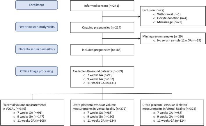
Methods: In 185 ongoing pregnancies from the VIRTUAL Placenta study, a subcohort of the ongoing prospective Rotterdam Periconception cohort, three-dimensional power Doppler ultrasounds of the placenta were obtained at 7-9-11 weeks gestational age (GA). The uPVV was measured as a parameter of volumetric development and reported the vascular quantity in cm3. The uPVS was generated as a parameter of morphologic (branching) development and reported the number of end-, bifurcation- crossing- or vessel points and total vascular length. At 11 weeks GA, maternal serum biomarkers suggested to reflect placental (vascular) development were assessed: placental growth factor (PlGF), soluble fms-like tyrosine kinase-1 (sFlt-1) and soluble endoglin (sEng). sFlt-1/PlGF and sEng/PlGF ratios were calculated. Multivariable linear regression with adjustments was used to estimate associations between serum biomarkers and uPVV and uPVS trajectories.

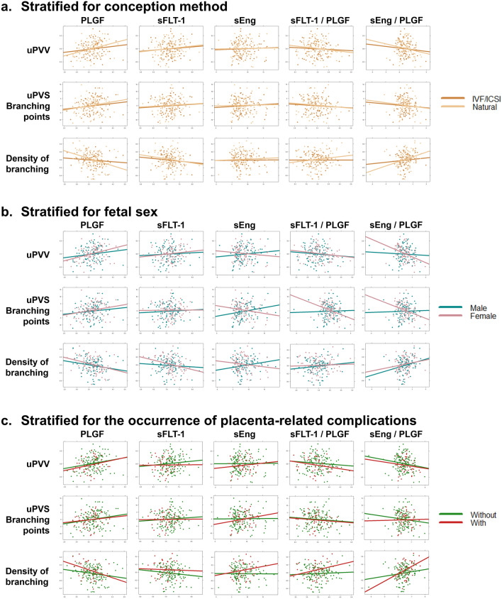
Results: Serum PlGF was positively associated with uPVV and uPVS development (uPVV: β = 0.39, 95% CI = 0.15;0.64; bifurcation points: β = 4.64, 95% CI = 0.04;9.25; crossing points: β = 4.01, 95% CI = 0.65;7.37; total vascular length: β = 13.33, 95% CI = 3.09;23.58, all p-values < 0.05). sEng/PlGF ratio was negatively associated with uPVV and uPVS development. We observed no associations between sFlt-1, sEng or sFlt-1/PlGF ratio and uPVV and uPVS development.

Conclusion(s): Higher first-trimester maternal serum PlGF concentration is associated with increased first-trimester utero-placental vascular development as reflected by uPVV and uPVS. Clinical trial registration number Dutch Trial Register NTR6854.

Keywords: Angiogenesis; Angiogenic factors; Placenta; Preeclampsia; Spiral artery remodeling; sFlt-1.


Conflict of interest statement

Declarations Competing interests The authors declare no competing interests. Ethical approval The authors report no conflicts of interest. This research was conducted in accordance with the ethical principles for medical research set out in the Declaration of Helsinki and was approved by the Institutional Review Board of the Erasmus Medical Centre on 2 June 2015 (MEC 2015–494).This study was registered at the Dutch Trial Register: NTR6854.

Figures

Fig. 1
Fig. 1
Pro-and anti-angiogenic factors that impact maternal vascular adaptation to pregnancy. PlGF stimulates angiogenesis in the utero-placental and feto-placental circulations by binding to the VEGF R1. TGFβ stimulates vasodilatation and angiogenesis via binding to the TGFβ R. sFlt-1, a soluble variant of VEGFR-1, binds to circulating PlGF thereby suppressing PlGF bioavailability and consequently inhibiting its proangiogenic functions. Similar to sFlt-1, sEng binds and neutralizes TGFβ. PlGF Placenta Growth Factor, TGFβ transforming growth factor beta, sFlt-1 soluble fms-like tyrosine kinase-1, sEng soluble endoglin, VEGF R1 vascular endothelial growth factor receptor-1, TGFβ R transforming growth factor beta receptor. This image was created in BioRender.com
Fig. 2
Fig. 2
Overview of utero-placental (vascular) imaging markers; the placental volume (PV), utero-placental vascular volume (uPVV) and utero-placental vascular skeleton (uPVS) of a first-trimester pregnancy. A: Utero-placental vascular volume (uPVV) and slice view of a three-dimensional (3D) power Doppler (PD) ultrasound of a first trimester pregnancy (bottom right). The yellow-highlighted section in the bottom right image indicates placental tissue, which is confined by the placental-myometrial interface on the outside and the gestational sac on the inside. These anatomical boundaries are used to calculate the placental volume (PV). Using virtual reality-based segmentation, all PD signals outside the PV-segment were erased. The uPVV is calculated by summation of the thresholded 3D PD voxels (3D pixels) within the PV-segment. B: Utero-placental vascular skeleton (uPVS) and slice view of a three-dimensional (3D) power Doppler (PD) ultrasound of a first trimester pregnancy (bottom right). A skeletonization algorithm has been applied to the uPVV (from panel A), resulting in the uPVS. The skeletonization algorithm repeatedly peels off the outermost layer of voxels from the uPVV, reducing the diameter of the PD signal at each point in the vascular network until one central voxel remains, thereby creating a network-like structure representing the vascular morphology. C: A magnified portion of the uPVS and overview of the uPVS including the magnified portion (yellow-highlighted section) at the bottom right. Each voxel of the uPVS is automatically assigned a morphologic characteristic based on the number of adjacent voxels: red = endpoint (1 adjacent voxel); white = vessel point (2 adjacent voxels); green = bifurcation point (3 adjacent voxels); blue = crossing point (4 adjacent voxels). Two other characteristics are derived from the uPVS: 1. Total network length was calculated by summation of the total number of voxels in the skeleton, multiplied by 1 voxel length (mm). 2. Average vessel thickness was calculated by the average number of voxel-layers that were peeled off from the uPVV to reach the central voxel of the uPVS, multiplied by 1 voxel length (mm)
Fig. 3
Fig. 3
Flowchart of participant selection
Fig. 4
Fig. 4
Correlation plot of maternal serum biomarkers at 11 weeks GA and imaging markers of the first-trimester utero-placental vascular development stratified for conception method, fetal sex and the occurrence of placenta-related complications. Unadjusted correlation plot. a. Stratified for conception method. b. Stratified for fetal sex. c. Stratified for the occurrence of placenta-related complications. Placenta-related complications are specified as PIH or PE and/or FGR, PTB and SGA. There are no statistically significant differences between the stratified groups. uPVV utero-placental vascular volume, uPVS utero-placental vascular skeleton, PlGF placenta growth factor, sFlt-1 soluble fms-like tyrosine kinase-1, sEng soluble endoglin, PIH pregnancy induced hypertension, PE preeclampsia, FGR fetal growth restriction, SGA small-for-gestational-age, PTB preterm birth

Similar articles

References

    1. Burton GJ, Fowden AL, Thornburg KL (2016) Placental origins of chronic disease. Physiol Rev 96(4):1509–1565. 10.1152/physrev.00029.2015 - PMC - PubMed
    1. Burton GJ, Fowden AL (2015) The placenta: a multifaceted, transient organ. Philos Trans R Soc Lond B Biol Sci 370(1663):20140066. 10.1098/rstb.2014.0066 - PMC - PubMed
    1. Junaid TO et al (2014) Fetoplacental vascular alterations associated with fetal growth restriction. Placenta 35(10):808–815. 10.1016/j.placenta.2014.07.013 - PubMed
    1. Sharma D, Shastri S, Sharma P (2016) Intrauterine growth restriction: antenatal and postnatal aspects. Clin Med Insights Pediatr 10:67–83. 10.4137/CMPed.S40070 - PMC - PubMed
    1. Sato Y (2020) Endovascular trophoblast and spiral artery remodeling. Mol Cell Endocrinol 503:110699. 10.1016/j.mce.2019.110699 - PubMed

Publication types

Substances

LinkOut - more resources