

Intraoperative computerised tomography scan for percutaneous fixation of the pelvis: a retrospective case series
- PMID: 39143425
- PMCID: PMC11422416
- DOI: 10.1007/s00264-024-06265-7
Intraoperative computerised tomography scan for percutaneous fixation of the pelvis: a retrospective case series
Abstract
Purpose: Fractures and dislocations of the pelvic ring are complex injuries that when treating require meticulous attention to detail and often specialized technical skill. These injuries can be the result of high-energy trauma, particularly in younger patients, or low energy trauma more often found in the elderly. Regardless of mechanism, these injuries lie on a spectrum of severity and can be treated conservatively or surgically. Percutaneous fixation under fluoroscopic guidance is the preferred standard technique when treating these fractures. This technique can be challenging for a variety of reasons including patient characteristics, intra-operative image quality, fracture morphology, among others.
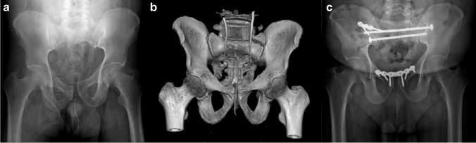
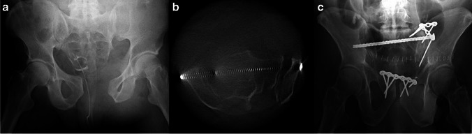
Methods: This retrospective study evaluated the use of intra-operative computed tomography (CT) using an O-arm imaging system for critical evaluation of fluoroscopic-guided screw placement in twenty-three patients. We retrospectively reviewed all cases of patients who were treated by three fellowship-trained orthopaedic traumatologists during a one-year span. Patients undergoing percutaneous pelvis fixation using both standard fluoroscopy and intraoperative CT with the Medtronic O-arm® (Minneapolis, MN) imaging system. Additionally, procedures performed included open reduction internal fixation (ORIF) of the pelvic ring, acetabulum, and associated extremity fractures.
Results: Twenty-three patients were included in this study. On average, the use of intraoperative CT added 24.4 min in operative time. Five patients (21.7%) required implant adjustment after O-arm spin. Fourteen patients underwent additional post-operative CT. No secondary revision surgeries were attempted after any post-operative CT.
Conclusions: Our study suggests that intra-operative CT scan, compared to post-operative CT scan, can be utilized to prevent take-back surgery for misplaced implants and allow for adjustment in real-time.
Keywords: CT scan; Fluoroscopy; Intra-operative; O-arm; Pelvis; Revision.
© 2024. The Author(s).
Conflict of interest statement
The authors declare they have no conflict of interest.
Figures
References
MeSH terms
LinkOut - more resources
Full Text Sources
Medical
