Observational Study
. 2024 Aug 14;25(1):308.
doi: 10.1186/s12931-024-02931-x.

The clinical impacts of lung microbiome in bronchiectasis with fixed airflow obstruction: a prospective cohort study

Affiliations
Observational Study

The clinical impacts of lung microbiome in bronchiectasis with fixed airflow obstruction: a prospective cohort study

Yen-Fu Chen et al. Respir Res. .

Abstract

Background: Airflow obstruction is a hallmark of disease severity and prognosis in bronchiectasis. The relationship between lung microbiota, airway inflammation, and outcomes in bronchiectasis with fixed airflow obstruction (FAO) remains unclear. This study explores these interactions in bronchiectasis patients, with and without FAO, and compares them to those diagnosed with chronic obstructive pulmonary disease (COPD).

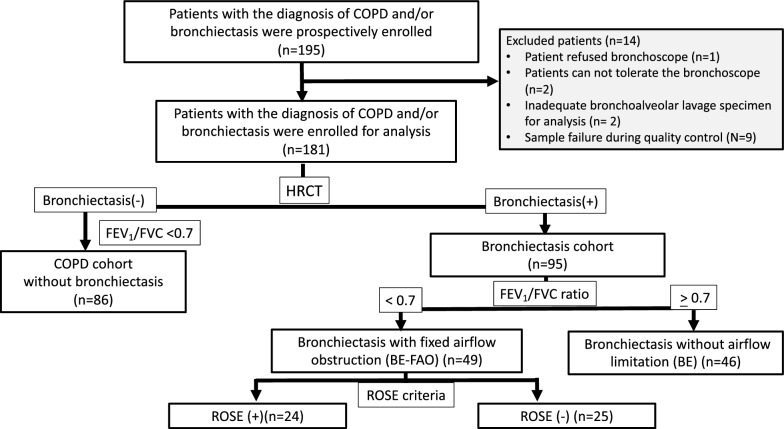
Methods: This prospective observational study in Taiwan enrolled patients with either bronchiectasis or COPD. To analyze the lung microbiome and assess inflammatory markers, bronchoalveolar lavage (BAL) samples were collected for 16S rRNA gene sequencing. The study cohort comprised 181 patients: 86 with COPD, 46 with bronchiectasis, and 49 with bronchiectasis and FAO, as confirmed by spirometry.

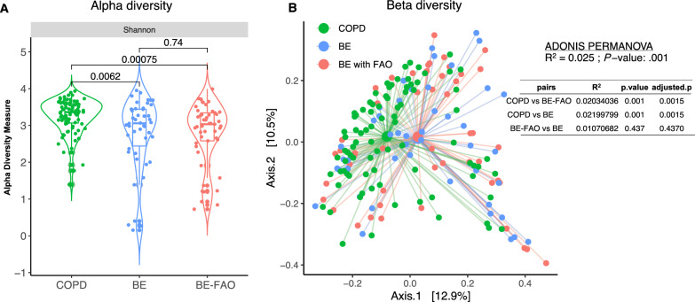
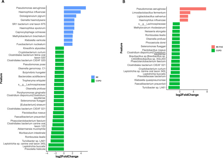
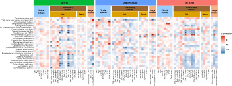
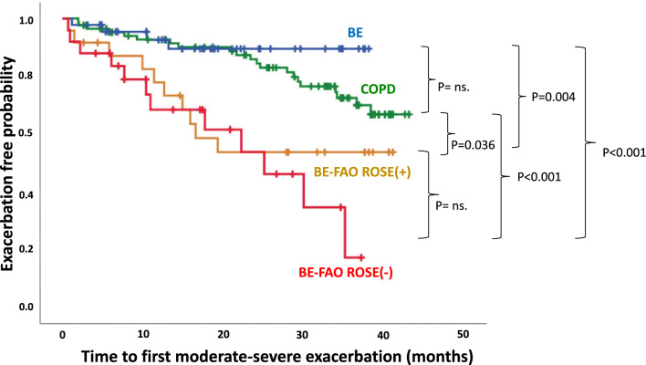
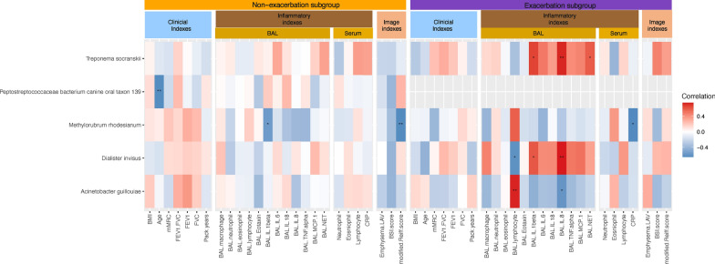
Results: Patients with bronchiectasis, with or without FAO, had similar microbiome profiles characterized by reduced alpha diversity and a predominance of Proteobacteria, distinctly different from COPD patients who exhibited more Firmicutes, greater diversity, and more commensal taxa. Furthermore, compared to COPD and bronchiectasis without FAO, bronchiectasis with FAO showed more severe disease and a higher risk of exacerbations. A significant correlation was found between the presence of Pseudomonas aeruginosa and increased airway neutrophilic inflammation such as Interleukin [IL]-1β, IL-8, and tumor necrosis factor-alpha [TNF]-α, as well as with higher bronchiectasis severity, which might contribute to an increased risk of exacerbations. Moreover, in bronchiectasis patients with FAO, the ROSE (Radiology, Obstruction, Symptoms, and Exposure) criteria were employed to classify individuals as either ROSE (+) or ROSE (-), based on smoking history. This classification highlighted differences in clinical features, inflammatory profiles, and slight microbiome variations between ROSE (-) and ROSE (+) patients, suggesting diverse endotypes within the bronchiectasis with FAO group.

Conclusion: Bronchiectasis patients with FAO may exhibit two distinct endotypes, as defined by ROSE criteria, characterized by greater disease severity and a lung microbiome more similar to bronchiectasis without FAO than to COPD. The significant correlation between Pseudomonas aeruginosa colonization and increased airway neutrophilic inflammation, as well as disease severity, underscores the clinical relevance of microbial patterns. This finding reinforces the potential role of these patterns in the progression and exacerbations of bronchiectasis with FAO.

Keywords: Bronchiectasis; Bronchoalveolar lavage; COPD; Fixed airflow obstruction; Lung microbiota; Neutrophilic inflammation; ROSE criteria.


Conflict of interest statement

The authors declare no competing interests.

Figures

Fig. 1
Fig. 1
The workflow of patients recruited in the study. BE Bronchiectasis without fixed airflow obstruction, BE-FAO Bronchiectasis with fixed airflow obstruction, COPD Chronic obstructive pulmonary disease, FEV1 forced expiratory volume in 1 s, FVC forced vital capacity, HRCT high-resolution computed tomography, ROSE Radiology, Obstruction, Symptoms, Exposure
Fig. 2
Fig. 2
Alpha diversity (A) and beta diversity (B) of BAL microbiome profiles in three groups. A Patients in BE and BE-FAO groups displayed similar Shannon diversity, which were significantly lower than those with COPD alone. B The pairwise values using Bray–Curtis distance and principal coordinates analysis (PCoA) to measure the beta diversity between COPD, BE-FAO and BE groups. BE Bronchiectasis without fixed airflow obstruction, BE-FAO Bronchiectasis with fixed airflow obstruction, COPD Chronic obstructive pulmonary disease
Fig. 3
Fig. 3
The differential abundance of lung microbiome analysis using DEseq2 in COPD, BE and BE-FAO groups (adjust gender and smoking status). The different taxonomic levels (adjusted P < 0.05 and fold change > 2.0) at species level in BE versus COPD groups (A) and in BE-FAO versus COPD groups (B). BE Bronchiectasis without fixed airflow obstruction, BE-FAO Bronchiectasis with fixed airflow obstruction, COPD Chronic obstructive pulmonary disease
Fig. 4
Fig. 4
Heatmap showing spearman correlation between clinical variables and microbiome in COPD, BE and BE-FAO groups. Clinical variables are grouped into three categories: clinical indexes, inflammatory indexes, and imaging indexes. Only those taxa that displayed at least one significant correlation (q < .01, following FDR correction) were selected. The color-coded matrix represents the Spearman correlation coefficient, with red indicating a positive correlation and blue indicating a negative correlation. FDRs are denoted: *q < 0.05; **q < 0.01; ***q < 0.001. BAL Bronchoalveolar lavage, BE Bronchiectasis without fixed airflow obstruction, BE-FAO Bronchiectasis with fixed airflow obstruction, BMI Body Mass Index, BSI Bronchiectasis severity index, CAT COPD Assessment Test, COPD Chronic obstructive pulmonary disease, CRP C-reactive protein, FDR False discovery rate, FEV1 forced expiratory volume in 1 s, FVC forced vital capacity, LAV low-attenuation volume, mMRC modified Medical Research Council, IL-1β interleukin [IL]-1β, IL-6 interleukin [IL]-6, IL-8 interleukin [IL]-8, IL-18 interleukin [IL]-18, MCP-1 Monocyte chemoattractant protein-1, NETs Neutrophil extracellular traps, TNF-α tumor necrosis factor [TNF]-α
Fig. 5
Fig. 5
Differences in airway inflammatory profiles based on BAL samples in patients with COPD, BE, BE-FAO ROSE (+), and BE-FAO ROSE (−). The bronchoalveolar lavage (BAL) samples from study subjects were applied for multiplex Immunoassays. (*P < 0.05, **P < 0.01, ***P < 0.005). BAL bronchoalveolar lavage, BE Bronchiectasis without fixed airflow obstruction, BE-FAO Bronchiectasis with fixed airflow obstruction, COPD Chronic obstructive pulmonary disease, IL-1 Interleukin [IL]-1β, IL-6 Interleukin [IL]-6, IL-18 Interleukin [IL]-18, IL-8 Interleukin [IL]-8, MCP-1 Monocyte chemoattractant protein-1, NETs neutrophil extracellular traps, ROSE Radiology, Obstruction, Symptoms, Exposure, TNF-α tumor necrosis factor [TNF]-α
Fig. 6
Fig. 6
Alpha diversity (A) and beta diversity (B) of patients based on lung microbiome profiles. A BE, BE-FAO ROSE (+), and BE-FAO ROSE (−) patients showed comparable alpha diversity levels. B Marked differences emerged in beta diversity between the BE-FAO ROSE (−) and COPD groups (adjusted P = 0.003), while the differences between BE-FAO ROSE (+) and COPD were less pronounced (adjusted P value = 0.068). BE Bronchiectasis without fixed airflow obstruction, BE-FAO Bronchiectasis with fixed airflow obstruction, COPD Chronic obstructive pulmonary disease, ROSE Radiology, Obstruction, Symptoms, Exposure
Fig. 7
Fig. 7
Time to first moderate-severe exacerbation: comparing COPD, BE, and BE-FAO (incorporating ROSE (+) and ROSE (−) subgroups). ns: not significant. BE Bronchiectasis without fixed airflow obstruction, BE-FAO Bronchiectasis with fixed airflow obstruction, COPD Chronic obstructive pulmonary disease, ROSE Radiology, Obstruction, Symptoms, Exposure
Fig. 8
Fig. 8
The correlation of clinical variables and lung microbiota in the bronchiectasis with FAO group. Heatmap showing spearman correlation between clinical variables and BAL microbiome in exacerbation subgroup and non-exacerbation subgroup. Clinical variables are grouped into three categories: clinical indexes, inflammatory indexes, and imaging indexes. Only those taxa that displayed at least one significant correlation (q < .01, following FDR correction) were selected. The color-coded matrix represents the Spearman correlation coefficient, with red indicating a positive correlation and blue indicating a negative correlation. FDRs are denoted: *q < 0.05; **q < 0.01; ***q < 0.001. The spearmans correlation revealed two oral taxa, Treponema socranskii and and Dialister invisus, in exacerbation group of BE-FAO were positively associated neutrophilic cytokines (BAL-IL 1β and BAL-IL 8). BAL Bronchoalveolar lavage, BE Bronchiectasis without fixed airflow obstruction, BE-FAO Bronchiectasis with fixed airflow obstruction, BMI Body Mass Index, BSI Bronchiectasis severity index, CAT COPD Assessment Test, COPD Chronic obstructive pulmonary disease, CRP C-reactive protein, FDR False discovery rate, FEV1 forced expiratory volume in 1 s, FVC forced vital capacity, LAV low-attenuation volume, mMRC modified Medical Research Council, IL-1β interleukin [IL]-1β, IL-6 interleukin [IL]-6, IL-8 interleukin [IL]-8, IL-18 interleukin [IL]-18, MCP-1 Monocyte chemoattractant protein-1, NETs Neutrophil extracellular traps, TNF-α tumor necrosis factor [TNF]-α

References

    1. Flume PA, Chalmers JD, Olivier KN. Advances in bronchiectasis: endotyping, genetics, microbiome, and disease heterogeneity. Lancet (London, England). 2018;392:880–90. 10.1016/S0140-6736(18)31767-7 - DOI - PMC - PubMed
    1. Polverino E, Goeminne PC, McDonnell MJ, Aliberti S, Marshall SE, et al. European Respiratory Society guidelines for the management of adult bronchiectasis. Eur Respir J. 2017;50:1700629. 10.1183/13993003.00629-2017 - DOI - PubMed
    1. Naidich DP, McCauley DI, Khouri NF, Stitik FP, Siegelman SS. Computed tomography of bronchiectasis. J Comput Assist Tomogr. 1982;6:437. 10.1097/00004728-198206000-00001 - DOI - PubMed
    1. Chalmers JD, Goeminne P, Aliberti S, McDonnell MJ, Lonni S, Davidson J, et al. The bronchiectasis severity index. An international derivation and validation study. Am J Respir Crit Care Med. 2014;189:576. 10.1164/rccm.201309-1575OC - DOI - PMC - PubMed
    1. Goeminne PC, Nawrot TS, Ruttens D, Seys S, Dupont LJ. Mortality in non-cystic fibrosis bronchiectasis: a prospective cohort analysis. Respir Med. 2014;108:287. 10.1016/j.rmed.2013.12.015 - DOI - PubMed

Publication types

LinkOut - more resources