. 2024 Aug 27;18(34):23445-23456.
doi: 10.1021/acsnano.4c06829. Epub 2024 Aug 15.

Multiplexed Nanoscopy via Buffer Exchange

Affiliations

Multiplexed Nanoscopy via Buffer Exchange

Ting-Jui Ben Chang et al. ACS Nano. .

Abstract

Understanding cellular functions, particularly in their intricate complexity, can greatly benefit from the spatial mapping of diverse molecules through multitarget single-molecule localization microscopy (SMLM). Existing methodologies, primarily restricting the encoding dimensions to color and lifetime or requiring cyclic staining, often involve broad chromatic detection, specialized optical configurations, or sophisticated labeling techniques. Here, we propose a simple approach called buffer-exchange stochastic optical reconstruction microscopy (beSTORM), which introduces an additional dimension to differentiate between single molecules irrespective of their spectral properties. This method leverages the distinguishable photoblinking responses to distinct buffer conditions, offering a straightforward yet effective means of fluorophore discrimination. Through buffer exchanges, beSTORM achieves multitarget SMLM imaging with minimal crosstalk. Direct integration with expansion microscopy (ExM) demonstrates its capability to resolve up to six proteins at the molecular level within a single emission color without chromatic aberration. Overall, beSTORM presents a highly compatible imaging platform, promising significant advancements in highly multiplexed nanoscopy for exploring multiple targets in biological systems with nanoscale precision.

Keywords: (d)STORM; SMLM; expansion microscopy (ExM); multicolor; multiplexed imaging; superresolution microscopy.


Conflict of interest statement

The authors declare no competing financial interest.

Figures

Figure 1
Figure 1
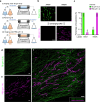
Principle of beSTORM. (a) Schematic of multitarget beSTORM imaging. fluorophores A and B are excited using a single laser at 637 nm. In the first channel, fluorophore A exhibits single-molecule blinking (ON state) while fluorophore B is in the OFF state in the buffer 1. Conversely, for the second channel, fluorophore B experiences a photoblinking process while fluorophore A is in the dark state in the buffer 2. A multitarget SMLM image is achieved by separately localizing fluorophores A and B in their respective channels. (b) Fluorescence of AF647 under different imaging conditions. AF647 showed repeated photoswitching when imaged in an imaging buffer containing thiols (TB); however, rapid photobleaching of AF647 occurred in PBS. (c) An increase in the pH value of the imaging buffer led to a significant decrease in the fluorescent intensity of HMSiR. (d) Reversibility of HMSiR fluorescence. The fluorescent states of HMSiR can be manipulated by exposing them to different buffer solutions. Scale bars, 5 μm (b, c).
Figure 2
Figure 2
beSTORM imaging with far-red dyes. (a) Schematic of the beSTORM setup and imaging procedures using a simple sample chamber with a one-way inlet and outlet. The beSTORM involves directly replacing different buffer conditions, as shown in steps (i) to (iii). (b) beSTORM using AF647 and HMSiR dyes. Two dyes specifically labeled for the outer mitochondrial membrane of RPE-1 cells were separately tested in two different buffer channels. AF647 was only detectable in the TB solution, while HMSiR was exclusively visible in PBS upon intense illumination. (c) Leakage fraction of localizations obtained from (b), indicating an extremely low level of crosstalk between the two buffer channels. (d–f) Dual-target beSTORM images showing AF647-labeled microtubules and HMSiR-labeled mitochondria in an RPE-1 cell. Individual images reveal distinct cellular organelles (d, e). Scale bars, 2 μm (b, d–f).
Figure 3
Figure 3
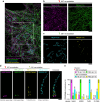
beSTORM validation for red-emitting dyes. (a–e) beSTORM utilizing red-emitting dyes (561 nm laser excitation). (a, b) Crosstalk assessment of the red-emitting dye pair, Cy3B and FLIP565. The outer mitochondrial membrane of RPE-1 cells was immunolabeled with either dye and examined separately in distinct beSTORM channels (a). The quantification of localization from (a) demonstrated minimal crosstalk between the two dyes (b). (c, d) beSTORM images acquired in different buffer channels revealing evident features of microtubules and mitochondrial membrane in a cell. (e) Composite beSTORM image from results (c, d). Scale bars, 2 μm (a, c–e).
Figure 4
Figure 4
Multiplexed beSTORM imaging. (a) Reconstruction of a four-target localization image using beSTORM with dyes spanning red to far-red emission. Dy654, HMSiR, Cy3B, and FLIP565 were used to respectively label vimentin filaments, microtubules, mitochondria, and peroxisomes in an RPE-1 cell. (b, c) Recording specific cellular molecules in each beSTORM channel demonstrating the capability for precise differentiation of the four dyes within the far-red (b) and red (c) emission spectra. (d) beSTORM revealing the exclusive ciliary compartments of a mammalian primary cilium, highlighting distinct protein localizations of the subdistal appendage (Centriolin), distal appendage (SCLT1), ciliary membrane (ARL13B), and transition zone (TMEM67). (e) Localization analyses indicating low crosstalk fractions of less than 1% across all four channels. Scale bars, 500 nm (a–d).
Figure 5
Figure 5
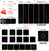
Extended multitarget molecular resolution imaging with expansion beSTORM. (a) Illustration depicting the localization-guided multitarget imaging of precharacterized cellular structures with Ex-SMLM, enabling molecular-level spatial identification of distinct proteins labeled with identical fluorophores. (b) Ex-dSTORM pinpointing colabeled SCLT1 and C2CD3, two centriolar proteins, as indicated by their radial distributions (dotted circle). (c) Seven-target Ex-beSTORM imaging of a primary cilium demonstrating four HMSiR-labeled proteins in the PBS channel and two Dy654-labeled proteins in the TB channel localized within distinct ciliary compartments: (i) Axo, axoneme; (ii) DA, distal appendage; (iii) sDA, subdistal appendage; (iv) Pro, proximal end. Additionally, the ciliary marker (Ac-Tub) was labeled with CF568. (d) Pseudocolored Ex-beSTORM results of the images from (c) revealing specific localizations of those proteins. (e) Ex-beSTORM imaging of two centriolar proteins (Ac-Tub and SCLT1, labeled with AF647) and three proteins (C2CD3, CEP90, and FBF1, labeled with HMSiR) in the TB and PBS channels, respectively. The result highlights nearly concentric 9-fold symmetric patterns with discernible radial arrangements. (f) Spatial partition for the images in (e) based on predetermined molecular distributions (dotted circles). (g) Five-target Ex-beSTORM imaging reconstruction of proteins spanning from the inner centriole wall to the outer DA, indicated by pseudocolor assignments. (h) Rotational averaging performed on signals from the centriole marker (Ac-Tub) in Ex-beSTORM, enhancing clarity in observing relative spatial relationships among proteins against the centriole. Scale bars, 500 nm (b–h).

References

    1. Lelek M.; Gyparaki M. T.; Beliu G.; Schueder F.; Griffié J.; Manley S.; Jungmann R.; Sauer M.; Lakadamyali M.; Zimmer C. Single-molecule localization microscopy. Nat. Rev. Methods Primers 2021, 1 (1), 3910.1038/s43586-021-00038-x. - DOI - PMC - PubMed
    1. Betzig E.; Patterson G. H.; Sougrat R.; Lindwasser O. W.; Olenych S.; Bonifacino J. S.; Davidson M. W.; Lippincott-Schwartz J.; Hess H. F. Imaging intracellular fluorescent proteins at nanometer resolution. Science 2006, 313 (5793), 1642–1645. 10.1126/science.1127344. - DOI - PubMed
    1. Hess S. T.; Girirajan T. P.; Mason M. D. Ultra-high resolution imaging by fluorescence photoactivation localization microscopy. Biophys. J. 2006, 91 (11), 4258–4272. 10.1529/biophysj.106.091116. - DOI - PMC - PubMed
    1. Rust M. J.; Bates M.; Zhuang X. Sub-diffraction-limit imaging by stochastic optical reconstruction microscopy (STORM). Nat. Methods 2006, 3 (10), 793–796. 10.1038/nmeth929. - DOI - PMC - PubMed
    1. Heilemann M.; Van De Linde S.; Schüttpelz M.; Kasper R.; Seefeldt B.; Mukherjee A.; Tinnefeld P.; Sauer M. Subdiffraction-resolution fluorescence imaging with conventional fluorescent probes. Angew. Chem., Int. Ed. 2008, 47 (33), 6172–6176. 10.1002/anie.200802376. - DOI - PubMed

Publication types

LinkOut - more resources