Review
. 2024 Aug 1;14(8):6108-6122.
doi: 10.21037/qims-24-477. Epub 2024 Jul 5.

Stages of benign thyroid nodules: principles and ultrasound signs

Affiliations
Review

Stages of benign thyroid nodules: principles and ultrasound signs

Andrey Valerievich Ushakov. Quant Imaging Med Surg. .

Abstract

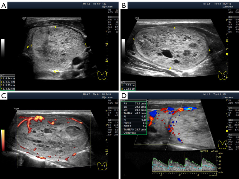
Benign thyroid nodules are significantly common and occur in 50-60% of the population. Therefore, differentiation from malignant nodes and the choice of treatment tactics in some cases of benign pathology remain relevant. Despite advances in the clinical evaluation of thyroid nodules, methodological challenges exist due to empirically simplistic understandings of the nodular process. Different opinions on the pathogenesis of thyroid nodules and the history of the formation of the idea of ​​the stages of nodules are considered. For the first time, based on natural principles and many years of ultrasound analysis of changes in benign thyroid nodules, three stages of the nodular process were identified: Development, Wasting and Scarring. The stage of exhaustion has three substages: Initial, Moderate and Significant Wasting. The principles of stage-by-stage changes in nodules are explained and their ultrasound signs are shown. The key principle of the stages of nodules is the ratio of the magnitudes of the processes of regeneration (proliferation) and destruction in the nodule. Separate stage changes may occur in node segments. In such cases, part of the segments may show signs of the Development stage, another part-Wasting, and the third part-Scarring. The different variants of thyroid nodules are explained in terms of stages. Practical recommendations for differentiating ultrasound signs of nodules associated with stages are proposed. Knowledge about the staged changes in thyroid nodules helps reduce the likelihood of diagnostic errors, better navigate the prognosis and choice of treatment tactics, and recommend preventive ultrasound examination of the thyroid.

Keywords: Thyroid nodules; nodules stage; thyroid ultrasound.


Conflict of interest statement

Conflicts of Interest: The author has completed the ICMJE uniform disclosure form (available at https://qims.amegroups.com/article/view/10.21037/qims-24-477/coif). The author has no conflicts of interest to declare.

Figures

Figure 1
Figure 1
Schematic diagram of the relationship between the severity of regeneration (proliferation) and destruction during the stages of the thyroid nodular process. The abscissa axis corresponds to time (years). The ordinate axis corresponds to the volume of the nodule and the severity of the relationship between regenerative (proliferative) and destructive changes. White columns correspond to regeneration (proliferation) of the node tissue. Black columns show the amount of node tissue destruction. The dotted line demonstrates the principle of change in the volume of the nodule during stages and substages. (A) Development stage. (B) Initial Wasting substage. (C) Moderate Wasting substage. (D) Significant Wasting substage. (E) Scarring stage.
Figure 2
Figure 2
Thyroid nodules at the Development stage. (A,B) Predominantly isoechogenicity tissue in the nodule (indicated by measurement marks). Patient, 42 years old, hypothyroidism, autoimmune thyroiditis. (C-F) Transition from the Development stage to the Initial Wasting substage. The nodule is dominated by slightly hypoechoic tissue (a sign of stromal edema) with a small amount of isoechoicity. Patient, 46 years old, euthyroidism. (C,D) In February 2020, the dimensions of the nodule are 30.1×25.6×26.1 mm. (E,F) Almost 3 years later (December 2022) the size of the nodule is 35.2×28.5×29 mm.
Figure 3
Figure 3
Thyroid nodule at the substage of Initial Wasting. The patient is 34 years old, euthyroidism. (A,B) The nodule contains isoechoic and slightly hypoechoic tissue, single hypoechoic lobules and an anechoic small area. Hyperechogenicity is determined dorsal to the nodule. (C,D) Moderate intensity of blood flow to the nodule (mainly along its perimeter).
Figure 4
Figure 4
Thyroid nodule at the substage of Initial Wasting. The patient is 39 years old, euthyroidism. Little hypoechoic tissue is visible in the nodule. Single small anechoic areas are noticeable. Hyperechogenicity is determined dorsal to the nodule. (A,B) In January 2019, the size of the nodule in the longitudinal projection is 13.8×12.1 mm. In the power Doppler mode, low active blood flow of the nodule is determined. (C,D) In January 2019, the nodule artery had a peak systolic velocity of 68 cm/s, and the superior thyroid artery had a peak systolic velocity of 29.3 cm/s. (E,F) In October 2020, the size of the nodule in the longitudinal projection is 16.2×13.3 mm. The blood flow of the nodule is poorly expressed.
Figure 5
Figure 5
Thyroid nodule at the substage of Moderate Wasting. The patient is 51 years old, hypothyroidism, autoimmune thyroiditis. During control measurements after 3 years, the size and structure of the nodule changed very little. Fine needle biopsy was performed twice. In both cases, benign signs with phenomena of destruction and proliferation were identified; Bethesda 3. (A,B) December 2020. Longitudinal and transverse projections of the left lobe of the gland in gray scale. In a nodule 26.2×20.5×15.4 mm, moderately hypoechoic tissue is visible, among which there are isolated significantly hypoechoic and isoechoic lobules. (C,D) January 2024. Nodule 26.3×19.2×14.5 mm. Longitudinal and transverse projections of the left lobe of the gland in gray scale. Moderately hypoechoic tissue is visible, among which there are isolated significantly hypoechoic and isoechoic lobules. (E,F) January 2024. Power Doppler mode. The blood flow of the nodule is inactive, mainly along the perimeter.
Figure 6
Figure 6
Thyroid nodule at the substage of Moderate Wasting. The patient is 47 years old, euthyroidism. (A,B) Isoechoic and slightly hypoechoic tissue of a large nodule is determined. It also contains a single small anechoic (fluid) area, an almost moderate number of small hypoechoic elements in the lobules, and single significantly hypoechoic medium-sized segments. (C) Power Doppler mode detects little active blood flow in the nodule. (D) The artery approaching the nodule has a peak systolic velocity of 71 cm/s.
Figure 7
Figure 7
Thyroid nodules at the substage of Significant Wasting. (A,B) The patient is 48 years old, euthyroidism. Inside the nodule, residual slightly hypoechoic tissue, hyperechoic arcuate elements, anechoic and significantly hypoechoic lobules are visible. There is hyperechogenicity dorsal to the nodule. (C,D) Patient, 65 years old, euthyroidism. A small amount of isoechoic tissue is present among the anechoic nodule. (E,F) Patient, 43 years old, euthyroidism. Microcystic thyroid nodule at the substage of Significant Wasting.
Figure 8
Figure 8
Thyroid nodules at the Scarring stage. (A,B) The patient is 70 years old, euthyroidism. In the right lobe there is a small nodule, in the tissue of which, mainly along the perimeter and partially inside, pinpoint moderately hyperechoic elements are identified. A significantly pronounced hypoechoic shadow extends dorsally from the nodule. (C,D) The patient is 55 years old, euthyroidism. There is a small nodule in the left lobe. Along the perimeter and inside the nodule, pinpoint significantly hyperechoic elements are visible, located together and separately. A slightly pronounced hypoechoic shadow extends dorsally from the nodule. (E,F) Patient, 52 years old, hypothyroidism. A small nodule with a significantly hyperechoic contour is identified in the right lobe. A significantly pronounced hypoechoic shadow extends dorsally from the nodule.

References

    1. Dean DS, Gharib H. Epidemiology of thyroid nodules. Best Pract Res Clin Endocrinol Metab 2008;22:901-11. 10.1016/j.beem.2008.09.019 - DOI - PubMed
    1. Guth S, Theune U, Aberle J, Galach A, Bamberger CM. Very high prevalence of thyroid nodules detected by high frequency (13 MHz) ultrasound examination. Eur J Clin Invest 2009;39:699-706. 10.1111/j.1365-2362.2009.02162.x - DOI - PubMed
    1. Gharib H, Papini E, Garber JR, Duick DS, Harrell RM, Hegedüs L, Paschke R, Valcavi R, Vitti P, AACE/ACE/AME Task Force on Thyroid Nodules . American association of clinical endocrinologists, American college of endocrinology, and associazione medici endocrinologi medical guidelines for clinical practice for the diagnosis and management of thyroid nodules--2016 update. Endocr Pract 2016;22:622-39. 10.4158/EP161208.GL - DOI - PubMed
    1. Grani G, Bruno R, Lucisano G, Costante G, Meringolo D, Puxeddu E, Torlontano M, Tumino S, Attard M, Lamartina L, Nicolucci A, Cooper DS, Filetti S, Durante C. Temporal Changes in Thyroid Nodule Volume: Lack of Effect on Paranodular Thyroid Tissue Volume. Thyroid 2017;27:1378-84. 10.1089/thy.2017.0201 - DOI - PubMed
    1. Brigante G, Monzani ML, Locaso M, Gnarini VL, Graziadei L, Kaleci S, De Santis MC, Tagliavini S, Simoni M, Rochira V, Madeo B. De novo Lesions Frequently Develop in Adult Normal Thyroid Over Almost Six Years. Front Endocrinol (Lausanne) 2020;11:18. 10.3389/fendo.2020.00018 - DOI - PMC - PubMed

LinkOut - more resources