. 2024 Aug 1;14(8):5420-5433.
doi: 10.21037/qims-23-1743. Epub 2024 Jul 12.

Deep learning-based detection of primary bone tumors around the knee joint on radiographs: a multicenter study

Affiliations

Deep learning-based detection of primary bone tumors around the knee joint on radiographs: a multicenter study

Danyang Xu et al. Quant Imaging Med Surg. .

Abstract

Background: Most primary bone tumors are often found in the bone around the knee joint. However, the detection of primary bone tumors on radiographs can be challenging for the inexperienced or junior radiologist. This study aimed to develop a deep learning (DL) model for the detection of primary bone tumors around the knee joint on radiographs.

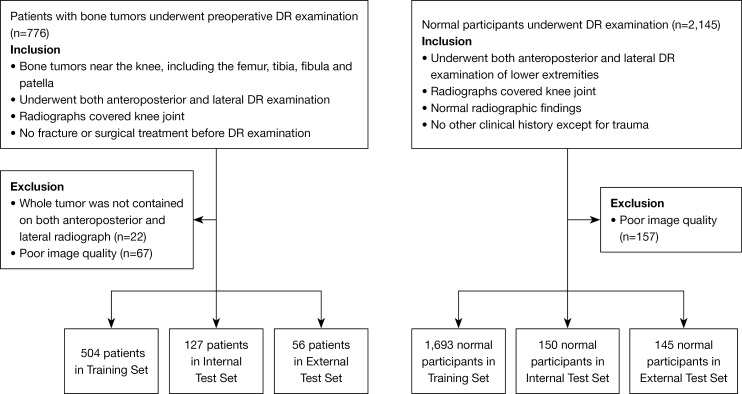
Methods: From four tertiary referral centers, we recruited 687 patients diagnosed with bone tumors (including osteosarcoma, chondrosarcoma, giant cell tumor of bone, bone cyst, enchondroma, fibrous dysplasia, etc.; 417 males, 270 females; mean age 22.8±13.2 years) by postoperative pathology or clinical imaging/follow-up, and 1,988 participants with normal bone radiographs (1,152 males, 836 females; mean age 27.9±12.2 years). The dataset was split into a training set for model development, an internal independent and an external test set for model validation. The trained model located bone tumor lesions and then detected tumor patients. Receiver operating characteristic curves and Cohen's kappa coefficient were used for evaluating detection performance. We compared the model's detection performance with that of two junior radiologists in the internal test set using permutation tests.

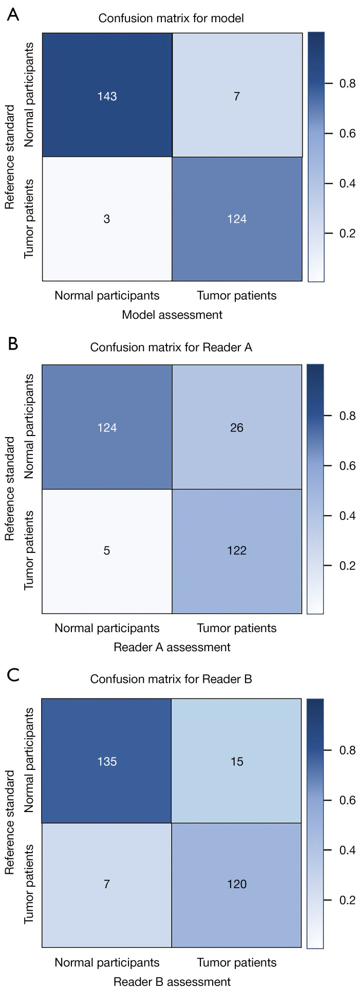
Results: The DL model correctly localized 94.5% and 92.9% bone tumors on radiographs in the internal and external test set, respectively. An accuracy of 0.964/0.920, and an area under the receiver operating characteristic curve (AUC) of 0.981/0.990 in DL detection of bone tumor patients were for the internal and external test set, respectively. Cohen's kappa coefficient of the model in the internal test set was significantly higher than that of the two junior radiologists with 4 and 3 years of experience in musculoskeletal radiology (Model vs. Reader A, 0.927 vs. 0.777, P<0.001; Model vs. Reader B, 0.927 vs. 0.841, P=0.033).

Conclusions: The DL model achieved good performance in detecting primary bone tumors around the knee joint. This model had better performance than those of junior radiologists, indicating the potential for the detection of bone tumors on radiographs.

Keywords: Bone neoplasms; deep learning (DL); knee joint; radiography.


Conflict of interest statement

Conflicts of Interest: All authors have completed the ICMJE uniform disclosure form (available at https://qims.amegroups.com/article/view/10.21037/qims-23-1743/coif). All authors report the funding from the Natural Science Foundation of Guangdong Province, China (No. 2022A1515011593) and the Medical Research Foundation of Guangdong Province, China (No. A2021010). The authors have no other conflicts of interest to declare.

Figures

Figure 1
Figure 1
Inclusion and exclusion criteria. DR, digital radiography.
Figure 2
Figure 2
Examples of the diagnostic reference standard for automatic bone tumor detection. (A,B) Anteroposterior (A) and lateral (B) radiographs of the knee of a patient with a giant cell tumor of bone. (C,D) Anteroposterior (C) and lateral (D) radiographs of the knee of a patient with osteosarcoma. Note that the reference standard was placed to fit the margin of the tumor. L, left; R, right.
Figure 3
Figure 3
ROC curve of the DL model for detecting bone tumors. ROC, receiver operating characteristic; AUC, area under the receiver operating characteristic curve; DL, deep learning.
Figure 4
Figure 4
Confusion matrices for the DL model and observer performance assessments in the internal independent test set. DL, deep learning.
Figure 5
Figure 5
Examples of anteroposterior and lateral radiographs of tibial osteosarcoma. (A) A bone tumor in the diagnostic reference standard box on the radiographs. (B) A tumor detected in the predicted box by the DL model. The values printed on top of the predicted boxes are the confidence scores for each predicted box. This bone tumor was not found with the naked eye by the two junior radiologists. L, left; R, right; DL, deep learning.
Figure 6
Figure 6
Anteroposterior (A) and lateral (B) radiographs of the right knee of a young normal participant. The area chosen with the bounding box on the lateral radiograph was misinterpreted as a tumor region by the junior radiologists and normal on the anteroposterior radiograph. Both the anteroposterior and lateral radiographs were diagnosed correctly by the DL model. R, right; DL, deep learning.
Figure 7
Figure 7
Benign tumors in the left tibia on radiographs were found by one of the two junior radiologists but missed by the other junior radiologist and were not detected by the DL model. (A) A bone tumor in the diagnostic reference standard box on the radiographs. (B) The tumor evaluated in the predicted box by an observer. L, left; DL, deep learning.
Figure 8
Figure 8
Two radiographs of normal knees diagnosed correctly by the two radiologists but misinterpreted as tumors by the DL model. The values on top of the predicted boxes are the confidence scores for the predicted box. R, right; DL, deep learning.

Similar articles

References

    1. Choi JH, Ro JY. The 2020 WHO Classification of Tumors of Bone: An Updated Review. Adv Anat Pathol 2021;28:119-38. 10.1097/PAP.0000000000000293 - DOI - PubMed
    1. Arora RS, Alston RD, Eden TO, Geraci M, Birch JM. The contrasting age-incidence patterns of bone tumours in teenagers and young adults: Implications for aetiology. Int J Cancer 2012;131:1678-85. 10.1002/ijc.27402 - DOI - PubMed
    1. Beird HC, Bielack SS, Flanagan AM, Gill J, Heymann D, Janeway KA, Livingston JA, Roberts RD, Strauss SJ, Gorlick R. Osteosarcoma. Nat Rev Dis Primers 2022;8:77. 10.1038/s41572-022-00409-y - DOI - PubMed
    1. Cole S, Gianferante DM, Zhu B, Mirabello L. Osteosarcoma: A Surveillance, Epidemiology, and End Results program-based analysis from 1975 to 2017. Cancer 2022;128:2107-18. 10.1002/cncr.34163 - DOI - PMC - PubMed
    1. He Y, Wang J, Zhang J, Yuan F, Ding X. A prospective study on predicting local recurrence of giant cell tumour of bone by evaluating preoperative imaging features of the tumour around the knee joint. Radiol Med 2017;122:546-55. 10.1007/s11547-017-0745-7 - DOI - PubMed

LinkOut - more resources