Application of the Kaiser score on contrast-enhanced mammography in the differential diagnosis of breast lesions: comparison with breast magnetic resonance imaging
- PMID: 39144044
- PMCID: PMC11320531
- DOI: 10.21037/qims-24-593
Application of the Kaiser score on contrast-enhanced mammography in the differential diagnosis of breast lesions: comparison with breast magnetic resonance imaging
Abstract
Background: The Kaiser score (KS) as a clinical decision rule has been proven capable of enhancing the diagnostic efficiency for suspicious breast lesions and obviating unnecessary benign biopsies. However, the consistency of KS in contrast-enhanced mammography (CEM-KS) and KS on magnetic resonance imaging (MRI-KS) is still unclear. This study aimed to evaluate and compare the diagnostic efficacy and agreement of CEM-KS and MRI-KS for suspicious breast lesions.
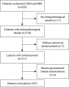
Methods: This retrospective study included 207 patients from April 2019 to June 2022. The radiologists assigned a diagnostic category to all lesions using the Breast Imaging Reporting and Data System (BI-RADS). Subsequently, they were asked to assign a final diagnostic category for each lesion according to the KS. The diagnostic performance was evaluated by the area under the receiver operating characteristic curve (AUC). The agreement in terms of the kinetic curve and the KS categories for CEM and MRI were evaluated via the Cohen kappa coefficient.
Results: The AUC was higher for the CEM-KS category assignment than for the CEM-BI-RADS category assignment (0.856 vs. 0.776; P=0.047). The AUC was higher for MRI-KS than for MRI-BI-RADS (0.841 vs. 0.752; P =0.015). The AUC of CEM-KS was not significantly different from that of MRI-KS (0.856 vs. 0.841; P=0.538). The difference between the AUCs for CEM-BI-RADS and MRI-BI-RADS was not statistically significant (0.776 vs. 0.752; P=0.400). The kappa agreement for the characterization of suspicious breast lesions using CEM-KS and MRI-KS was 0.885.
Conclusions: The KS substantially improved the diagnostic performance of suspicious breast lesions, not only in MRI but also in CEM. CEM-KS and MRI-KS showed similar diagnostic performance and almost perfect agreement for the characterization of suspicious breast lesions. Therefore, CEM holds promise as an alternative when breast MRI is not available or contraindicated.
Keywords: Breast neoplasms; clinical decision support system; contrast-enhanced mammography (CEM); diagnosis; magnetic resonance imaging (MRI).
2024 Quantitative Imaging in Medicine and Surgery. All rights reserved.
Conflict of interest statement
Conflicts of Interest: All authors have completed the ICMJE uniform disclosure form (available at https://qims.amegroups.com/article/view/10.21037/qims-24-593/coif). The authors have no conflicts of interest to declare.
Figures


References
-
- Fallenberg EM, Schmitzberger FF, Amer H, Ingold-Heppner B, Balleyguier C, Diekmann F, Engelken F, Mann RM, Renz DM, Bick U, Hamm B, Dromain C. Contrast-enhanced spectral mammography vs. mammography and MRI - clinical performance in a multi-reader evaluation. Eur Radiol 2017;27:2752-64. 10.1007/s00330-016-4650-6 - DOI - PubMed
-
- Kim EY, Youn I, Lee KH, Yun JS, Park YL, Park CH, Moon J, Choi SH, Choi YJ, Ham SY, Kook SH. Diagnostic Value of Contrast-Enhanced Digital Mammography versus Contrast-Enhanced Magnetic Resonance Imaging for the Preoperative Evaluation of Breast Cancer. J Breast Cancer 2018;21:453-62. 10.4048/jbc.2018.21.e62 - DOI - PMC - PubMed
LinkOut - more resources
Full Text Sources
Miscellaneous
