. 2024 Jul 20;10(15):e34961.
doi: 10.1016/j.heliyon.2024.e34961. eCollection 2024 Aug 15.

ELOVL1 is upregulated and promotes tumor growth in hepatocellular carcinoma through regulating PI3K-AKT-mTOR signaling

Affiliations

ELOVL1 is upregulated and promotes tumor growth in hepatocellular carcinoma through regulating PI3K-AKT-mTOR signaling

Liang Qin et al. Heliyon. .

Abstract

Background: The functions of the ELOVLs are mainly involved in the elongation of saturated and polyunsaturated fatty acids, thus influencing the metabolism of fatty acids. Abnormal lipid metabolism may result in NAFLD and NASH, which may lead to cirrhosis and liver cancer. These results suggest that ELOVLs-mediated metabolism might be involved in the development of HCC. The purpose of this study was to study the expression and function of ELOVL1 in human liver cancer.

Method: Using TCGA, GEPIA and other databases, we analyzed the relationship between the expression of ELOVL1 and liver cancer. The expression of ELOVL1 was detected by immunohistochemical method and Western blot method in hepatic carcinoma and hepatic carcinoma cells. Then, the effects of ELOVL1 on proliferation, apoptosis and invasion in vitro and in vivo were investigated by means of different methods.

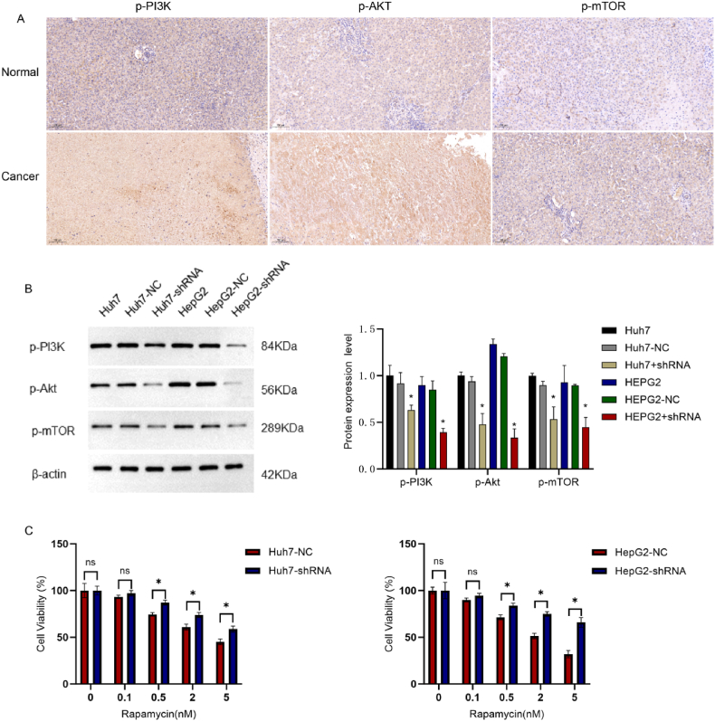
Result: Our results indicate that ELOVL1 is more highly expressed in liver cancer than in normal tissues. Survival analysis showed that OS and DSS were shorter in patients with high ELOVL1 expression than in those with low expression. Multivariate Cox analysis further demonstrated that over-expression of ELOVL1 was an independent risk factor for overall survival in HCC. The results of ROC also confirmed the value of ELOVL1 in the diagnosis of liver cancer. The results of KEGG enrichment and GSEA indicate that ELOVL1 is associated with lipid metabolism and NAFLD, as well as PPAR, PI3K-AKT-mTOR. Compared with the control group, it was found that silencing ELOVL1 in Huh7 and HepG2 cells could inhibit the growth of cells, promote the apoptosis and decrease the metastasis and invasion. Changes in ELOVL1 induced cell proliferation and metastasis may be related to PI3K/AKT/mTOR. Low expression of ELOVL1 inhibited the growth of xenograft tumors in hepatocellular carcinoma xenograft model.

Conclusion: Our data indicate that the activation of PI3K/AKT/mTOR pathway in HCC may contribute to the promotion of cancer. Thus, ELOVL1 may be a promising therapeutic target for HCC.

Keywords: ELOVL1; HCC; PI3K-AKT-mTOR; Tumor promotion.


Conflict of interest statement

The authors declare the following financial interests/personal relationships which may be considered as potential competing interests:Zi-Li Chen reports financial support was provided by Guizhou Medcial University. If there are other authors, they declare that they have no known competing financial interests or personal relationships that could have appeared to influence the work reported in this paper.

Figures

Fig. 1
Fig. 1
Expression of ELOVL1 in Pan-Cancer and hepatocellular carcinoma. (A) The differential expression of ELOVL1 in various tumor tissues and normal tissues; (B) The relative expression level of ELOVL1 in hepatocellular carcinoma tissues and normal tissues; (C)The relative expression level of ELOVL1 in hepatocellular carcinoma tissues and paired adjacent non-cancerous tissues; (D) Verification of ELOVL1 expression in hepatocellular carcinoma by GEO database; (E) Diagnostic ROC curve.
Fig. 2
Fig. 2
GEPIA database analysis of the survival outcomes of hepatocellular carcinoma patients related to ELOVL1. (A) Overall survival rate of hepatocellular carcinoma patients with high expression of ELOVL1 compared to low expression patients. (B) Disease-free survival rate of hepatocellular carcinoma patients with high expression of ELOVL1 compared to low expression patients.(C) ELOVL1 and T stage of hepatocellular carcinoma.(D) ELOVL1 and N stageof hepatocellular carcinoma.(E) ELOVL1 and M stage of hepatocellular carcinoma.(F) ELOVL1 and pathologic stage of hepatocellular carcinoma.(G) ELOVL1 and histologic stage of hepatocellular carcinoma.(H) ELOVL1 and residual stage of hepatocellular carcinoma.
Fig. 3
Fig. 3
Expression of ELOVL1 in hepatocellular carcinoma tissues and liver cancer cell lines (A and B) ELOVL1 was detected by immunohistochemistry in HPA database; (C and D) ELOVL1 expression was confirmed by immunohistochemistry in clinical liver cancer and paired adjacent tissue; (E) ELOVL1 mRNA was detected by RT-PCR in normal hepatic (LO2) and hepatic carcinoma (Huh7, HepG2); (F) Western blot was used to detect the expression of ELOVL1 in normal hepatic (LO2) and hepatic carcinoma (Huh7, HepG2).
Fig. 4
Fig. 4
Transfection and screening of interfering lentivirus. (A and B) Using fluorescence microscopy to observe the transfection of empty and interfering lentiviruses in HCC cells; (C and E) Western blotting to detect the protein expression of ELOVL1 in HCC cells after lentivirus transfection; (D and F) RT-PCR to detect the mRNA expression of ELOVL1 in HCC cells after lentivirus transfection.
Fig. 5
Fig. 5
ELOVL1 expression affects the cell cycle and apoptosis of HCC cells. (A and B) The silence of ELOVL1 on the cell cycle of HCC; (C and D) Cell apoptosis experiment validates the effect of high or low expression of ELOVL1 on HCC cells.
Fig. 6
Fig. 6
The expression of ELOVL1 influences the biological behavior of HCC cells. (A and B) Scratch test was used to detect the migration ability of HCC cells after silencing ELOVL1; (C and D) After silencing ELOVL1, the migration and invasion abilities of HCC cells were detected through Transwell; (E and F) Cell clone experiment verified the changes in the proliferation and growth ability of HCC cells after silencing ELOVL1.
Fig. 7
Fig. 7
Enrichment Analysis of ELOVL1 Function in Hepatocellular Carcinoma. (A)KEGG enrichment analysis; (B)Nonalcoholic fatty liver disease; (C)Fatty acid metabolism; (D)Focal adhesion PI3K-AKT-mTOR signaling pathway; (E)PI3K-AKT signaling in cancer; (F)Wnt signaling.
Fig. 8
Fig. 8
ELOVL1 inhibits tumor growth and metastasis by regulating the PI3K-AKT-mTOR signaling pathway. (A) Expression of phosphorylated protein of PI3K/AKT/mTOR pathway in hepatocellular carcinoma (B) After silencing ELOVL1, the related proteins of the PI3K/AKT/mTOR pathway were detected by Western blotting; (C) The cytotoxic effect of the mTOR inhibitor rapamycin at different concentrations on ELOVL1-silenced HCC cells was detected by CCK8 experiment.
Fig. 9
Fig. 9
Effect of rapamycin on biological behavior of hepatocellular carcinoma cells. (A) Effect of rapamycin on invasion ability of hepatocellular carcinoma cells; (B) Effect of rapamycin on migration ability of hepatocellular carcinoma cells.
Fig. 10
Fig. 10
Low expression of ELOVL1 inhibits tumor growth in vivo. (A) Tumor-bearing nude mice euthanized by cervical dislocation method; (B) Dissected tumor tissue; (C and D) Comparison of tumor growth curve and tumor weight; (E)Expression of ELOVL1, p-PI3K, p-AKT and p-mTOR in transplanted tumor.

References

    1. Llovet J.M., Zucman-Rossi J., Pikarsky E., Sangro B., Schwartz M., Sherman M., Gores G. Hepatocellular carcinoma. Nat. Rev. Dis. Prim. 2016;2 - PubMed
    1. Craig A.J., von Felden J., Garcia-Lezana T., Sarcognato S., Villanueva A. Tumour evolution in hepatocellular carcinoma. Nat. Rev. Gastroenterol. Hepatol. 2020;17(3):139–152. - PubMed
    1. Yang J.D., Hainaut P., Gores G.J., Amadou A., Plymoth A., Roberts L.R. A global view of hepatocellular carcinoma: trends, risk, prevention and management. Nat. Rev. Gastroenterol. Hepatol. 2019;16(10):589–604. - PMC - PubMed
    1. Peneau C., Imbeaud S., La Bella T., Hirsch T.Z., Caruso S., Calderaro J., Paradis V., Blanc J.F., Letouze E., Nault J.C., et al. Hepatitis B virus integrations promote local and distant oncogenic driver alterations in hepatocellular carcinoma. Gut. 2022;71(3):616–626. - PMC - PubMed
    1. Llovet J.M., Kelley R.K., Villanueva A., Singal A.G., Pikarsky E., Roayaie S., Lencioni R., Koike K., Zucman-Rossi J., Finn R.S. Hepatocellular carcinoma. Nat. Rev. Dis. Prim. 2021;7(1):6. - PubMed

LinkOut - more resources