. 2024 Jul 31;13(7):3299-3317.
doi: 10.21037/tcr-23-2274. Epub 2024 Jul 26.

Pan-cancer bioinformatics analysis of hepatic leukemia factor and further validation in colorectal cancer

Affiliations

Pan-cancer bioinformatics analysis of hepatic leukemia factor and further validation in colorectal cancer

Yirong Lin et al. Transl Cancer Res. .

Abstract

Background: Hepatic leukemia factor (HLF) is associated with cancer onset, growth, and progression; however, little is known regarding its biological role in pan-cancer. In order to further evaluate the diagnostic and prognostic value of HLF in pan-cancer and colorectal cancer (CRC), we performed comprehensive bioinformatics analyses of the molecular mechanism of HLF in pan-cancer, with subsequent verification in CRC.

Methods: We downloaded data (gene expression, clinical data, follow-up duration, and immune-related data) related to 33 solid tumor types from UCSC Xena (University of California Santa Cruz cancer database, https://xena.ucsc.edu/). HLF expression was analyzed in pan-cancer, and its diagnostic efficacy, prognostic value, and correlation with pathological stage and cancer immunity were determined. We also analyzed gene alterations in HLF and biological processes involved in its regulation in pan-cancer. Using CRC data in The Cancer Genome Atlas (TCGA), we assessed correlations between HLF and CRC diagnosis, prognosis, and drug sensitivity and performed functional enrichment analyses. Moreover, we constructed an HLF-related ceRNA regulatory network. Finally, we externally validated HLF expression and diagnostic and prognostic value in CRC using Gene Expression Omnibus (GEO) database, as well as by performing in vitro experiments.

Results: HLF expression was downregulated in most tumors, and HLF showed good predictive potential for pan-cancer diagnosis and prognosis. It was closely related to the clinicopathological stages of pan-cancer. Further, HLF was associated with tumor microenvironment and immune cell infiltration in many tumors. Analyses involving cBioPortal revealed changes in HLF amplifications and mutations in most tumors. HLF was also closely associated with microsatellite instability and tumor mutational burden in pan-cancer and involved in regulating various tumor-related pathways and biological processes. In CRC, HLF expression was similarly downregulated, with implications for CRC diagnosis and prognosis. Functional enrichment analysis indicated the association of HLF with many cancer-related pathways. Further, HLF was associated with drug (e.g., oxaliplatin) sensitivity in CRC. The ceRNA regulatory network showed multigene regulation of HLF in CRC. External validation involving GEO databases and quantitative real-time polymerase chain reaction (qRT-PCR) data substantiated these findings.

Conclusions: HLF expression generally exhibited downregulation in pan-cancer, contributing to tumor occurrence and development by regulating various biological processes and affecting tumor immune characteristics. HLF was also closely related to CRC occurrence and development. We believe HLF can serve as a reliable diagnostic, prognostic, and immune biomarker for pan-cancer.

Keywords: Hepatic leukemia factor (HLF); cancer immunity; diagnosis; pan-cancer; prognosis.


Conflict of interest statement

Conflicts of Interest: All authors have completed the ICMJE uniform disclosure form (available at https://tcr.amegroups.com/article/view/10.21037/tcr-23-2274/coif). The authors have no conflicts of interest to declare.

Figures
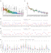
Figure 1
Figure 1
HLF gene expression and activity. Relationship between HLF expression and clinicopathological stage. (A) HLF gene expression in tumor tissues. (B) HLF gene expression between human normal and tumor tissues. (C) HLF activity in tumor tissues. (D) HLF activity between human normal and tumor tissues. (E) Relationship between HLF expression and clinicopathological stage. HLF, hepatic leukemia factor; LIHC, liver hepatocellular carcinoma; LGG, brain lower grade glioma; PCPG, pheochromocytoma and paraganglioma; KIRP, kidney renal papillary cell carcinoma; THYM, thymoma; KIRC, kidney renal clear cell carcinoma; ACC, adrenocortical carcinoma; PRAD, prostate adenocarcinoma; CHOL, cholangiocarcinoma; THCA, thyroid carcinoma; LUAD, lung adenocarcinoma; GBM, glioblastoma multiforme; LUSC, lung squamous cell carcinoma; KICH, kidney chromophobe; PAAD, pancreatic adenocarcinoma; SARC, sarcoma; TGCT, testicular germ cell tumors; ESCA, esophageal carcinoma; BRCA, breast invasive carcinoma; CESC, cervical squamous cell carcinoma and endocervical adenocarcinoma; HNSC, head and neck squamous cell carcinoma; STAD, stomach adenocarcinoma; UCS, uterine carcinosarcoma; UVM, uveal melanoma; MESO, mesothelioma; COAD, colon adenocarcinoma; UCEC, uterine corpus endometrial carcinoma; READ, rectum adenocarcinoma; OV, ovarian serous cyst adenocarcinoma; SKCM, skin cutaneous melanoma; DLBC, lymphoid neoplasm diffuse large B cell lymphoma; BLCA, bladder urothelial carcinoma; LAML, acute myeloid leukemia.
Figure 2
Figure 2
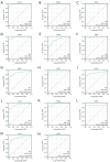
Diagnostic capability of HLF. (A) Diagnostic capability of HLF in BLCA. (B) Diagnostic capability of HLF in BRCA. (C) Diagnostic capability of HLF in CESC. (D) Diagnostic capability of HLF in CHOL. (E) Diagnostic capability of HLF in COAD. (F) Diagnostic capability of HLF in GBM. (G) Diagnostic capability of HLF in HNSC. (H) Diagnostic capability of HLF in KICH. (I) Diagnostic capability of HLF in LUAD. (J) Diagnostic capability of HLF in LUSC. (K) Diagnostic capability of HLF in READ. (L) Diagnostic capability of HLF in SARC. (M) Diagnostic capability of HLF in THCA. (N) Diagnostic capability of HLF in UCEC. HLF, hepatic leukemia factor; AUC, area under the curve; CI, confidence interval; FPR, false positive rate; TPR, true positive rate; BLCA, bladder urothelial carcinoma; BRCA, breast invasive carcinoma; CESC, cervical squamous cell carcinoma and endocervical adenocarcinoma; CHOL, cholangiocarcinoma; COAD, colon adenocarcinoma; GBM, glioblastoma multiforme; HNSC, head and neck squamous cell carcinoma; KICH, kidney chromophobe; LUAD, lung adenocarcinoma; LUSC, lung squamous cell carcinoma; READ, rectum adenocarcinoma; SARC, sarcoma; THCA, thyroid carcinoma; UCEC, uterine corpus endometrial carcinoma.
Figure 3
Figure 3
Relationship between HLF expression in pan-cancer and its implications for prognosis. (A) In COAD, HNSC, and KIRC, overall survival between the low HLF expression group and the high HLF expression group. (B) In MESO, PAAD, and SARC, disease-free survival between the low HLF expression group and the high HLF expression group. (C) In COAD, HNSC, and KIRC, disease-specific survival between the low HLF expression group and the high HLF expression group. (D) In HNSC, KIRC, and LGG, progression-free survival between the low HLF expression group and the high HLF expression group. HLF, hepatic leukemia factor; COAD, colon adenocarcinoma; HNSC, head and neck squamous cell carcinoma; KIRC, kidney renal clear cell carcinoma; MESO, mesothelioma; PAAD, pancreatic adenocarcinoma; SARC, sarcoma; LGG, brain lower grade glioma.
Figure 4
Figure 4
Correlation of HLF with ESTIMATE, immune, and stromal scores and with tumor purity. (A) Correlation of HLF with ESTIMATE score, stromal score and tumor purity in BLCA. (B) Correlation of HLF with ESTIMATE score, immune score and tumor purity in LGG. (C) Correlation of HLF with ESTIMATE score, stromal score and tumor purity in PRAD. (D) Correlation of HLF with ESTIMATE score, immune score and tumor purity in THCA. (E) Correlation of HLF with immune score in THYM. HLF, hepatic leukemia factor; ESTIMATE, Estimation of STromal and Immune cells in MAlignant Tumor tissues using Expression data; BLCA, bladder urothelial carcinoma; LGG, brain lower grade glioma; PRAD, prostate adenocarcinoma; THCA, thyroid carcinoma; THYM, thymoma.
Figure 5
Figure 5
Correlation of HLF with immune infiltration. (A) Correlation of HLF with mast cells resting in LUAD. (B) Correlation of HLF with macrophages M0 in PAAD. (C) Correlation of HLF with B cells naive in TGCT. (D) Correlation of HLF with NK cells activated in TGCT.HLF, hepatic leukemia factor; LUAD, lung adenocarcinoma; PAAD, pancreatic adenocarcinoma; TGCT, testicular germ cell tumors.
Figure 6
Figure 6
Correlation of HLF with TMB and MSI. cBioPortal analysis of HLF. (A) cBioPortal analysis of HLF. (B) Correlation of HLF with TMB. (C) Correlation of HLF with TMB and MSI. *, P<0.05; **, P<0.01; ***, P<0.001. CNA, copy number alteration; BLCA, bladder urothelial carcinoma; ACC, adrenocortical carcinoma; BRCA, breast invasive carcinoma; CESC, cervical squamous cell carcinoma and endocervical adenocarcinoma; CHOL, cholangiocarcinoma; COAD, colon adenocarcinoma; DLBC, lymphoid neoplasm diffuse large B cell lymphoma; ESCA, esophageal carcinoma; GBM, glioblastoma multiforme; HNSC, head and neck squamous cell carcinoma; KICH, kidney chromophobe; KIRC, kidney renal clear cell carcinoma; KIRP, kidney renal papillary cell carcinoma; LAML, acute myeloid leukemia; LGG, brain lower grade glioma; LIHC, liver hepatocellular carcinoma; LUAD, lung adenocarcinoma; LUSC, lung squamous cell carcinoma; MESO, mesothelioma; OV, ovarian serous cyst adenocarcinoma; PAAD, pancreatic adenocarcinoma; PCPG, pheochromocytoma and paraganglioma; PRAD, prostate adenocarcinoma; READ, rectum adenocarcinoma; SARC, sarcoma; SKCM, skin cutaneous melanoma; STAD, stomach adenocarcinoma; TGCT, testicular germ cell tumors; THCA, thyroid carcinoma; THYM, thymoma; UCEC, uterine corpus endometrial carcinoma; UCS, uterine carcinosarcoma; UVM, uveal melanoma; HLF, hepatic leukemia factor; TMB, tumor mutation burden; MSI, microsatellite instability.
Figure 7
Figure 7
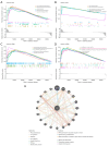
Functional enrichment analysis of HLF in pan-cancer. (A) KEGG pathway enrichment analysis of HLF in ACC. (B) GO pathway enrichment analysis of HLF in ACC. (C) KEGG pathway enrichment analysis of HLF in STAD. (D) GO pathway enrichment analysis of HLF in STAD. (E) GeneMANIA results of HLF. ACC, adrenocortical carcinoma; KEGG, Kyoto Encyclopedia of Genes and Genomes; STAD, stomach adenocarcinoma; HLF, hepatic leukemia factor.
Figure 8
Figure 8
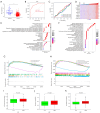
Correlation analysis between HLF and CRC in TCGA. Clinical relative analysis between HLF and CRC (A-C). The top ten genes associated with HLF (D). GO, KEGG, GSEA analysis of HLF (E-H). Drug sensitivity of 5-fluorouracil, irinotecan, and oxaliplatin (I-K). ***, P<0.001. HLF, hepatic leukemia factor; AUC, area under the curve; CI, confidence interval; BP, biological process; CC, cell component; MF, molecular function; KEGG, Kyoto Encyclopedia of Genes and Genomes; GO, Gene Ontology; CRC, colorectal cancer; TCGA, The Cancer Genome Atlas; GSEA, gene set enrichment analysis.
Figure 9
Figure 9
Expression differences and diagnostic ability of HLF in GSE87211 and GSE106582; ceRNA regulatory network of HLF; qRT-PCR results showing HLF expression in CRC and adjacent normal colorectal tissues. (A) In GSE87211, HLF gene expression between human normal and tumor tissues. (B) In GSE106582, HLF gene expression between human normal and tumor tissues. (C) Diagnostic ability of HLF in GSE87211. (D) Diagnostic ability of HLF in GSE106582. (E) ceRNA regulatory network of HLF. (F) qRT-PCR results showing HLF expression being downregulated in CRC tissues. ***, P<0.001. HLF, hepatic leukemia factor; AUC, area under the curve; CRC, colorectal cancer; ceRNA, competing endogenous ribonucleic acids; qRT-PCR, quantitative real-time polymerase chain reaction.

References

    1. Sung H, Ferlay J, Siegel RL, et al. Global Cancer Statistics 2020: GLOBOCAN Estimates of Incidence and Mortality Worldwide for 36 Cancers in 185 Countries. CA Cancer J Clin 2021;71:209-49. 10.3322/caac.21660 - DOI - PubMed
    1. Siegel RL, Miller KD, Wagle NS, et al. Cancer statistics, 2023. CA Cancer J Clin 2023;73:17-48. 10.3322/caac.21763 - DOI - PubMed
    1. Chen F, Fan Y, Cao P, et al. Pan-Cancer Analysis of the Prognostic and Immunological Role of HSF1: A Potential Target for Survival and Immunotherapy. Oxid Med Cell Longev 2021;2021:5551036. 10.1155/2021/5551036 - DOI - PMC - PubMed
    1. Lulli M, Napoli C, Landini I, et al. Role of Non-Coding RNAs in Colorectal Cancer: Focus on Long Non-Coding RNAs. Int J Mol Sci 2022;23:13431. 10.3390/ijms232113431 - DOI - PMC - PubMed
    1. Huang X, Chen Z, Xiang X, et al. Comprehensive multi-omics analysis of the m7G in pan-cancer from the perspective of predictive, preventive, and personalized medicine. EPMA J 2022;13:671-97. 10.1007/s13167-022-00305-1 - DOI - PMC - PubMed

LinkOut - more resources