. 2024 Aug 4;8(9):bvae142.
doi: 10.1210/jendso/bvae142. eCollection 2024 Jul 26.

Identification of Succinate Dehydrogenase Gene Variant Carriers by Blood Biomarkers

Affiliations

Identification of Succinate Dehydrogenase Gene Variant Carriers by Blood Biomarkers

Marcel Gebhardt et al. J Endocr Soc. .

Abstract

Background: Carriers of germline pathogenic variants (PVs) in succinate dehydrogenase genes (SDHx) are at risk of developing tumors, including paragangliomas, gastrointestinal stromal tumors, and renal cell carcinomas. Early tumor detection is paramount for improved clinical outcome. Blood-based biomarkers could aid in identifying individuals with PVs early and provide functional evidence in patients with variants of unknown significance.

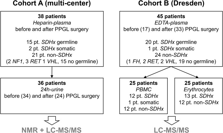
Methods: Blood plasma, urine, peripheral blood mononuclear cells, and erythrocytes from patients with and without SDHx PVs were investigated for central carbon metabolites. These were measured by liquid chromatography-tandem mass spectrometry and nuclear magnetic resonance spectroscopy and included among others, succinate, fumarate, α-ketoglutarate, and lactate.

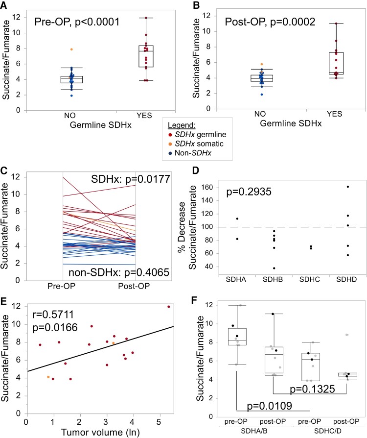
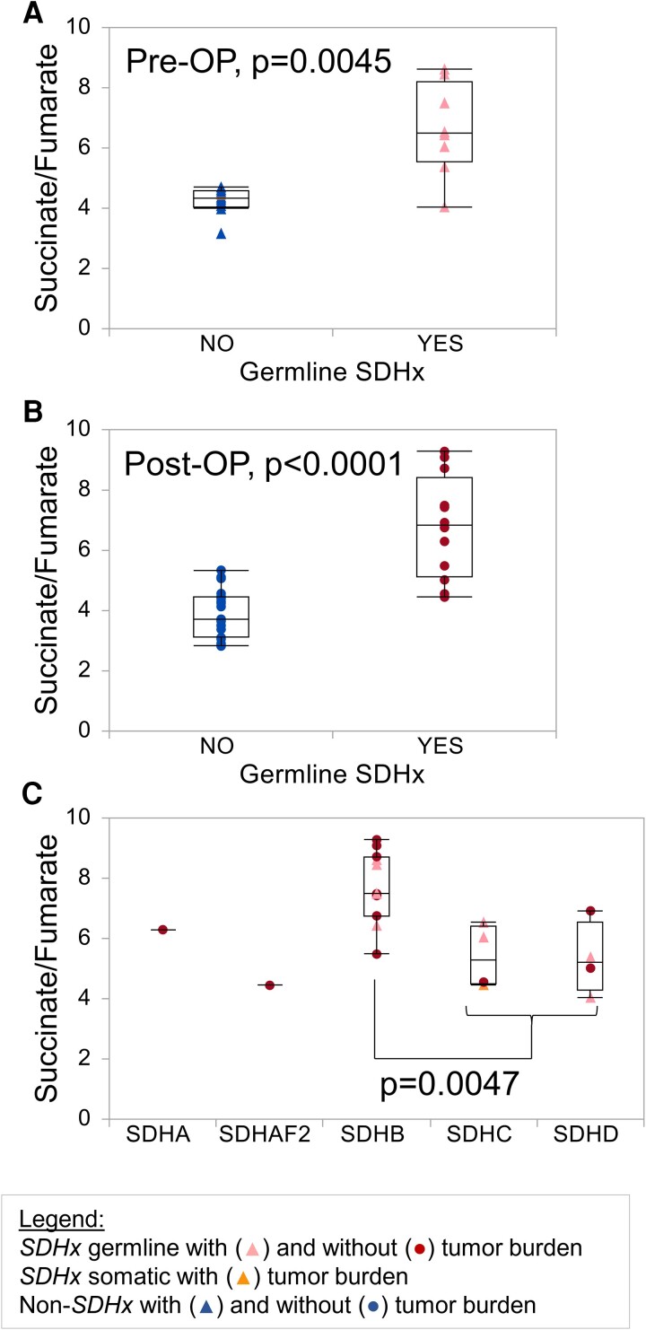
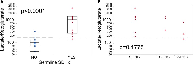
Results: Plasma succinate to fumarate ratios effectively distinguished tumor-bearing and asymptomatic patients with and without SDHx PV with promising diagnostic performance (areas under the receiver operating characteristic curve 0.86-0.95), although higher levels were noted in individuals with SDHB PV. Metabolites in urine and in peripheral blood mononuclear cell extracts were largely similar between groups. Erythrocytes showed strong metabolic alterations in patients with SDHx PV compared to controls, with 8 of 13 low-molecular organic acids being significantly different (P < .05). The lactate-α-ketoglutarate-ratio of erythrocytes identified individuals with SDHx PV equally well as plasma, with a sensitivity and specificity of 92% (AUC 0.97).

Conclusion: Blood biomarkers have been underutilized for identifying carriers of SDHx PV or to validate variants of unknown significance. Our findings advocate for further investigation into a combined approach involving plasma and erythrocytes for future diagnostic strategies.

Keywords: PBMC; blood plasma; erythrocytes; metabolism; succinate dehydrogenase gene variants; urine.


Figures

Figure 1.
Figure 1.
Overview of patient (pt.) cohorts and available material for LC-MS/MS and NMR. A detailed breakdown of patients can be found in Supplementary Tables 1-7 [14]. Abbreviations: PPGL, pheochromocytoma/paraganglioma; SDHx, pathogenic variant in a succinate dehydrogenase gene; FH, pathogenic variant in a fumarate hydratase gene; PBMC, peripheral mononuclear cells.
Figure 2.
Figure 2.
Succinate to fumarate ratio determined in heparin plasma by LC-MS/MS. Plasma was collected from patients with PPGL (cohort A) either preoperatively (Pre-OP, A) or postoperatively (Post-OP, B). C) Paired analysis between pre-OP and post-OP samples; P value represents Wilcoxon signed-rank test (1-tailed for SDHx, 2-tailed for non-SDHx). D) Decrease in the succinate to fumarate ratio according to genes in % (Kruskal-Wallis test). E) Correlation between pre-OP succinate to fumarate ratios and tumor volume (natural logarithm of value in cm3 displayed). F. Succinate to fumarate ratio according to the affected gene comparing patients with germline SDHA (black dots) and SDHB (gray dots) PVs to patients with SDHC (black dots) and SDHD (gray dots) PVs (Kruskal-Wallis test).
Figure 3.
Figure 3.
Succinate to fumarate ratio determined in EDTA plasma by LC-MS/MS. Plasma was collected from patients of cohort B either preoperatively (Pre-OP, n = 18, A) or in patients without tumor burden (Post-OP, n = 31, removal of a high succinate outlier in the group of patients with SDHx PV, B). Tumor burden refers to the presence of primary tumors or metastases and was determined by anatomical and/or functional imaging. C) Succinate-fumarate-ratios are displayed according to the affected gene. Triangles depict patients with a PPGL, circles show patients without tumor burden. Patients with SDHx germline PV, SDHx somatic PV, and without SDHx PV are shown in red, orange, and blue, respectively.
Figure 4.
Figure 4.
Krebs cycle metabolites in erythrocytes from patients with and without SDHx PVs. A-I) Erythrocytes were collected from patients (cohort B) in whom PPGL was present (triangles, light red) or absent (circles, dark red). Patients with SDHx germline PV and without SDHx PV are shown in red and blue, respectively.
Figure 5.
Figure 5.
The lactate/α-ketoglutarate-ratio from erythrocytes separates patients with SDHx PVs. A) Erythrocytes were collected from patients (cohort B) in whom PPGL was present (triangles, light red) or absent (circles, dark red). Patients with SDHx germline PV and without SDHx PV are shown in red and blue, respectively. Gray dashed lines indicate cutoff according to Youden index (186). As ratios of lactate/ketoglutarate and lactate/2-hydroxyglutarate show similar results, only the first one is displayed here. B) Lactate/α-ketoglutarate does not significantly differ between patients with different SDHx genes. Since the lactate/2-hydroxyglutarate ratio shows exactly the same results as lactate/α-ketoglutarate, only the latter is displayed in this figure.
Figure 6.
Figure 6.
SDHB protein levels in erythrocytes. Membrane fractions of erythrocytes were collected from carriers of SDHx PV and controls (cohort B), isolated and analyzed for protein levels by Western blot. One lane represents results for one patient. Full blots and additional patients are available in the supplement (Supplementary Fig. S1 and S2 [14]). Densitometry calculations are provided on the bottom. M—Marker.

References

    1. MacFarlane J, Seong KC, Bisambar C, et al. A review of the tumour spectrum of germline succinate dehydrogenase gene mutations: beyond phaeochromocytoma and paraganglioma. Clin Endocrinol. 2020;93(5):528‐538. - PubMed
    1. Amar L, Pacak K, Steichen O, et al. International consensus on initial screening and follow-up of asymptomatic SDHx mutation carriers. Nat Rev Endocrinol. 2021;17(7):435‐444. - PMC - PubMed
    1. Buffet A, Ben Aim L, Leboulleux S, et al. Positive impact of genetic test on the management and outcome of patients with paraganglioma and/or pheochromocytoma. J Clin Endocrinol Metab. 2019;104(4):1109‐1118. - PubMed
    1. Davidoff DF, Benn DE, Field M, et al. Surveillance improves outcomes for carriers of sdhb pathogenic variants: a multicenter study. J Clin Endocrinol Metab. 2022;107(5):e1907‐e1916. - PMC - PubMed
    1. Benn DE, Zhu Y, Andrews KA, et al. Bayesian approach to determining penetrance of pathogenic SDH variants. J Med Genet. 2018;55(11):729‐734. - PMC - PubMed

LinkOut - more resources