. 2024 Aug 15;134(16):e168393.
doi: 10.1172/JCI168393.

MNK-driven eIF4E phosphorylation regulates the fibrogenic transformation of mesenchymal cells and chronic lung allograft dysfunction

Affiliations

MNK-driven eIF4E phosphorylation regulates the fibrogenic transformation of mesenchymal cells and chronic lung allograft dysfunction

Natalie M Walker et al. J Clin Invest. .

Abstract

Tissue fibrosis remains unamenable to meaningful therapeutic interventions and is the primary cause of chronic graft failure after organ transplantation. Eukaryotic translation initiation factor (eIF4E), a key translational regulator, serves as convergent target of multiple upstream profibrotic signaling pathways that contribute to mesenchymal cell (MC) activation. Here, we investigate the role of MAP kinase-interacting serine/threonine kinase-induced (MNK-induced) direct phosphorylation of eIF4E at serine 209 (Ser209) in maintaining fibrotic transformation of MCs and determine the contribution of the MNK/eIF4E pathway to the pathogenesis of chronic lung allograft dysfunction (CLAD). MCs from patients with CLAD demonstrated constitutively higher eIF4E phosphorylation at Ser209, and eIF4E phospho-Ser209 was found to be critical in regulating key fibrogenic protein autotaxin, leading to sustained β-catenin activation and profibrotic functions of CLAD MCs. MNK1 signaling was upregulated in CLAD MCs, and genetic or pharmacologic targeting of MNK1 activity inhibited eIF4E phospho-Ser209 and profibrotic functions of CLAD MCs in vitro. Treatment with an MNK1/2 inhibitor (eFT-508) abrogated allograft fibrosis in an orthotopic murine lung-transplant model. Together these studies identify what we believe is a previously unrecognized MNK/eIF4E/ATX/β-catenin signaling pathway of fibrotic transformation of MCs and present the first evidence, to our knowledge, for the utility of MNK inhibitors in fibrosis.

Keywords: Cell migration/adhesion; Extracellular matrix; Pulmonology; Translation; Transplantation.


Conflict of interest statement

Conflict of interest: The authors have declared that no conflict of interest exists.

Figures

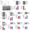
Figure 1
Figure 1. Phosphorylation of eIF4E (Ser209) is MNK1 dependent but mTORC-independent in fibrotic MCs derived from lung-transplant patients.
(A) Protein expression of phosphorylated and total forms of eIF4E and MNK1 was measured by Western blot analysis in MCs derived from normal (non-Fib-MCs) or fibrotic (Fib-MCs) human-lung allografts. Densitometry analyses of phospho-eIF4E to total eIF4E are shown with Fib-MCs for RAS (circles) and BOS (triangles). (B) Lentiviral infections were performed using empty pLenti-LoxEV vector or vector expressing active MNK1 (T344D) in nonfibrotic MCs or kinase-dead MNK1 (D191A) in Fib-MCs. Western blotting and corresponding densitometry analyses were performed for phospho-eIF4E and total eIF4E. (C) Fibrotic MCs treated with eFT-508 (10 μM, 24 hours) were subjected to m7GDP cap pulldown assay followed by Western blotting analyses. (D) Fib-MCs were transfected with RPTOR-specific or scrambled siRNA, and protein lysates were analyzed by Western blotting. Representative immunoblots and corresponding densitometry are shown for phospho-eIF4E and total eIF4E. (E and F) Fib-MCs were treated with ATP-competitive mTORC inhibitors (AZD8055: 250 nM; rapamycin: 250 nM) for 24 hours and analyzed for phospho-eIF4E and total eIF4E by Western blotting and densitometry. (G) Fib-MCs were transfected with MNK1-specific or scrambled siRNA. Protein lysates were immunoblotted for mTORC1/2 substrates. Representative immunoblots and corresponding densitometry are shown for phosphorylated and total forms of 4E-BP1, p70S6K1, and AKT. Data are represented as means ± SEM. **P < 0.01; ***P < 0.001; ****P < 0.0001, unpaired t test.
Figure 2
Figure 2. MNK/eIF4E (Ser209) activation is mediated by upstream JNK signaling.
(AC) MCs derived from fibrotic human lung allografts (Fib-MCs) were treated with pharmacologic inhibitors against MEK1/2 (U0126), JNK (broad spectrum effect — SP600125), and p38 MAPK (SB203580) at 10 μM for 2 hours (A), treated with the indicated doses of irreversible JNK1/2/3 inhibitor (JNK-IN-8) (B, 2 hours), or subjected to lentiviral infections of empty pLenti-LoxEV vector or vector expressing constitutively active JNK1, followed by treatment with eFT-508 (C; 10 μM, 2 hours) or AZD8055 (C; 250 nM, 2 hours). Protein lysates were analyzed by Western blotting. Representative immunoblots and corresponding densitometry are shown for phospho-eIF4E and total eIF4E. (D) MCs derived from normal human lung allografts (non-Fib-MCs) were infected with lentiviral particles containing constitutively active JNK1 for 48 hours, followed by infection with particles containing rLuc-PolIRES-fLuc cap-translation luciferase vector. Twenty-four hours later, lysates were collected and readings for Renilla and Firefly luminescence were performed. Data are represented as means ± SEM. **P < 0.01; ***P < 0.001; ****P < 0.0001, 1-way ANOVA; post hoc test: Bonferroni’s test (AC); 2-way ANOVA; post hoc test: Bonferroni’s test (D).
Figure 3
Figure 3. MNK/eIF4E (Ser209) activation drives profibrotic phenotypes in lung MCs.
(A) MCs isolated from fibrotic (Fib-MCs) and normal human lung allografts (non-Fib-MCs) were infected with lentiviral particles containing pLenti-LoxEV–eIF4E (S209A) and pLenti-LoxEV–eIF4E (S209D), respectively, and then cultured in Matrigel-coated Transwells. Cell migration was assessed by MTT assay. (B) Fib-MCs were transfected with EIF4E or scrambled control siRNA followed by lentiviral infection of empty vector or vector expressing S209A or S209D. Protein lysates were immunoblotted against collagen I. Representative immunoblots and corresponding densitometry are shown. (C and D) Fib-MCs were transfected with scrambled or MNK1 siRNA (C) or treated with eFT-508 (10 μM, 2 hours) (D). Protein lysates were immunoblotted against collagen I and SPARC. Representative immunoblots and corresponding densitometry are shown. (E) Proliferation was assessed in Fib-MCs treated with eFT-508 (10 μM, 72 hours) using Cyquant Proliferation Assay Kit. Protein lysates were immunoblotted for cyclin D1 as well as phosphorylated and total eIF4E. Data are represented as means ± SEM. **P < 0.01; ***P < 0.001; ****P < 0.0001, unpaired t test (A, C, D, and E); 1-way ANOVA; post hoc test: Bonferroni’s test (B).
Figure 4
Figure 4. MNK/eIF4E (Ser209) positively regulates ATX expression and activity in fibrotic lung MCs.
(A) MCs isolated from fibrotic (Fib-MCs) or normal (non-Fib-MCs) human lung allografts were infected with lentiviral vectors containing pLenti-LoxEV–eIF4E (S209A) and pLenti-LoxEV–eIF4E (S209D) plasmids, respectively. Protein lysates were analyzed by Western blotting for cellular and secreted ATX expression. Representative immunoblots and densitometry for cellular ATX are shown. (B) Immunoblots for CD44 protein expression in lentiviral-infected MCs described in A. (C and D) Fib-MCs were transfected with MNK1 or scrambled siRNA (C) or treated with eFT-508 (10 μM, 2 hours; D). Protein lysates were analyzed by Western blotting. Representative immunoblots of phospho-eIF4E and total forms of eIF4E, and expression of cellular ATX and CD44 are shown. (E) Conditioned media of Fib-MCs treated with eFT-508 (10 μM, 24 hours) were measured for ATX activity utilizing the fluorogenic substrate FS-3. n = 9. (F) Cultured mouse lung fibroblasts from WT, Eif4eSer209A/A, and Mnk1/2-KO mice were assessed by immunoblotting for cellular ATX, MNK1, phospho-eIF4E, and total eIF4E. (G) Secreted ATX levels were analyzed in the conditioned media by sandwich ELISA. Data are represented as means ± SEM. *P < 0.05; **P < 0.01; ***P < 0.001; ****P < 0.0001, unpaired t test (A and B); simple linear regression (E); 1-way ANOVA; post hoc test: Bonferroni’s test (G).
Figure 5
Figure 5. MNK/eIF4E (Ser209)/ATX signaling drives β-catenin expression in lung MCs.
(A and B) MCs derived from fibrotic human-lung allografts (Fib-MCs) were transfected with MNK1 or scrambled control siRNA. (A) Nuclear extracts were analyzed by immunoblotting for β-catenin expression. Representative image from 6 cell lines. (B) Whole-cell lysates were assessed for active, nonphosphorylated β-catenin by immunoblotting and densitometry. (C) Fib-MCs were treated with eFT-508 (10 μM). Nuclear extracts were assessed for β-catenin expression by immunoblotting and densitometry. (D) Nonfibrotic MCs (non-Fib-MCs) were subjected to lentiviral infection with pLenti-LoxEV–eIF4E S209D plasmid. Protein lysates were assessed for active β-catenin expression. (E) Non-Fib-MCs were silenced with scrambled or ATX siRNA followed by lentiviral expression of active mutant pLenti-LoxEV–MNK1 (T344D). Western blotting analyses were performed for active β-catenin, MNK1, and phospho-eIF4E. Data are represented as means ± SEM. *P < 0.05; **P < 0.01; ****P < 0.0001, unpaired t test (B and D); 1-way ANOVA; post hoc test: Bonferroni’s test (C and E).
Figure 6
Figure 6. Pharmacologic MNK inhibition utilizing eFT-508 decreases lung allograft fibrosis and ATX expression in a murine orthotopic left lung–transplant model of RAS.
(A) Experimental schematic. B6D2F1/J donor lungs were transplanted into C57BL/6J recipient mice, followed by treatment with eFT-508 (5 mg/kg/d; oral gavage) between days 7 and 28. (B) Histopathology examination of formalin-fixed, paraffin-embedded tissues — isografts (Iso), RAS allografts (Allo), and eFT-508-treated RAS allografts (Allo+eFT-508) at day 28 using H&E staining (top panels) and Masson’s trichrome staining (bottom panels). Scale bars: 100 μm. V, vessel; AW, airway. (C) Acid-digested lung homogenates were assessed for collagen content by hydroxyproline assay. n = 6–8. Data are represented as means ± SEM. ***P < 0.001; ****P < 0.0001, 1-way ANOVA; post hoc test: Bonferroni’s test. (D) Gli1CreERT2/WT;Rosa26mTmG/WT murine allograft controls or treated with eFT-508 were harvested at day 14 after transplant. Top panels: dual-labeling against GFP (green) and α-smooth muscle actin (α-SMA) (red). Nuclear counterstaining was by DAPI. Original magnification, ×400. Scale bars: 50 μm. Bottom panels: Masson’s trichrome staining in contiguous sections indicating collagen deposition (blue). (E) eFT-508 therapeutic treatment regimen from days 28 to 40 after transplant resulted in reduced collagen content as measured by hydroxyproline assay. *P < 0.05, unpaired t test. (F and G) Supernatants from day 28–transplanted lung homogenates were analyzed for ATX levels by sandwich ELISA (F) and ATX activity utilizing FS-3 fluorogenic substrate (G). n = 7–9. Data are represented as means ± SEM. **P < 0.01; ****P < 0.0001, simple linear regression.
Figure 7
Figure 7. Genetic deficiency of Eif4eSer209A/A and Mnk1/2 prevents collagen deposition and ATX synthesis in bleomycin-injured mice.
(A) Representative immunoblot and densitometric analysis of phospho-eIF4E (Ser209) from lung homogenates of mice 21 days after bleomycin administration. n = 5. Unpaired t test. (B) Quantitation of collagen content in lung homogenates of C57BL/6J, Eif4eSer209A/A, and Mnk1/2-KO by hydroxyproline assay. n = 4 in saline control groups; n = 9–15 in bleomycin-treated groups. One-way ANOVA; post hoc test: Bonferroni’s test. (C) ATX protein expression levels in saline- and bleomycin-treated lung homogenates, as measured by sandwich ELISA. n = 4–8. One-way ANOVA; post hoc test: Bonferroni’s test. Data are represented as means ± SEM. ***P < 0.001; ****P < 0.0001. (D) Representative images of trichrome staining of day-21 tissue sections of C57BL/6J, Eif4eSer209A/A, and Mnk1/2-KO mice administered bleomycin or saline control. Original magnification, ×200. n = 6 mice for each group.

References

    1. Chambers DC, et al. The International Thoracic Organ Transplant Registry of the International Society for Heart and Lung Transplantation: Thirty-eighth adult lung transplantation report - 2021; Focus on recipient characteristics. J Heart Lung Transplant. 2021;40(10):1060–1072. doi: 10.1016/j.healun.2021.07.021. - DOI - PMC - PubMed
    1. Verleden GM, et al. Chronic lung allograft dysfunction: Definition, diagnostic criteria, and approaches to treatment-A consensus report from the Pulmonary Council of the ISHLT. J Heart Lung Transplant. 2019;38(5):493–503. doi: 10.1016/j.healun.2019.03.009. - DOI - PubMed
    1. Cao P, et al. Autocrine lysophosphatidic acid signaling activates β-catenin and promotes lung allograft fibrosis. J Clin Invest. 2017;127(4):1517–1530. doi: 10.1172/JCI88896. - DOI - PMC - PubMed
    1. Geng Y, et al. PD-L1 on invasive fibroblasts drives fibrosis in a humanized model of idiopathic pulmonary fibrosis. JCI Insight. 2019;4(6):e125326. doi: 10.1172/jci.insight.125326. - DOI - PMC - PubMed
    1. Lam AP, Gottardi CJ. β-catenin signaling: a novel mediator of fibrosis and potential therapeutic target. Curr Opin Rheumatol. 2011;23(6):562–567. doi: 10.1097/BOR.0b013e32834b3309. - DOI - PMC - PubMed

Substances