. 2024 Aug 15;134(16):e174374.
doi: 10.1172/JCI174374.

Posttranslationally modified self-peptides promote hypertension in mouse models

Affiliations

Posttranslationally modified self-peptides promote hypertension in mouse models

Nathaniel Bloodworth et al. J Clin Invest. .

Abstract

Posttranslational modifications can enhance immunogenicity of self-proteins. In several conditions, including hypertension, systemic lupus erythematosus, and heart failure, isolevuglandins (IsoLGs) are formed by lipid peroxidation and covalently bond with protein lysine residues. Here, we show that the murine class I major histocompatibility complex (MHC-I) variant H-2Db uniquely presents isoLG-modified peptides and developed a computational pipeline that identifies structural features for MHC-I accommodation of such peptides. We identified isoLG-adducted peptides from renal proteins, including sodium glucose transporter 2, cadherin 16, Kelch domain-containing protein 7A, and solute carrier family 23, that are recognized by CD8+ T cells in tissues of hypertensive mice, induce T cell proliferation in vitro, and prime hypertension after adoptive transfer. Finally, we find patterns of isoLG-adducted antigen restriction in class I human leukocyte antigens that are similar to those in murine analogs. Thus, we have used a combined computational and experimental approach to define likely antigenic peptides in hypertension.

Keywords: Antigen; Cardiology; Hypertension; Immunology; MHC class 1.


Conflict of interest statement

Conflict of interest: The authors have declared that no conflict of interest exists.

Figures

Figure 1
Figure 1. Presentation of IsoLG-adducted peptides is H-2Db restricted.
(A) Transgenic mice develop hypertension in response to angiotensin II (Ang II) (n = 4). **P < 0.01, ***P < 0.001 by Student’s t test. (B) Transgenic mice expressing soluble forms of H-2Db or H-2Kb were treated with Ang II to induce hypertension before splenocyte harvesting and culture. Shed MHC-I was adsorbed onto Ni-agarose beads, cocultured with T cells from WT mice, and T cell proliferation measured with serial dye dilution and flow cytometry. (C) CD8+ T cells proliferate if exposed to bead-bound H-2Db, but not H-2Kb (n = 4). ***P < 0.001 by Student’s t test. (D) CD8+ T cell proliferation is only observed if both soluble H-2Db and T cells are isolated from Ang II–treated animals, and treating transgenic mice with the IsoLG scavenger 2-HOBA or blocking T cell–MHC-I interactions with the anti-IsoLG antibody D11 inhibits CD8+ T cell activation (n = 4–11). APCs, antigen-presenting cells. **P < 0.01 vs. No Treatment Ang II APCs + Ang II T cells by 2-way ANOVA and Holm-Šidák post hoc test. (E) After treating mouse DCs with tBHP to induce IsoLG adduct formation, cells were stained with antibodies for MHC-I and IsoLG conjugated to a complementary FRET fluorophore pair. FRET signal was observed when staining for H-2Db and IsoLG in tBHP-treated DCs, but not untreated cells or when staining for H-2Kb (n = 3). All data are presented as mean ± SD. ***P < 0.001 by Student’s t test.
Figure 2
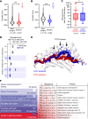
Figure 2. A computational modeling pipeline predicts IsoLG-adducted peptides presented by H-2Db.
(A and B) Rosetta scores are more favorable (more negative) for peptides known to bind to H-2Kb (A) and H-2Db (B) when compared with known nonbinders. ***P < 0.001 by Mann-Whitney test. (C) When lysine-containing peptides are adducted with IsoLG in silico, Rosetta predicts smaller (more favorable) score changes if those peptides are bound to H-2Db compared with H-2Kb (solid bars = mean, dashed line = median, box boundaries = interquartile range, error bars ± min/max values). *P < 0.05 by Mann-Whitney test. (D) Rosetta score changes after in silico IsoLG adduction for all nonanchoring residues in lysine-containing H-2Db–bound epitopes predict residue sites 4, 6, and 7 as energetically favorable positions (positions at which multiple peptides modeled have no score increase after adduction. Black bars indicate mean Δ Rosetta score). (E) These residues correspond to areas recognized by T cell receptors interacting with H-2Db epitopes. (F) Strategy for identifying a library of peptide candidates to screen in vitro and in vivo.
Figure 3
Figure 3. A subset of candidate IsoLG-adducted peptides are recognized by CD8+ T cells enriched in the aortas of hypertensive mice and induce CD8+ T cell proliferation in vitro.
(A) Seven candidate IsoLG-adducted peptides induce the proliferation of T cells isolated from the bone marrow of hypertensive mice, while unadducted peptides do not (n = 7–9, mean ± SD). *P < 0.05 by Student’s t test. (B) Nine IsoLG-adducted peptides identify a population of peptide-specific CD8+ T cells that are enriched in the aortas of hypertensive mice (n = 3–9, mean ± SD). *P < 0.05, **P < 0.01, ***P < 0.001, ****P < 0.0001 by 2-way ANOVA and Holm-Šidák post hoc test. Six candidates are both recognized by CD8+ T cells and induce CD8+ T cell proliferation in vitro (highlighted in red). (C) Representative histograms illustrating proliferation of CD8+ T cells after exposure to each of these 6 candidate IsoLG-adducted peptides and (D) flow plots illustrating the increase in peptide-specific CD8+ T cells in the aorta.
Figure 4
Figure 4. IsoLG-adducted peptide–specific CD8+ T cells are enriched in the kidneys of hypertensive animals.
(A) Candidate peptides adducted with IsoLG are recognized by CD8+ T cells in the kidney and enriched after hypertension is induced with Ang II infusion (n = 3–8, mean ± SD). *P < 0.05; **P < 0.01; ***P < 0.001; ****P < 0.0001 by 2-way ANOVA and Holm-Šidák post hoc test. (B) Representative flow plots illustrating the increase in peptide-specific CD8+ T cells in the kidneys during hypertension.
Figure 5
Figure 5. CD8+ T cells recognizing IsoLG-adducted peptides are predominantly memory T cells in the aorta and kidney.
Staining for memory T cell markers CD44 (all memory cells) and CD62L (central memory cells) reveals that IsoLG-adducted peptide–specific CD8+ T cells are primarily effector memory cells in the kidney (A) and in the aorta (B) (n = 3–8, mean ± SD). FMO, fluorescence-minus-one control. *P < 0.05; **P < 0.01; ***P < 0.001 by 1-way ANOVA and Holm-Šidák post hoc test.
Figure 6
Figure 6. IsoLG-adducted peptides induce hypertension in mice.
(A) Experimental diagram. IsoLG-adducted peptide candidates and their unadducted counterparts were loaded onto DCs and adoptively transferred prior to 14 days of treatment with a subpressor dose of Ang II. (B) Four of the 6 candidates tested induced a significant increase in blood pressure following adoptive transfer, while unadducted peptides did not (n = 4–5, mean ± SD). *P < 0.05; **P < 0.01; ***P < 0.001 by 2-way ANOVA and Holm-Šidák post hoc test. (C) Of the original 13 candidates screened, 4 are recognized by CD8+ T cells, induce CD8+ T cell activation, and induce hypertension in mice following adoptive transfer.
Figure 7
Figure 7. IsoLG-adducted peptides are preferentially displayed by certain HLA molecules.
(A) Treating K562 cells expressing single HLA alleles with tBHP induces a significant increase in the HLA-IsoLG FRET proximity mean fluorescence intensity (MFI) for all alleles screened, excepting the HLA-null control. However, there are certain alleles with significantly higher FRET MFI compared with the lowest measured in each allele class (AC) (n = 4, mean ± SD). *P < 0.05 vs. lowest average MFI for corresponding allele class by 2-way ANOVA and Holm-Šidák post hoc test. (B) Treatment with the dicarbonyl scavenger ethyl-2-HOBA significantly reduces FRET MFI compared with tBHP-only-treated cells (n = 4). #P < 0.05 vs. corresponding tBHP-treated group by 2-way ANOVA with Holm-Šidák post hoc test. (C) Example FRET signal for all HLA alleles tested. “High-presenting” HLA-A and HLA-B alleles are highlighted in blue.

References

    1. Norlander AE, et al. The immunology of hypertension. J Exp Med. 2018;215(1):21–33. doi: 10.1084/jem.20171773. - DOI - PMC - PubMed
    1. Madhur MS, et al. Hypertension: do inflammation and immunity hold the key to solving this epidemic? Circ Res. 2021;128(7):908–933. doi: 10.1161/CIRCRESAHA.121.318052. - DOI - PMC - PubMed
    1. Guzik TJ, et al. Role of the T cell in the genesis of angiotensin II induced hypertension and vascular dysfunction. J Exp Med. 2007;204(10):2449–2460. doi: 10.1084/jem.20070657. - DOI - PMC - PubMed
    1. Rudemiller N, et al. CD247 modulates blood pressure by altering T-lymphocyte infiltration in the kidney. Hypertension. 2014;63(3):559–564. doi: 10.1161/HYPERTENSIONAHA.113.02191. - DOI - PMC - PubMed
    1. Itani HA, et al. Activation of human T cells in hypertension: studies of humanized mice and hypertensive humans. Hypertension. 2016;68(1):123–132. doi: 10.1161/HYPERTENSIONAHA.116.07237. - DOI - PMC - PubMed