. 2024 Aug 27;43(8):114622.
doi: 10.1016/j.celrep.2024.114622. Epub 2024 Aug 14.

RPL22 is a tumor suppressor in MSI-high cancers and a splicing regulator of MDM4

Affiliations

RPL22 is a tumor suppressor in MSI-high cancers and a splicing regulator of MDM4

Hannah N W Weinstein et al. Cell Rep. .

Abstract

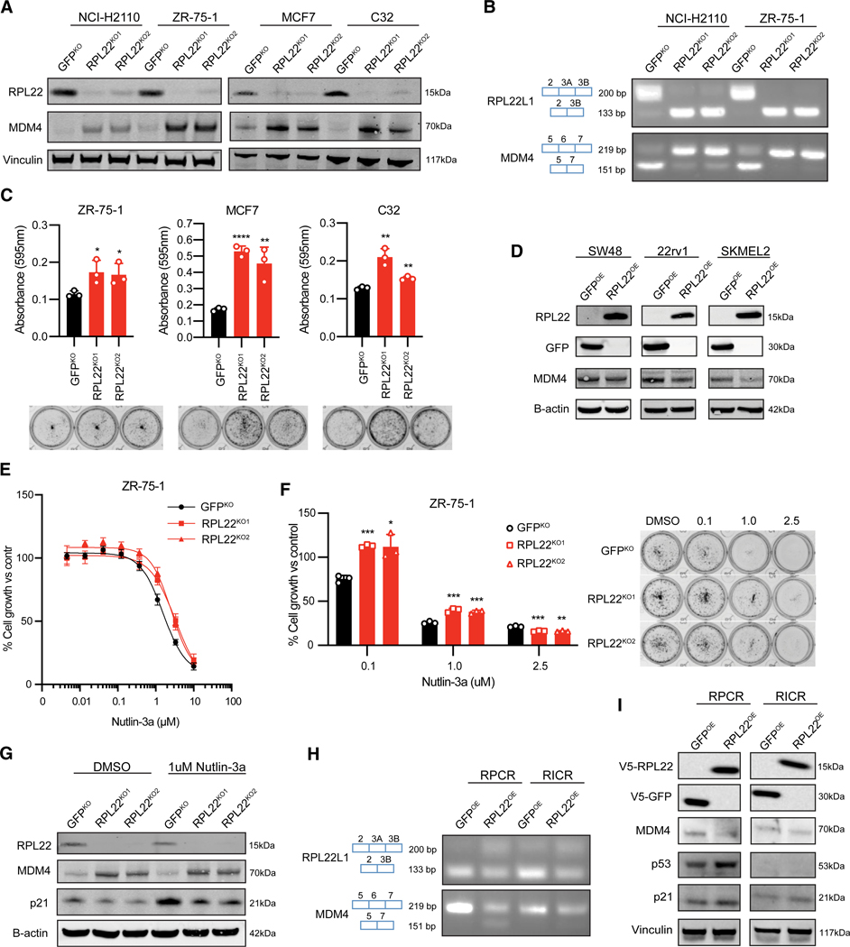
Microsatellite instability-high (MSI-H) tumors are malignant tumors that, despite harboring a high mutational burden, often have intact TP53. One of the most frequent mutations in MSI-H tumors is a frameshift mutation in RPL22, a ribosomal protein. Here, we identified RPL22 as a modulator of MDM4 splicing through an alternative splicing switch in exon 6. RPL22 loss increases MDM4 exon 6 inclusion and cell proliferation and augments resistance to the MDM inhibitor Nutlin-3a. RPL22 represses the expression of its paralog, RPL22L1, by mediating the splicing of a cryptic exon corresponding to a truncated transcript. Therefore, damaging mutations in RPL22 drive oncogenic MDM4 induction and reveal a common splicing circuit in MSI-H tumors that may inform therapeutic targeting of the MDM4-p53 axis and oncogenic RPL22L1 induction.

Keywords: CP: Cancer; CP: Molecular biology; MDM4; MDM4 exon 6 inclusion; MSI-H; RPL22; RPL22 p.K15fs; alternative splicing; microsattelite instability-high; p53; ribosomal proteins; tumor suppressor.


Conflict of interest statement

Declaration of interests F.V. receives research support from the Dependency Map Consortium, Riva Therapeutics, Bristol Myers Squibb, Merck, Illumina, and Deerfield Management. F.V. is on the scientific advisory board of GSK, is a consultant and holds equity in Riva Therapeutics, and is a co-founder and holds equity in Jumble Therapeutics.

Figures

Figure 1.
Figure 1.. RPL22 genomic alterations are common in cancer and associated with changes to specific transcripts of MDM4 and RPL22L1 mRNA
(A) Frequencies of RPL22 p.K15fs frameshift mutation in microsatellite instability-high (MSI-H) cell lines in the Cancer Cell Line Encyclopedia (CCLE) and MSI-H tumors in The Cancer Genome Atlas (TCGA) (COAD, colon adenocarcinoma; STAD, stomach adenocarcinoma; UCEC, uterine corpus endometrial carcinoma). (B) Proportion of RPL22 copy-number loss and truncating mutations across TCGA tissue types. ΔCN, copy-number loss, single (—1) or biallelic (—2). (C) Association between decreased inclusion of MDM4 exon 6 and deleterious alterations to RPL22 in TCGA samples. (D) Association between decreased inclusion of RPL22L1 exon 3A and deleterious alterations to RPL22 in TCGA samples. (E) Schematic of splicing switches responsible for the major MDM4 (top) and RPL22L1 (bottom) isoforms. (F) Association between RPL22L1 dependency and deleterious alterations to RPL22 in CCLE samples. (G) Focus formation assay of MSI-H RPL22 mutant cell lines with short hairpin RNA knockdown of RPL22L1. p values: SW48 RPL22L1KD1 = 0.0008, SW48 RPL22L1KD2 = 0.0025, LNCaP RPL22L1KD1 = 0.0400, and LNCaP RPL22L1KD2 = 0.0155. Error bars represent the mean ± standard deviation (SD) of three replicates. *p < 0.05, **p < 0.01, ***p < 0.001, and ****p < 0.0001.
Figure 2.
Figure 2.. MDM4 and RPL22L1 are splicing targets of RPL22
(A) Total RPL22L1 mRNA expression across experiments. Bars represent the mean of three replicates (**p < 0.01). (B) Common differentially expressed genes across RPL22 knockout experiments. The columns and numbers indicate the total counts of differentially expressed genes in each combination of intersections between the different experiments. (C) Inclusion proportions of RPL22L1 exon 3A across experiments. RPL22 knockdown reduces RPL22L1 exon 3A inclusion. Bars represent the mean of three replicates (**p < 0.01). PSI, percent spliced in. (D) Changes in splicing modes across experiments as indicated by significant differentially spliced variants. The numbers in parentheses indicate the total counts of differential splicing events in each experiment. A3SS, alternative 3 splicing site; A5SS, alternative 5 splicing site; MXE, mutually exclusive exons; RI, retained intron; SE, skipped exon. (E) Common splicing changes across RPL22 knockout experiments. The columns and numbers indicate the total counts of differentially expressed exons in each combination of intersections between the different experiments. (F) Inclusion levels of MDM4 exon 6 across experiments. Bars represent the mean of three replicates (**p < 0.01).
Figure 3.
Figure 3.. Loss of RPL22 increases MDM4 expression and augments resistance to Nutlin-3a
(A) Immunoblot of MSS cell lines with CRISPR-Cas9 knockout of RPL22. (B) RT-PCR analysis of MSS cell lines with CRISPR-Cas9 knockout of RPL22. (C) Focus formation assay of MSS cell lines with CRISPR-Cas9 knockout of RPL22. p values: ZR-75–1 RPL22KO1 = 0.0377, ZR-75–1 RPL22KO2 = 0.0464, MCF7 RPL22KO1 < 0.0001, MCF7 RPL22KO2 = 0.0089, C32 RPL22KO1 = 0.0033, and C32 RPL22KO2 = 0.0029. Error bars represent the mean ± SD of three replicates. (D) Immunoblot of RPL22 mutant MSI-H cell lines with overexpression of RPL22. (E) Nutlin-3a treatment of MSS cell lines (ZR-75–1) with CRISPR-Cas9 knockout of RPL22. Error bars represent the mean ± SD of six replicates. Half-maximal inhibitory concentrations (IC50) were as follows: GFPKO = 1.566 μM, RPL22KO1 = 3.297 μM, and RPL22KO2 = 2.574 μM. (F) Focus formation assay with Nutlin-3a treatment of MSS cell lines (ZR-75–1) with CRISPR-Cas9 knockout of RPL22. p values: 0.1 μM Nutlin-3a: ZR-75–1 RPL22KO1 = 0.0001 and ZR-75–1 RPL22KO2 = 0.0126; 1.0 μM Nutlin-3a: ZR-75–1 RPL22KO1 = 0.0008 and ZR-75–1 RPL22KO2 = 0.0008; and 2.5 μM Nutlin-3a: ZR-75–1 RPL22KO1 = 0.0010 and ZR-75–1 RPL22KO2 = 0.0015. Error bars represent the mean ± SD of three replicates. (G) Immunoblot of MSS cell line (ZR-75–1) with CRISPR-Cas9 knockout of RPL22 with immunostaining for p21 after 1 week of 1 μM Nutlin-3a treatment. (H) RT-PCR analysis of RPL22 mutant MSI-H cell lines with overexpression of RPL22. RPCR cells are TP53 wild type, while RICR cells are TP53 null. (I) Immunoblot of RPL22 mutant MSI-H cell lines with overexpression of RPL22.
Figure 4.
Figure 4.. Characterization of RPL22 binding preference with CLIP-seq
(A) Binding of RPL22 to the 3 UTR and exon 6 of MDM4 as determined by peak calling on CLIP-seq measurements from ZR-75–1 cells. p = 0.05. (B) Left: peak frequencies relative to the transcription termination site (TTS). Right: breakdown of peak-containing regions. (C) Gene set enrichment analysis of RPL22-bound peaks. (D) Overlaps between genes containing RPL22-bound peaks and differentially spliced genes across RPL22 modulation experiments. Total number of genes and hypergeometric test for each cell line: LNCaP: 15,602 genes, p = 1.536 × 10⁻⁴; NCI-H2110: 15,226 genes, p = 6.682 × 10⁻⁹; and ZR75–1: 15,690 genes, p = 8.901 × 10⁻⁶. (E) Schematic model of loss of RPL22 and recurrent RPL22 frameshift mutations in MSI-H tumors that results in a splicing-mediated switch to the functional isoforms of MDM4 and RPL22L1. The functional isoform of MDM4 inhibits p53 activity and increased levels of RPL22L1 compensate for RPL22 reduction in the ribosome and perform additional extraribosomal functions.

Update of

References

    1. Maruvka YE, Mouw KW, Karlic R, Parasuraman P, Kamburov A, Polak P, Haradhvala NJ, Hess JM, Rheinbay E, Brody Y, et al. (2017). Analysis of somatic microsatellite indels identifies driver events in human tumors. Nat. Biotechnol 35, 951–959. 10.1038/nbt.3966. - DOI - PMC - PubMed
    1. Eso Y, Shimizu T, Takeda H, Takai A, and Marusawa H. (2020). Microsatellite instability and immune checkpoint inhibitors: toward precision medicine against gastrointestinal and hepatobiliary cancers. J. Gastroenterol 55, 15–26. 10.1007/s00535-019-01620-7. - DOI - PMC - PubMed
    1. Cortes-Ciriano I, Lee S, Park W-Y, Kim T-M, and Park PJ (2017). A molecular portrait of microsatellite instability across multiple cancers. Nat. Commun 8, 15180. 10.1038/ncomms15180. - DOI - PMC - PubMed
    1. Baugh EH, Ke H, Levine AJ, Bonneau RA, and Chan CS (2018). Why are there hotspot mutations in the TP53 gene in human cancers? Cell Death Differ. 25, 154–160. 10.1038/cdd.2017.180. - DOI - PMC - PubMed
    1. Ghandi M, Huang FW, Jané-Valbuena J, Kryukov GV, Lo CC, Mcdonald ER, Barretina J, Gelfand ET, Bielski CM, Li H, et al. (2019). Next-generation characterization of the Cancer Cell Line Encyclopedia. Nature 569, 503–508. 10.1038/s41586-019-1186-3. - DOI - PMC - PubMed

MeSH terms