. 2024 Aug 15;20(8):e1012468.
doi: 10.1371/journal.ppat.1012468. eCollection 2024 Aug.

Rapid adaptive evolution of avian leukosis virus subgroup J in response to biotechnologically induced host resistance

Affiliations

Rapid adaptive evolution of avian leukosis virus subgroup J in response to biotechnologically induced host resistance

Magda Matoušková et al. PLoS Pathog. .

Abstract

Genetic editing of the germline using CRISPR/Cas9 technology has made it possible to alter livestock traits, including the creation of resistance to viral diseases. However, virus adaptability could present a major obstacle in this effort. Recently, chickens resistant to avian leukosis virus subgroup J (ALV-J) were developed by deleting a single amino acid, W38, within the ALV-J receptor NHE1 using CRISPR/Cas9 genome editing. This resistance was confirmed both in vitro and in vivo. In vitro resistance of W38-/- chicken embryonic fibroblasts to all tested ALV-J strains was shown. To investigate the capacity of ALV-J for further adaptation, we used a retrovirus reporter-based assay to select adapted ALV-J variants. We assumed that adaptive mutations overcoming the cellular resistance would occur within the envelope protein. In accordance with this assumption, we isolated and sequenced numerous adapted virus variants and found within their envelope genes eight independent single nucleotide substitutions. To confirm the adaptive capacity of these substitutions, we introduced them into the original retrovirus reporter. All eight variants replicated effectively in W38-/- chicken embryonic fibroblasts in vitro while in vivo, W38-/- chickens were sensitive to tumor induction by two of the variants. Importantly, receptor alleles with more extensive modifications have remained resistant to the virus. These results demonstrate an important strategy in livestock genome engineering towards antivirus resistance and illustrate that cellular resistance induced by minor receptor modifications can be overcome by adapted virus variants. We conclude that more complex editing will be necessary to attain robust resistance.


Conflict of interest statement

The authors have declared that no competing interests exist.

Figures
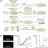
Fig 1
Fig 1. Detection of adapted RCASBP(J)GFP variants that overcome W38-/- CEF resistance.
(A) Preparation of adapted variants: schematic representation of the workflow and timeline. (B) Representative images of W38-/- fibroblasts observed by fluorescence microscope 2 d.p.i. with (P+) or without (P-) polybrene support are shown. Cell nuclei were stained with DAPI. (C) Outcome of the primary infection: six RCASBP(J)GFP virus stocks were prepared by transfection of DF-1 cells with pRCASBP(J)GFP plasmid in six parallels. Virus was collected 1 week after transfection. W38-/- CEF infections performed either with support of polybrene (black lines) or without (gray lines) are shown. The virus spread in the cell culture was assayed as the percentage of GFP-positive cells by FACS for 14 days. (D) Comparison of W38-/- CEF primary and secondary infection efficacy with (black dots) or without (gray dots) polybrene support 2 d.p.i. Figure was created with Biorender.com.
Fig 2
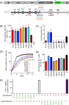
Fig 2. Characterization of adapted ALV-J variants.
(A) Scheme of RCASBP(J)GFP and Env glycoprotein domain structure with positions of all amino acid substitutions detected in adapted variants (black triangles). Host-range regions (hr1/2) and variable regions (vr1/2/3) in the SU are depicted as dark gray boxes. Heptad repeats (HR1/2) in the TM are shown as light gray boxes. env was amplified by depicted primers (blue arrows) from RCASBP(J)GFP infected W38-/- fibroblasts. (B) W38-/- CEFs and DF-1 cells were infected with wt RCASBP(J)GFP and eight cloned variants each carrying a single adaptive mutation. Cells were analyzed 2 d.p.i. by FACS. The percentage of GFP-positive W38-/- fibroblasts was normalized to the percentage of GFP-positive DF-1 cells infected in parallel. Each infection was performed in triplicate and means ± SD are shown. Student’s T-test comparison is shown for comparison with the A432T variant. T-test comparisons for all variants are shown in Supplementary Information (S2 Table). (C) Thermosensitivity of the RCASBP(J)GFP variants. Triplicates of RCASBP(J)GFP variants were incubated at 42°C or kept on ice for 3 hours. DF-1 cells were infected with these viruses and the GFP positivity was determined 3 d.p.i. by FACS. The results are presented as the ratio of GFP-positive DF-1 cells infected by the virus kept at 42°C and GFP-positive DF-1 cells infected by the virus kept on ice. Each value represents the mean percentage ± SD. (D) Virus spread at 37 and 42°C. DF-1 cells were infected with all virus variants and GFP positive cells were sorted. The infected cells were mixed with fresh DF-1 cells in the ratio 1:19. The virus spread was calculated as the percentage of GFP-positive cells by FACS at 4, 7, 9, and 11 days of co-cultivation. (E) Difference of virus spread in DF-1 cells at 42°C normalized to the percentage of GFP positive cells cultured at 37°C. The GFP positivity was measured 4 days after mixing with fresh cells. T-test comparisons for all variants are shown in supplementary information (S3 Table). (F) Superinfection interference of RCASBP(J)GFP variants (green labels of substitutions) with the wt RCASBP(J)dsRED (red wt). RCASBP(B)GFP (green B) was used as a negative control of infection interference. The results are shown as mean percentages of GFP-positive/dsRed-positive cells measured in triplicate. Figure was created with Biorender.com.
Fig 3
Fig 3. Sensitivity of W38-/- chickens to selected adapted RCASBP(J)GFP variants.
V-src-transducing virus pseudotyped with selected variants of J envelope was used to induce tumor growth in W38-/- (A) and in control W38+/+ chickens (B). Tumor growth was inspected 10, 14, 21 and 35 d.p.i. Each dot represents the percentage of chickens infected with a particular virus variant carrying a tumor at the time of inspection. W38-/- chickens and control W38+/+ chickens were infected by each virus at least in triplicate. n indicates numbers of chickens used for infection by individual variants of pseudotyped viruses. Figure was created with Biorender.com.
Fig 4
Fig 4. Sensitivity of modified DF-1 cell lines and primary fibroblasts to RCASBP(J)GFP variants.
(A) Fibroblasts prepared from selected galliform species and DF-1 cell lines with partial or complete deletion of ALV-J receptor NHE1 were infected in triplicates by wt RCASBP(J)GFP and variants with mutations N311D and A432T. In the left column, the source of primary fibroblasts or NHE1 modification in DF-1 cells is described. The middle column shows alignment of the crucial part of the first extracellular loop of NHE1 molecule involved in ALV-J infection. Amino acids matching the sequence of the original DF-1 cell line are on a gray background. The W38 tryptophan is highlighted in red. Graph on the right side shows the percentage of GFP-positive cells analyzed by FACS 3 d.p.i. with the respective virus (mean ± SD). (B) Individual GFP-positive DF-1 ΔNHE1 cells infected with adapted RCASBP(J)GFP variants were sorted. Collected virus was used for secondary infection of DF-1 ΔNHE1 and DF-1. Representative photos of infection with a RCASBP(J)GFP variant carrying N311K substitution are shown. Other examples are shown in S2 Fig. Figure was created with Biorender.com.

References

    1. Gao F, Li P, Yin Y, Du X, Cao G, Wu S, et al.. Molecular breeding of livestock for disease resistance. Virology. 2023;587: 109862. doi: 10.1016/j.virol.2023.109862 - DOI - PubMed
    1. Whitworth KM, Rowland RRR, Ewen CL, Trible BR, Kerrigan MA, Cino-Ozuna AG, et al.. Gene-edited pigs are protected from porcine reproductive and respiratory syndrome virus. Nat Biotechnol. 2016;34: 20–22. doi: 10.1038/nbt.3434 - DOI - PubMed
    1. Koslová A, Trefil P, Mucksová J, Reinišová M, Plachý J, Kalina J, et al.. Precise CRISPR/Cas9 editing of the NHE1 gene renders chickens resistant to the J subgroup of avian leukosis virus. Proc Natl Acad Sci U S A. 2020;117: 2108–2112. doi: 10.1073/pnas.1913827117 - DOI - PMC - PubMed
    1. Koslová A, Trefil P, Mucksová J, Krchlíková V, Plachý J, Krijt J, et al.. Knock-Out of Retrovirus Receptor Gene Tva in the Chicken Confers Resistance to Avian Leukosis Virus Subgroups A and K and Affects Cobalamin (Vitamin B12)-Dependent Level of Methylmalonic Acid. Viruses. 2021;13. doi: 10.3390/v13122504 - DOI - PMC - PubMed
    1. Sheppard CM, Goldhill DH, Swann OC, Staller E, Penn R, Platt OK, et al.. An Influenza A virus can evolve to use human ANP32E through altering polymerase dimerization. Nat Commun. 2023;14: 6135. doi: 10.1038/s41467-023-41308-4 - DOI - PMC - PubMed

Substances

LinkOut - more resources