. 2024 Aug 16;385(6710):eadh7814.
doi: 10.1126/science.adh7814. Epub 2024 Aug 16.

Hebbian instruction of axonal connectivity by endogenous correlated spontaneous activity

Affiliations

Hebbian instruction of axonal connectivity by endogenous correlated spontaneous activity

Naoyuki Matsumoto et al. Science. .

Abstract

Spontaneous activity refines neural connectivity prior to the onset of sensory experience, but it remains unclear how such activity instructs axonal connectivity with subcellular precision. We simultaneously measured spontaneous retinal waves and the activity of individual retinocollicular axons and tracked morphological changes in axonal arbors across hours in vivo in neonatal mice. We demonstrate that the correlation of an axon branch's activity with neighboring axons or postsynaptic neurons predicts whether the branch will be added, stabilized, or eliminated. Desynchronizing individual axons from their local networks, changing the pattern of correlated activity, or blocking N-methyl-d-aspartate receptors all significantly altered single-axon morphology. These observations provide the first direct evidence in vivo that endogenous patterns of correlated neuronal activity instruct fine-scale refinement of axonal processes.


Conflict of interest statement

Competing interests: The authors declare that they have no competing interests.

Figures

Fig. 1.
Fig. 1.. Synchronized firing between individual RGC axons and retinal waves instructs axon branch dynamics.
(A) Schematic of the in vivo two-photon (2P) imaging approach for tracking axon branch dynamics of a single RGC and simultaneous dual-color calcium imaging of the RGC axon activity and retinal waves in the SC of awake, head-restrained mice at P8 to P9. Stochastic expression of Cre in a few RGCs was achieved by intravitreal injections of AAV2/1-TRE-Cre in Ai162 mice, which harbor CAG-LSL-tTA2 and TRE2-LSL-GCaMP6s alleles. AAV2/2-CAG-FLEX-EGFP and AAV2/1-Syn-jRGECO1a were also injected intravitreally. As a result, Cre+ RGCs expressed GFP, GCaMP6s, and jRGECO1a, whereas Cre RGCs only expressed jRGECO1a. The blue-boxed area is magnified in (B). Scale bar, 500 μm. PMT, photomultiplier tube; Ti:Sapphire, Ti:Sapphire laser. (B) In vivo two-photon imaging of an entire single RGC axon within a depth of 200 μm below the surface of the SC at P8. Directions R, L, M, and C correspond to rostral, lateral, medial, and caudal in the SC, respectively, unless otherwise stated. Scale bar, 100 μm. (C) Experimental timeline. Z stacks of a single–RGC axon arbor were acquired at a 2-hour interval, and dual-color calcium imaging was performed for 90 min in between taking the z stacks. (D) Traced z projection of axon branches from a single RGC within ±20 μm of the optical plane for dual-color calcium imaging. Orange and red circles indicate central and distal regions of interest (ROIs) for calculating the retinal wave traces shown in (F), respectively. (E) Example ΔF/F (fractional change in fluorescence) montages of single-axon firing (GCaMP6s) and of retinal waves (jRGECO1a). In montage 1, the axon did not fire when a retinal wave partially overlapped with its axon arbors. In montage 2, the axon fired when a retinal wave passed the center of the axon arbor. Orange and red circles indicate ROIs in the central and distal regions of the axon, respectively. Scale bar, 100 μm. (F) A GCaMP6s signal trace (ΔF/F) from the single RGC axon and jRGECO1a signal traces (ΔF/F) from the ROIs indicated in (D). The periods (1 and 2) depicted in montages (E) are shown in gray. (G) The fraction of synchronization between single-axon firing and retinal waves at central regions of the axons was significantly higher than that at distal regions. Data points from the same axon were paired (central, 0.92 ± 0.02; distal, 0.49 ± 0.05; **P = 0.008, one-tailed Wilcoxon signed-rank test, n = 7 axons from 7 animals). (H and I) Z projection of a single RGC axon at 0 (H) and 2 hours (I) of in vivo two-photon imaging. The orange-boxed areas in (H) are magnified in (K). Scale bar, 100 μm. (J) A single reconstructed RGC axon. The behavior of individual branches over the 2-hour interval were categorized as added (green), eliminated (red), extended (cyan), and retracted (magenta). (K) Zoomed-in images [corresponding to numbered orange boxes in (H)] show changes of axon branches over the 2-hour imaging session. The left and middle columns show projected images of two to three optical sections with 2-μm intervals at 0 and 2 hours. The right column displays changes of branch dynamics in 2 hours. Scale bar, 10 μm. (L) Terminals of stable (white), eliminated (red), extended (cyan), and retracted (magenta) axon branches at 2 hours were labeled on a z projection of the single–RGC axon arbor. The positions from which newly added branches emerged were also labeled (green). Scale bar, 100 μm. (M) A spatial map of Pearson’s correlation coefficients (r) between the activity of the single RGC axon and retinal waves. (N) Stable and eliminated branch terminals and positions where newly added branches emerged from were plotted on the correlation map. (O) Means of correlation coefficients [stable, 0.34 ± 0.01; added, 0.42 ± 0.01, P = 0.0004; eliminated, 0.26 ± 0.03, P = 0.001; extended, 0.33 ± 0.02, P = 0.999; retracted, 0.31 ± 0.03, P = 0.36; ns, not significant; one-way analysis of variance (ANOVA) with Dunnett’s multiple comparison test, n = 7 axons from 7 animals]. (P to S) Simultaneous in vivo two-photon imaging of single–RGC axon branch dynamics and dual-color calcium imaging of axonal activity and postsynaptic waves in the SC. Stochastic expression of Cre in a few RGCs was achieved by intravitreal injections of AAV2/1-TRE-Cre in Ai162 mice, which harbor CAG-LSL-tTA2 and TRE-LSL-GCaMP6s alleles, and expression of jRGECO1a in neurons of the SC was achieved by injection of AAV2/9-Syn-NES-jRGECO1a into the SC one day after eye injection. After the AAV injections, only a few RGC axons expressed GFP and GCaMP6, and most of SC neurons expressed jRGECO1a. (P) Terminals of stable, eliminated, extended, and retracted axon branches at 2 hours were labeled on a z projection of the single RGC axon arbor. The positions from which newly added branches emerged were also labeled. Scale bar, 100 mm. (Q) A spatial map of Pearson’s correlation coefficients between presynaptic activity of the single–RGC axon arbor and postsynaptic waves. (R) Stable and eliminated branch terminals and positions from where newly added branches emerged were plotted on the correlation map. (S) Mean correlation coefficients (stable, 0.52 ± 0.03; added, 0.57 ± 0.03, P = 0.0097; eliminated, 0.44 ± 0.03, P = 0.0002; extended, 0.49 ± 0.03, P = 0.29; retracted, 0.51 ± 0.03, P = 0.88, one-way ANOVA with Dunnett’s multiple comparison test, n = 6 axons from 6 animals). (T) Schematic model of Hebbian axon remodeling by endogenous patterns of spontaneous activity. Synchronized presynaptic inputs (retinal waves) excite postsynaptic cells (i and ii). When an RGC axon takes part in firing a SC neuron repeatedly or persistently (i), the axon forms new branches near the SC neurons to increase their efficacy (iii). By contrast, when an RGC axon does not participate in making an SC neuron fire repeatedly or persistently (ii), branches of the axon near the SC neuron are eliminated (iii). For all the box plots, the central line indicates the median, and the bottom and top edges indicate the 25th and 75th percentiles of the data across animals, respectively. **P < 0.01; ***P < 0.001; ns, not significant. Data are mean ± SEM.
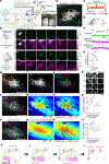
Fig. 2.
Fig. 2.. Decoupling synchronization between retinal waves and individual axon firing results in random distribution of added and eliminated axon branches.
β2-nAChR was knocked out in only a few RGCs by intravitreal injections of AAV2/1-TRE-Cre in Ai162; β2fl/− mice. (A) ΔF/F montages of single-axon firing (GCaMP6s) and of retinal waves (jRGECO1a) in Ai162; β2fl/−. In montage 1, a β2-nAChR–knockout axon did not fire when a retinal wave covered the center of the axon arbor, but it did fire in montage 2. Orange circles correspond to a ROI in the central region of the axon for traces in (B). Scale bars, 100 μm. (B) GCaMP6s and jRGECO1a signal traces (ΔF/F) of the ROI indicated in (A) from an Ai162; β2fl/− mouse. The periods (1 and 2) depicted in montages (A) are shown in gray. (C) The fraction of retinal waves and single-axon firing that are synchronized in β2-nAChR–knockout (Ai162; β2fl/−) and control axons (Ai162; β2fl/+) (β2fl/+, 0.91 ± 0.03; β2fl/−, 0.14 ± 0.09; **P = 0.003, one-tailed Wilcoxon rank sum test). (D) The frequency of retinal waves was not altered (β2fl/+, 0.45 ± 0.06; β2fl/−, 0.45 ± 0.05; P = 0.28, one-tailed Wilcoxon rank sum test). (E) The frequency of spontaneous firing was reduced in β2-nAChR–knockout axons (β2fl/+, 0.46 ± 0.06; β2fl/−, 0.07 ± 0.04; P = 0.003, one-tailed Wilcoxon rank sum test). (F) The frequency of single-axon firing in the absence of retinal waves was not altered (β2fl/+, 0.017 ± 0.008; β2fl/−, 0.010 ± 0.009; P = 0.28, one-tailed Wilcoxon rank sum test). [(C) to (F)] β2fl/+, n = 4 axons from 4 animals at P8; β2fl/−, n = 7 axons from 7 animals at P8. (G) Terminals of stable (white), eliminated (red), extended (cyan), and retracted (magenta) axon branches at 2 hours were labeled on a z projection of a β2-nAChR–knockout RGC axon arbor. The positions from which newly added branches emerged were also plotted (green). Scale bar, 100 μm. (H) A spatial map of Pearson’s correlation coefficient between the firing of the β2-nAChR–knockout RGC axon and retinal waves. (I) Stable and eliminated branch terminals and the positions from which added branches emerged were plotted on the correlation map. (J) Distances from the center of single-axon arbors to their stable and eliminated branch terminals and added branch points. Distances were normalized by the average distance from the center to each branch. The normalized values would be one if axon branch positions were randomly distributed and would be smaller than one if axon branch positions were distributed near the center (stable: β2fl/+, 1.00 ± 0.02; β2fl/−, 1.03 ± 0.03; P = 0.47. added: β2fl/+, 0.78 ± 0.04; β2fl/−, 1.03 ± 0.06; P = 0.015. eliminated: β2fl/+, 1.24 ± 0.04; β2fl/−, 0.89 ± 0.04; P = 0.002. β2fl/+, n = 5 axons from 5 animals at P8; β2fl/−, n = 6 axons from 6 animals at P8; one-tailed Wilcoxon rank sum test). (K) Axon branch terminals (green) and covariance ellipses with a 90% confidence interval (red) were overlaid on the z projection of a single RGC axon from an Ai162; β2fl/+ control mouse (left) and from an Ai162; β2fl/− mouse (right) at P8. Scale bar, 100 μm. (L) The areas of covariance ellipses were enlarged in β2-nAChR–knockout axons (β2fl/+, 0.023 ± 0.002 mm2, n = 5 axons from 5 animals at P8; β2fl/−, 0.037 ± 0.005 mm2, n = 6 axons from 6 animals at P8; P = 0.04; one-tailed Wilcoxon rank sum test). *P < 0.05; **P < 0.01; ***P < 0.001; ns, not significant. Data are mean ± SEM.
Fig. 3.
Fig. 3.. Patterns of retinal waves are related to the direction of RGC axonal extension.
(A) Schematic illustration of distinct directions of RGC axonal extension in the midline region (1, green; within 200 μm from the midline) and the lateral region (2, red; at least 400 μm away from the midline) of the SC. (B) Z projections of single RGC axons with rostrocaudal extension in the midline region (1) and with mediolateral extension in the lateral region (2). Scale bars, 100 μm. (C) Ellipse fitting for a mediolaterally extended RGC axon in the lateral region of the SC. A covariance ellipse with a 90% confidence interval (red) was calculated from axon branch terminal positions (green) by using principal component analysis (PCA). The eigenvectors with the length corresponding to the eigenvalues are also shown (blue). Scale bar, 100 μm. (D) Orientation angles of the major axes of covariance ellipses at P8 to P9 (midline region, 77.4 ± 3.8°, n = 4 axons from 4 animals; lateral region, 18.7 ± 3.7°, n = 7 axons from 7 animals; P = 0.003, one-tailed Wilcoxon rank sum test). (E to G) In vivo wide-field single-photon calcium imaging of retinal waves in the SC at P8 to P9. GCaMP6s expression in RGCs was achieved by intravitreal injections of AAV2/1-Syn-GCaMP6s at P0 to P1. (E) ΔF/F montages of a retinal wave propagating in the lateral region (top) and of flashlike spontaneous activity in the midline area (bottom). Scale bar, 500 μm. (F) Examples of seed-based correlation maps of retinal waves, with the seed located in the lateral (top) and the midline regions (bottom), respectively. Black dots indicate seed locations. Scale bar, 500 μm. (G) Orientation angles of the area where correlation coefficient values in a correlation map were above the threshold (4 SD + mean) (midline, 80.0 ± 2.3°; lateral, 17.7 ± 2.7°; P = 0.001, one-tailed Wilcoxon signed-rank test, n = 4 animals at P9). (H and I) In vivo wide-field single-photon calcium imaging of RGC axons in the SC of FRMD7tm mice. (H) Examples of seed-based correlation maps when seed locations were in the lateral region of the SC in a control mouse (top) and in a FRMD7 mutant mouse (bottom). Black dots indicate seed locations. Scale bar, 500 μm. (I) The ratio of the mediolateral axis length over the rostrocaudal axis length for the high correlation area was reduced in FRMD7 mutant mice (control, 1.58 ± 0.15, n = 4 animals at P9; FRMD7, 1.18 ± 0.04, n = 4 animals at P9; P = 0.03, one-tailed Wilcoxon rank sum test). A ratio of more than 1 suggests that the high correlation area is mediolaterally extended. (J to M) Single-axon morphology in FRMD7tm mice. Sparse GFP expression in RGCs was achieved by intravitreal injections of AAV2/1-TRE-Cre and AAV2/2-CAG-FLEX-EGFP in Ai162; FRMD7tm mice. (J) Axon branch terminals (green) and their covariance ellipses with a 90% confidence interval (red) were plotted over the z projections of a single RGC axon in Ai162; FRMD7tm (right) and its control (left) at P9. Scale bar, 100 μm. (K) Lengths of the major axes of covariance ellipses with a 90% confidence interval calculated from axon branch terminal positions of individual axon arbors (control, 221.9 ± 12.1 μm; FRMD7, 186.5 ± 8.2 μm; P = 0.024, one-tailed Wilcoxon rank sum test). (L) Lengths of minor axes of covariance ellipse (control, 119.6 ± 5.9 μm; FRMD7, 135.3 ± 7.6 μm; P = 0.13, one-tailed Wilcoxon rank sum test). (M) The ratio of the length of the major axis over the length of the minor axis of the covariance ellipse (control, 1.88 ± 0.13; FRMD7, 1.40 ± 0.07; P = 0.015, one-tailed Wilcoxon rank sum test). [(K) to (M)] All axons were from lateral regions. Control, n = 5 axons from 5 animals at P9; FRMD7, n = 7 axons from 7 animals at P9. (N) Terminals of stable (white), eliminated (red), extended (cyan), and retracted (magenta) axon branches at 2 hours were labeled on a z projection of a single–RGC axon arbor in an Ai162; FRMD7tm mouse. Positions from which newly added branches emerged were also plotted (green). Scale bar, 100 μm. (O) A spatial map of Pearson’s correlation coefficients between the activity of a single RGC axon and retinal waves in a FRMD7 mutant. (P) Stable and eliminated branch terminals and positions from which added branches emerged were plotted on the correlation map. (Q) Mean correlation coefficients for axon branches exhibiting different dynamics were significantly different in FRMD7 mutant mice (stable, 0.32 ± 0.03; added, 0.37 ± 0.03, P = 0.002; eliminated, 0.28 ± 0.03, P = 0.016; extended, 0.32 ± 0.03, P = 0.999; retracted, 0.31 ± 0.02, P = 0.682; one-way ANOVA with Dunnett’s multiple comparison test, n = 6 axons from 6 animals at P9). (R) The ratio of the mediolateral axis length over the rostrocaudal axis length for area with correlation coefficients of more than 2.5 SD + mean in a spatial map of Pearson’s correlation coefficients between the activity of a single RGC axon and retinal waves (control, 1.72 ± 0.08, n = 7 axons from 7 animals; FRMD7, 1.37 ± 0.11, n = 6 axons from 6 animals; P = 0.01, one-tailed Wilcoxon rank sum test). A ratio >1 suggests that the high correlation area is mediolaterally extended. *P < 0.05, **P < 0.01. Data are mean ± SEM.
Fig. 4.
Fig. 4.. Presynaptic release sites are related to the position of added and eliminated branches at the single-axon level.
In vivo two-photon imaging combining time-lapse imaging of axon branch dynamics and imaging of glutamate release from a single RGC in the SC of awake mice at P9. Stochastic expression of Cre in a few RGCs was achieved by intravitreal injections of AAV2/1-TRE-Cre and AAV2/2-CAG-FLEX-tTA2 in Ai9 mice, which harbor CAG-LSL-tdTomato. AAV2/1-hSyn-FLEX-iGluSnFR3-PDGFR was also injected intravitreally. As a result, only Cre+ RGCs expressed tdTomato and iGluSnFR3. (A) Experimental timeline. Z stacks of a single-RGC axon arbor were acquired at a 2-hour interval. In vivo two-photon glutamate imaging was performed for 6 min after taking the z stacks. (B) Z projection of a single RGC axon labeled by tdTomato and imaged in vivo at P9. The orange-boxed area is magnified in (C). Scale bar, 100 μm. (C and D) Optical sections of the single RGC axon at 0 (C) and 2 hours (D). The orange-boxed areas in (D) are magnified in (J). Scale bar, 100 μm. (E) The behavior of individual branches over the 2-hour interval was categorized as added (green), eliminated (red), extended (cyan), or retracted (magenta). (F) Example ΔF/F montages of single-axon glutamate release (iGluSnFR3). Scale bar, 100 μm. (G) Maximum values of ΔF/F of the example field of view. (H) GRSs (magenta) are overlaid on the optical section (white). GRSs were defined as axon segments with significant iGluSnFR signal (ΔF/F more than mean + 2 SD) and containing at least six pixels (1 μm2). (I) iGluSnFR signal traces (ΔF/F) corresponding to the GRSs indicated in (H). The period depicted in montages (F) are shown in gray. (J) Zoomed-in images corresponding to the numbered orange boxes in (D) that show changes of example axon branches over the 2-hour imaging session. (Left) Maximum values of ΔF/F (iGluSnFR3). (Middle) Projected images of three optical sections with 2-μm intervals at 0 and 2 hours (tdTomato). (Right) Added branches (green) in 2 hours. Scale bar, 10 μm. (K) GRSs with newly added branch points are shown in green. Scale bar, 100 μm. (L) The positions of added (green), eliminated (red), and persisting (white) branch points are overlaid on GRSs (magenta). (M) The fraction of branch points with GRS (persisting branch points, 0.38 ± 0.06; newly added branch points, 1.00 ± 0.00; P = 0.002, paired t test). (N) Means of ΔF/F were calculated from GRSs without added branch points (others, 1.1 ± 0.2) or from GRSs with newly added branch points (added, 1.3 ± 0.2) (P = 0.005, paired t test). (O) Means of ΔF/F were calculated from GRSs with persisting branch points (persisting, 0.9 ± 0.2) or from GRSs with newly added branch points (added, 1.3 ± 0.2) (P = 0.007, paired t test). (P) Means of numbers of GRSs within a 5-μm radius from persisting (persisting, 1.3 ± 0.1) or eliminated branch points (eliminated, 0.6 ± 0.1) (P = 0.009, paired t test). n = 4 axons from 4 animals. **P < 0.01. Data are mean ± SEM.
Fig. 5.
Fig. 5.. NMDAR is required for the instructive roles of retinal waves in axon branch dynamics.
MK-801 was intraperitoneally injected into mice 30 min prior to imaging sessions. Z stacks of a single–RGC axon arbor were acquired at a 2-hour interval, and dual-color calcium imaging of single RGC axons (GCaMP6s) and retinal waves of bulk RGC axons (jRGECO1a) was performed for 90 min in between taking the z stacks. (A) ΔF/F montage of single-axon firing (GCaMP6s) and of a retinal wave (jRGECO1a) after MK-801 injection. Orange circles correspond to a central ROI for traces in (B). Scale bar, 100 μm. (B) GCaMP6s and jRGECO1a signal traces (ΔF/F) corresponding to the ROIs in (A). Gray area (1) indicates the period depicted in the montage in (A). (C) The fraction of retinal waves and single-axon firing that were synchronized in MK-801–treated and control mice (control, 0.93 ± 0.03; MK-801, 0.88 ± 0.04; P = 0.18, one-tailed Wilcoxon rank sum test). (D) Frequency of retinal waves (control, 0.47 ± 0.03; MK-801, 0.51 ± 0.06; P = 0.24, one-tailed Wilcoxon rank sum test). (E) Frequency of single-axon firing (control, 0.48 ± 0.04; MK-801, 0.49 ± 0.05; P = 0.44, one-tailed Wilcoxon rank sum test). [(C) to (E)] Control, n = 5 axons from 5 animals at P8 to P9; MK-801, n = 7 axons from 7 animals at P8 to P9. (F) Axon branch terminals of stable (white), eliminated (red), extended (cyan), and retracted (magenta) branches at 2 hours were labeled on a z projection of a single–RGC axon arbor in a MK-801–treated mouse. Positions from which newly added branches emerged were also plotted (green). Scale bar, 100 μm. (G) A spatial map of Pearson’s correlation coefficients between the activity of a single RGC axon and retinal waves. (H) Stable and eliminated branch terminals and positions from which added branches emerged were plotted on the correlation map. (I) Mean correlation coefficients for axon branches exhibiting different dynamics were not significantly different (stable, 0.34 ± 0.04; added, 0.34 ± 0.05, P = 0.99; eliminated, 0.35 ± 0.05, P = 0.61; extended, 0.35 ± 0.04, P = 0.78; retracted, 0.32 ± 0.04, P = 0.65; one-way ANOVA with Dunnett’s multiple comparison test, n = 6 axons from 6 MK-801–treated animals at P8 to P9). (J) Distances from the center of single axons to stable and eliminated branch terminals and to the points where new axon branches were added. Distances were normalized by the average distance from the center to each branch. Stable: control, 1.00 ± 0.02; MK-801, 1.01 ± 0.05; P = 0.27. Added: control, 0.78 ± 0.05; MK-801, 0.91 ± 0.03; P = 0.037. Eliminated: control, 1.16 ± 0.06; MK-801, 0.90 ± 0.04; P = 0.004. Control, n = 7 axons from 7 animals at P8 to P9; MK-801, n = 6 axons from 6 animals at P8 to P9; one-tailed Wilcoxon rank sum test. (K to M) MK-801 or saline (control) was intraperitoneally injected into mice every 24 hours from P5 to P8. Z stacks of single–RGC axon arbors were acquired at P8. Axon branch terminals (green) and covariance ellipses with a 90% confidence interval (red) were overlaid on the z projection of the single RGC axon from a mouse treated with saline (K) and from a mouse treated with MK-801 (L). Scale bar, 100 μm. (M) The areas of covariance ellipses were enlarged in axons of mice injected with MK-801 (control, 0.023 ± 0.002 mm2, n = 5 axons from 5 animals at P8; MK-801, 0.037 ± 0.003 mm2, n = 5 axons from 5 animals at P8; P = 0.008; one-tailed Wilcoxon rank sum test). *P < 0.05, **P < 0.01, ***P < 0.001. Data are mean ± SEM.

Comment in

References

    1. Katz LC, Shatz CJ, Synaptic activity and the construction of cortical circuits. Science 274, 1133–1138 (1996). doi: 10.1126/science.274.5290.1133 - DOI - PubMed
    1. Huberman AD, Feller MB, Chapman B, Mechanisms underlying development of visual maps and receptive fields. Annu. Rev. Neurosci. 31, 479–509 (2008). doi: 10.1146/annurev.neuro.31.060407.125533 - DOI - PMC - PubMed
    1. Serizawa S et al. , A neuronal identity code for the odorant receptor-specific and activity-dependent axon sorting. Cell 127, 1057–1069 (2006). doi: 10.1016/j.cell.2006.10.031 - DOI - PubMed
    1. Nicol X et al. , cAMP oscillations and retinal activity are permissive for ephrin signaling during the establishment of the retinotopic map. Nat. Neurosci. 10, 340–347 (2007). doi: 10.1038/nn1842 - DOI - PubMed
    1. Cynader M, Berman N, Hein A, Cats reared in stroboscopic illumination: Effects on receptive fields in visual cortex. Proc. Natl. Acad. Sci. U.S.A. 70, 1353–1354 (1973). doi: 10.1073/pnas.70.5.1353 - DOI - PMC - PubMed

Publication types

Substances

LinkOut - more resources