. 2024 Nov 1;42(11):1966-1975.
doi: 10.1097/HJH.0000000000003823. Epub 2024 Aug 13.

CD8 + T-cell deficiency protects mice from abdominal aortic aneurysm formation in response to calcium chloride 2

Affiliations

CD8 + T-cell deficiency protects mice from abdominal aortic aneurysm formation in response to calcium chloride 2

Zhuo Lin et al. J Hypertens. .

Abstract

Objective: Abdominal aortic aneurysm (AAA) is an aneurysm-like dilated and highly fatal cardiovascular disease. CD8 + T cells have been shown to be critical for vascular pathological processes, but the contribution of these lymphocytes to vascular diseases remains elusive.

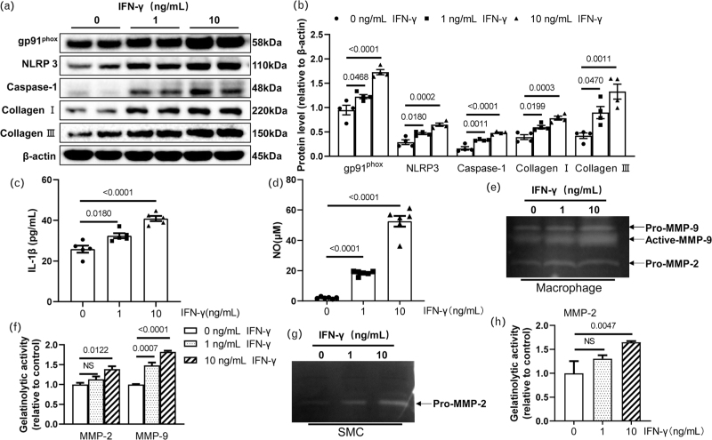
Methods and results: Eight-week-old male wildtype (CD8 +/+ ) and Cd8a knockout (CD8 -/- ) mice were used in a calcium chloride 2 (CaCl 2 )-induced experimental AAA model. At 6 weeks after surgery, CD8 + T-cell deletion prevented the formation of AAA, accompanied by reductions of the levels of inflammatory (interferon-γ [IFN-γ], interleukin-1β, monocyte chemoattractant protein-1, intracellular adhesion molecule-1, vascular cell adhesion molecule-1, NOD-like receptor protein 3, caspase-1), oxidative stress [NADPH oxidase and gp91 phox ], and proteolysis (cathepsin S, cathepsin K, matrix metalloproteinase-2 [MMP-2] and MMP-9) proteins and/or genes in plasma and/or AAA tissues. Immunoreactivities of MMP-2 and MMP-9 were observed in macrophages. An injection of IFN-γ and adoptive transfer of CD8 + T cells of IFN-γ +/+ mice diminished CD8 -/- -mediated vasculoprotective actions in the AAA mice. In vitro, IFN-γ enhanced MMP-2 and MMP-9 gelatinolytic activities in macrophage and/or vascular smooth muscle cells.

Conclusion: The vasculoprotective effects of CD8 + T-cell deletion in a mouse CaCl 2 -induced AAA model were likely attributable to, at least in part, the attenuation of IFN-γ-dependent inflammation action, oxidative stress production, and proteolysis, suggesting a novel therapeutic target for AAA formation by regulating CD8 + T-cell-derived IFN-γ secretion.


Conflict of interest statement

The authors declare that they have no known competing financial interests or personal relationships or conflicts of interests that could have appeared to influence the work reported in this article.

Figures

None
Graphical abstract
FIGURE 1
FIGURE 1
CD8+ T-cell deletion mitigated the CaCl2-induced AAA formation and the investigated protein levels in aortic tissues. A,B: Representative photographs and data of the maximal external aortic diameter in CD8+/+ and CD8−/− mice treated with NaCl or CaCl2, respectively (n = 8/group). C–F: Representative western blotting images and quantitative data for the levels of NLRP3, caspase-1, gp91phox, collagen I, and collagen III proteins in four experimental groups (n = 4/group). G,H: The ELISA results showing the serum levels of IFN-γ and IL-1β in the four groups (n = 6/group).
FIGURE 2
FIGURE 2
CD8+ T-cell deficiency alleviated the histopathological changes and recruitment of inflammatory cells. A: The histopathological analysis (H&E, EVG, and Masson trichrome staining) of abdominal aortas of CD8+/+ and CD8−/− mice treated with NaCl or CaCl2, respectively. Scale bar: upper layer 100 μm, lower layer 50 μm. B: Quantification of the collagen deposition area of the abdominal aortas (n = 5/group). C: Quantification of the elastin degradation score of the abdominal aortas (n = 5/group). D-F: Representative images of IFN-γ+/CD8a+ T cell and MMP-2+/ and MMP-9+/CD68+ macrophage in the adventitia of CD8+/+ and CD8−/− AAA mice (n = 5/group). Scale bar: 50 μm. L, Lumen.
FIGURE 3
FIGURE 3
IFN-γ loading accelerated the CaCl2-induced AAA formation in the CD8−/− mice. A,B: Representative photographs and quantitative data of the maximal external aortic diameter in the PBS-injected (Vehicle) or IFN-γ-injected CD8−/− mice after CaCl2 aneurysm induction (n = 8/group). C,D: Representative immunoblotting images and quantitative data for the levels of NLRP3, caspase-1, gp91phox, collagen I, and collagen III proteins in the two groups (n = 3/group). E: The ELISA results showing the serum levels of IL-1β in both groups (n = 6/group).
FIGURE 4
FIGURE 4
IFN-γ accelerated the histopathological changes and the recruitment of inflammatory cells in the CaCl2-induced AAAs in the CD8−/− mice. A: The histopathological analysis (H&E, EVG, and Masson trichrome staining) of abdominal aortas of PBS-injected (Vehicle) and IFN-γ-injected CD8−/− mice after CaCl2 aneurysm induction. Scale bar: upper layer 100 μm, lower layer 50 μm. B: Quantification of the collagen deposition area of the abdominal aortas (n = 5/group). C: Quantification of the elastin degradation score of the abdominal aortas (n = 6/group). D-F: Representative images of IFN-γ+/CD8a+ T cell and MMP-2+/ and MMP-9+/CD68+ macrophage in the adventitia of CD8−/− AAA mice treated with PBS and IFN-γ (n = 5/group). Scale bar: 50 μm. L, lumen.
FIGURE 5
FIGURE 5
IFN-γ enhanced the inflammasome-related protein expressions and gelatinase activities in the macrophages and smooth muscle cells (SMCs). The macrophages and SMCs were treated with 0, 1, or 10 ng/mL of IFN-γ for 24 hr, and the lysates were then subjected to western blotting; the conditioned media were collected for the gelatin zymography assays. A,B: Representative western blotting images and quantitative data for NLRP3, caspase-1, gp91phox, collagen I, and collagen III (n = 4/group). C,D: IFN-γ increased IL-1β levels and nitric oxide (NO) release (n = 5–6/group). E-H: Representative images and the quantitative data of the gelatin zymography assays of MMP-2 and/or MMP-9 in the condition medium of macrophages and SMCs (n = 3/group).

Similar articles

Cited by

References

    1. Lu Y, Ma Q, Tan H, Li X, Zhang X, Tie Y. Specific inhibition of SHP2 suppressed abdominal aortic aneurysm formation in mice by augmenting the immunosuppressive function of MDSCs. Life Sci 2021; 265:118751. - PubMed
    1. Xu B, Li G, Guo J, Ikezoe T, Kasirajan K, Zhao S, et al. . Angiotensin-converting enzyme 2, coronavirus disease 2019, and abdominal aortic aneurysms. J Vasc Surg 2021; 74:1740–1751. - PMC - PubMed
    1. Sakalihasan N, Michel JB, Katsargyris A, Kuivaniemi H, Defraigne JO, Nchimi A, et al. . Abdominal aortic aneurysms. Nat Rev Dis Primers 2018; 4:34. - PubMed
    1. Jiang H, Sasaki T, Jin E, Kuzuya M, Cheng XW. Inflammatory cells and proteases in abdominal aortic aneurysm and its complications. Curr Drug Targets 2018; 19:1289–1296. - PubMed
    1. Koh CH, Lee S, Kwak M, Kim BS, Chung Y. CD8 T-cell subsets: heterogeneity, functions, and therapeutic potential. Exp Mol Med 2023; 55:2287–2299. - PMC - PubMed