. 2024 Sep 5;32(9):1404-1418.e7.
doi: 10.1016/j.str.2024.07.014. Epub 2024 Aug 14.

Engineering of pH-dependent antigen binding properties for toxin-targeting IgG1 antibodies using light-chain shuffling

Affiliations

Engineering of pH-dependent antigen binding properties for toxin-targeting IgG1 antibodies using light-chain shuffling

Tulika Tulika et al. Structure. .

Abstract

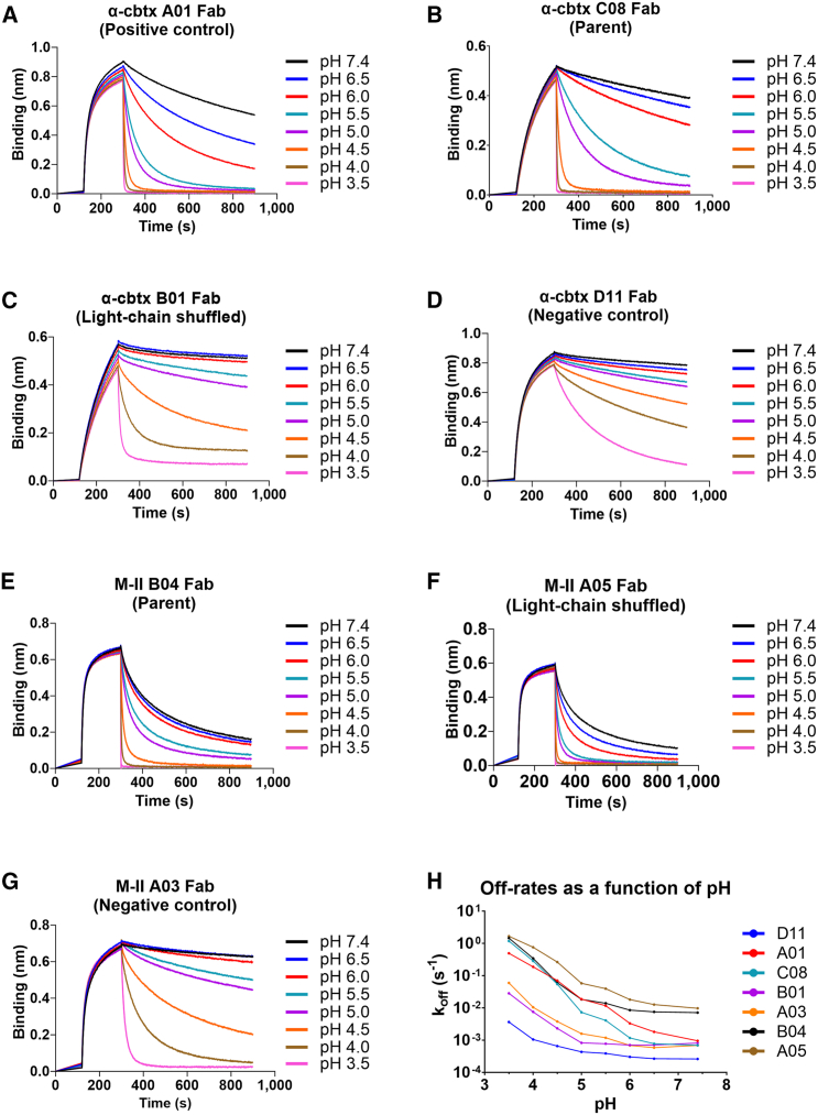
Immunoglobulin G (IgG) antibodies that bind their cognate antigen in a pH-dependent manner (acid-switched antibodies) can release their bound antigen for degradation in the acidic environment of endosomes, while the IgGs are rescued by the neonatal Fc receptor (FcRn). Thus, such IgGs can neutralize multiple antigens over time and therefore be used at lower doses than their non-pH-responsive counterparts. Here, we show that light-chain shuffling combined with phage display technology can be used to discover IgG1 antibodies with increased pH-dependent antigen binding properties, using the snake venom toxins, myotoxin II and α-cobratoxin, as examples. We reveal differences in how the selected IgG1s engage their antigens and human FcRn and show how these differences translate into distinct cellular handling properties related to their pH-dependent antigen binding phenotypes and Fc-engineering for improved FcRn binding. Our study showcases the complexity of engineering pH-dependent antigen binding IgG1s and demonstrates the effects on cellular antibody-antigen recycling.

Keywords: Antibody recycling; FcRn; HERA; acid-switched antibodies; human endothelial cell-based recycling assay; light-chain shuffling; myotoxin II; pH-dependent antigen binding properties; phage display technology; snake venom; α-cobratoxin.


Conflict of interest statement

Declaration of interests The authors declare no competing interests.

Figures

None
Graphical abstract
Figure 1
Figure 1
Phage display selection and binding of reformatted Fab fragments to snake toxins (A–E) Schematic illustration of the (A) light-chain shuffling and (B) phage display selection campaigns for isolation of scFvs with pH-dependent antigen binding properties derived from the B04, B12, and C08 light-chain shuffled libraries. (1) scFv-displaying phages were incubated with biotinylated antigen at pH 5.5, which was followed by capture and removal of phages that bound the antigen at low pH using streptavidin-coated magnetic beads. (2) The phages that did not bind to the antigen at pH 5.5 were collected and incubated with biotinylated antigen at pH 7.4 prior to capture on streptavidin-coated magnetic beads. (3) The beads were washed to remove unspecific phages. (4) The bound phages were then eluted using a pH 5.5 buffer and (5) amplified for the next round of selection. Three consecutive rounds of selection were performed with the generated libraries. Polyclonal phage ELISA of the phage outputs from the three rounds of selection performed with libraries (C) B04, (D) B12, and (E) C08 showing that the outputs bind to their respective cognate antigens and that minimal binding to streptavidin is detected. (F–H) (F) Schematic illustration of the off-rate screening approach using BLI, where the association was performed at pH 7.4, which was followed by dissociation for 300 s at pH 7.4 or pH 5.5. BLI sensorgrams of Fab-containing supernatant from CHO cell expression experiments of (G) B01 and (H) A05 showing binding to α-cbtx and M-II, respectively, at pH 7.4, followed by dissociation at pH 7.4 or 5.5. (SN, supernatant).
Figure 2
Figure 2
Dissociation of the Fabs measured through a pH gradient using BLI (A–G) Sensorgrams for the α-cbtx-targeting Fabs: (A) Fab A01 (positive control for pH-dependent antigen binding properties), (B) parental Fab C08, (C) light-chain shuffled Fab B01, and (D) Fab D11 (negative control with non-pH-dependent antigen binding properties). Sensorgrams for the M-II-targeting Fabs: (E) Parental Fab B04, (F) light-chain shuffled Fab A05, and (G) Fab A03 (negative control with non-pH-dependent antigen binding properties). Off-rates were determined at pH 3.5, 4.0, 4.5, 5.0, 5.5, 6.0, 6.5, and 7.4. The negative Fab controls showed similar off-rates at pH 5.5 and 7.4. (H) pH versus off-rate (koff) plot for the assessed Fabs.
Figure 3
Figure 3
The engineered IgG1 variants show distinct hFcRn binding properties in the absence and presence of their antigens (A–E) (A) Schematic illustration of the ELISA setup used to detect binding between the IgG1 variants and biotinylated hFcRn. Binding of biotinylated hFcRn to WT and YTE-containing IgG1 variants targeting α-cbtx (B and C) and M-II (D and E) at pH 5.5 and pH 7.4. Data shown as mean ± SD of duplicates. (F–O) (F) Schematic illustration of the hFcRn affinity chromatography experiment, where the pH varies from 5.5 to 8.8, used to assess the release of IgG1s from the receptor. Elution profiles of WT and YTE-containing IgG1 variants targeting (G) α-cbtx and (H) M-II from the hFcRn-coupled column are shown as relative absorbance units as a function of a pH gradient. Elution profiles of WT and YTE-containing the IgG1 variants when pre-incubated with (I–L) α-cbtx or (M–O) M-II at pH 5.5 are shown as the relative absorbance as a function of pH. The pH is plotted on the right Y axis (dotted line). Figures (A) and (F) were created with BioRender.
Figure 4
Figure 4
Cellular transport properties of the IgG1 variants in the absence and presence of antigen (A) Schematic overview of the HERA protocol. The IgG1 variants were pre-incubated with or without their cognate antigen and added to the hFcRn-expressing HMEC-1 cells (1). After incubation for 3 h to allow for cellular uptake of the IgG1s (2), the medium was removed, and the cells were washed prior to lysis and collection of samples. In parallel, cells were washed, and fresh medium was added, which was followed by a 3-h incubation step to allow recycling and release into the medium. Medium was then collected, and cells were lysed (3). A two-way anti-Fc ELISA was used to measure the presence of IgGs in the collected medium and lysate samples (4). The figure was created with BioRender. ELISA quantification of the amounts of IgG1 taken up, recycled, or accumulated in the absence and presence of cognate antigen for (B–D) anti-α-cbtx IgG1 and (E–G) anti-M-II IgG1 variants. Data shown as mean ± SD of one representative experiment with triplicates (n = 3 per data point). p > 0.05, ∗∗p > 0.01, ∗∗∗p > 0.001, ∗∗∗∗p > 0.0001 (unpaired Student’s t test).
Figure 5
Figure 5
Tracking of antigen-bound IgG1 variants following cellular transport in HERA (A) Schematic overview of the protocol for measuring IgG1 variants bound to their cognate antigen during cellular uptake, recycling, and termination of HERA. (1) IgG1 variants were pre-incubated with or without biotinylated antigens and (2) analyzed with HERA. (3) IgG1s present in the collected lysates and recycling media were quantified by an ELISA, where biotinylated antigens were captured on streptavidin-coated wells followed by detection of bound IgG1 with an anti-Fc antibody. The figure was created with BioRender. ELISA results showing the uptake, recycling, and accumulation of antigen-bound (B–F) anti-α-cbtx IgG1 and (G–J) anti-M-II IgG1 variants. For each IgG1-antigen complex, the relative uptake, recycling, and residue amounts were normalized to the obtained uptake value of the complex. Data shown as mean ± SD of one representative experiment with triplicates (n = 3 per data point).

References

    1. Kaplon H., Chenoweth A., Crescioli S., Reichert J.M. Antibodies to watch in 2022. mAbs. 2022;14 doi: 10.1080/19420862.2021.2014296. - DOI - PMC - PubMed
    1. Carter P.J., Lazar G.A. Next generation antibody drugs: pursuit of the “high-hanging fruit.”. Nat. Rev. Drug Discov. 2018;17:197–223. doi: 10.1038/nrd.2017.227. - DOI - PubMed
    1. Carter P.J., Rajpal A. Designing antibodies as therapeutics. Cell. 2022;185:2789–2805. doi: 10.1016/j.cell.2022.05.029. - DOI - PubMed
    1. Lu R.-M., Hwang Y.-C., Liu I.-J., Lee C.-C., Tsai H.-Z., Li H.-J., Wu H.-C. Development of therapeutic antibodies for the treatment of diseases. J. Biomed. Sci. 2020;27:1. doi: 10.1186/s12929-019-0592-z. - DOI - PMC - PubMed
    1. Ward E.S., Ober R.J. Targeting FcRn to generate antibody-based therapeutics. Trends Pharmacol. Sci. 2018;39:892–904. doi: 10.1016/j.tips.2018.07.007. - DOI - PMC - PubMed

Publication types

MeSH terms

LinkOut - more resources