. 2024 Oct:374:550-562.
doi: 10.1016/j.jconrel.2024.08.017. Epub 2024 Aug 30.

Development of an engineered extracellular vesicles-based vaccine platform for combined delivery of mRNA and protein to induce functional immunity

Affiliations

Development of an engineered extracellular vesicles-based vaccine platform for combined delivery of mRNA and protein to induce functional immunity

Xin Luo et al. J Control Release. 2024 Oct.

Abstract

mRNA incorporated in lipid nanoparticles (LNPs) became a new class of vaccine modality for induction of immunity against COVID-19 and ushered in a new era in vaccine development. Here, we report a novel, easy-to-execute, and cost effective engineered extracellular vesicles (EVs)-based combined mRNA and protein vaccine platform (EVX-M+P vaccine) and explore its utility in proof-of-concept immunity studies in the settings of cancer and infectious disease. As a first example, we engineered EVs, natural nanoparticle carriers shed by all cells, to contain ovalbumin mRNA and protein (EVOvaM+P vaccine) to serve as cancer vaccine against ovalbumin-expressing melanoma tumors. EVOvaM+P administration to mice with established melanoma tumors resulted in tumor regression associated with effective humoral and adaptive immune responses. As a second example, we generated engineered EVs that contain Spike (S) mRNA and protein to serve as a combined mRNA and protein vaccine (EVSpikeM+P vaccine) against SARS-CoV-2 infection. EVSpikeM+P vaccine administration in mice and baboons elicited robust production of neutralizing IgG antibodies against RBD (receptor binding domain) of S protein and S protein specific T cell responses. Our proof-of-concept study describes a new platform with an ability for rapid development of combination mRNA and protein vaccines employing EVs for deployment against cancer and other diseases.


Conflict of interest statement

Declaration of competing interest MD Anderson Cancer Center and RK have filed patent applications and patents issued in the area of exosome biology, and some of them are licensed to PranaX, Inc. for non-cancer related use. MD Anderson and RK hold equity in PranaX and RK serves as an advisor on non-cancer related matters.

Figures
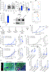
Fig. 1.
Fig. 1.
EVOvaM+P elicits antigen specific immune responses to prevent tumor growth. (a) NTA of wide type 293F EV and EVOvaM+P (EVs carrying OVA mRNA and protein). TEM images of EVs (inset). Scale bar, 100 nm. (b-c) EVs production from cells quantified by NTA (b) and microBCA assay (c) (n = 3). (d) Flow cytometry analysis of EVs markers CD9, CD47, CD63, and CD81 (n = 3). (e) Quantification of OVA mRNA from EVOvaM+P. Data are shown as 1/∆CT (n = 3). n.d., not detected. (f) Representative western blot of OVA protein and CD81 expression in EVOvaM+P (n = 3). (g) Schematic illustration of vaccination and tumor challenge schedule with EVOvaM+P in mice. C57BL/6J mice were intramuscularly injected with EVs, EVOvaM+P, or recombinant OVA protein (OvaP). (h) Quantification of anti-OVA IgG antibodies from vaccinated C57BL/6J mice by ELISA (n = 6 for EV vaccination, n = 8 for EVOvaM+P, n = 7 for OVAP). (i) Tumor volume and weight measurement of wide type (WT) and OVA tumor in EVOvaM+P vaccinated mice (n=8 per group). (j) Tumor volume and weight measurement of WT and OVA tumor in EV vaccinated mice (n = 6 per group for tumor volume, n = 5 per group for EV tumor weight). (k) Tumor volume and weight measurement of WT and OVA tumor in OVAP vaccinated mice (n = 7 per group). (l) Frequency of CD11b+ cells out of CD45+ cells from tumor tissue (n = 5 for EV vaccination, n = 7 for EVOvaM+P, n = 7 for OVAP). (m) Frequency of NK1.1+ cells out of CD45+ cells from tumor tissue (n = 5 for EV vaccination, n = 7 for EVOvaM+P, n = 7 for OVAP). (n) Representative images of CD4+ T cell and CD4+ T cell proliferation by TSA staining in WT tumor and OVA tumor tissue from EVOvaM+P vaccinated mice. Scale bar, 100 µm. (o) Quantification of CD4+ T and proliferative CD4+ T cells (Ki67+) of images in (n). Data are presented as mean ± s.e.m. Significance was determined by unpaired t-test in b-c, the right panel of i-k, l-m, o, two-way ANOVA with Tukey’s multiple comparisons test in h, mixed-effects analysis with Bonferroni’s multiple comparisons test in the left panel of i, and two-way ANOVA with Bonferroni’s multiple comparisons test in the left panel of j-k. * P < 0.05, ** P < 0.01, *** P < 0.001, **** P < 0.0001, ns: not significant.
Fig. 2.
Fig. 2.
Generation and validation of EVSpikeM+P. (a) NTA of wild type 293F EV, EV-SC2S (EVs carrying Spike mRNA and protein), and EVSpikeM+P (EVs carrying Spike-2P mRNA and protein). TEM images of EVs (inset). Scale bar, 100 nm. (b-c) EVs production from cells quantified by NTA (b) and microBCA assay (c) (n = 6). (d) Flow cytometry analysis of EVs markers CD9, CD47, CD63, and CD81 (n = 3). (e) Quantification of S mRNA from EVSpikeM+P. Data are shown as 1/ΔCT (n = 3). n.d., not detected. (f) Representative western blot of S protein, syntenin-1, and CD81 expression in EV-SC2S and EVSpikeM+P (n = 3). Data are presented as mean ± s.e.m. Significance was determined by one-way ANOVA with Dunnett’s multiple comparisons test in b-c. ns: not significant.
Fig. 3.
Fig. 3.
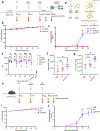
EVSpikeM+P vaccination elicits humoral and cellular responses in mice. (a) Schematic illustration of vaccination schedule with EVSpikeM+P in mice. Balb/c mice were intramuscularly injected with EVs, EV-SC2S and EVSpikeM+P. (b) Body weight change of Balb/c mice during vaccination expressed as percentage of starting weight (n = 6 per group). (c) Quantification of anti-S-RBD IgG antibodies from vaccinated Balb/c mice by ELISA (n = 6 per group). (d) Schematic illustration of the pseudovirus neutralization assay. (e) Neutralizing capacity of the antibodies from vaccinated mice evaluated with pseudovirus neutralization assay (n = 6 per group). Data are expressed as relative luminescence units (RLU). SFM + virus, serum-free media with pseudovirus; SFM, serum-free media alone. (f-g) S protein-specific CD4+ T cells in Balb/c mice splenocytes showing T cell activation (f) and Th1 response (g) by intracellular cytokine staining (n = 6 for PBS, n = 5 for EV vaccination, n = 5 for EVSpikeM+P). Splenocytes were re-stimulated with S peptide pool. (h) Schematic illustration of vaccination schedule with EVSpikeM+P in mice. C57BL/6J mice were intramuscularly injected with PBS, EV, EVSpikeM+P. (i) Body weight change of C57BL/6J mice during vaccination expressed as percentage of starting weight (n = 10 per group). (j) Quantification of anti-S-RBD IgG antibodies from vaccinated C57BL/6J mice by ELISA (n = 10 per group). Data are presented as mean ± s.e.m. Significance was determined by mixed-effects analysis with Tukey’s multiple comparisons test in c and j, two-way ANOVA with Tukey’s multiple comparisons test in e, unpaired t-test in f, and Mann-Whitney test in g. * P < 0.05, ** P < 0.01, *** P < 0.001, ns: not significant.
Fig. 4.
Fig. 4.
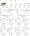
EVSpikeM+P vaccinated baboons develop antibodies and T cell responses against S protein. (a) Schematic illustration of vaccination schedule with EVSpikeM+P of 3 dosages in baboons. (b) Quantification of anti-S-RBD IgG antibodies from vaccinated baboons by ELISA (n = 3 per group). (c) Titration of anti-S-RBD IgG antibodies at different time points plotted as log AUC (area under curve, n = 3 per group). (d-f) Quantification of cytokines (IL-2, IFNy, and TNFa) releasing spots from S protein-specific T cells in circulation re-stimulated with S peptide pool (d) or recombinant S protein (e-f) by ELISpot (n = 3 per group). Data are shown as SFCs per 2 × 10⁶ cells. (g-i) Frequency of S protein-specific CD4+ T cells in circulation producing single (IL-2 (g), IFNy (h)) and two of Th1 cytokines (IL-2, IFNy, and TNFa, (i)) determined by intracellular cytokine staining (n = 3 per group). PBMCs were re-stimulated with S peptide pool. (j-k) Frequency of S protein-specific CD4+ T cells in circulation producing single (TNFa (j)) or three (k) of Th1 cytokines (IL-2, IFNy, and TNFa) determined by intracellular cytokine staining (n = 3 per group). PBMCs were re-stimulated with S recombinant protein. Data are presented as mean ± s.e.m. was determined by two-way ANOVA with Tukey’s multiple comparisons test in b-c, and by unpaired t-test in d-k. * P < 0.05, ** P < 0.01, **** P < 0.0001, ns: not significant, or exact p-values reported.

References

    1. Kalluri R, LeBleu VS, The biology, function, and biomedical applications of exosomes, Science 367 (2020) 6478. - PMC - PubMed
    1. Kalluri R, McAndrews KM, The role of extracellular vesicles in cancer, Cell 186 (8) (2023) 1610–1626. - PMC - PubMed
    1. Yáñez-MÓ M, Siljander PR-M, Andreu Z, Bedina Zavec A, Borrás FE, Buzas EI, Buzas K, Casal E, Cappello F, Carvalho J, Biological properties of extracellular vesicles and their physiological functions, J. Extracell. Vesicles 4 (1) (2015) 27066. - PMC - PubMed
    1. Van Niel G, d’Angelo G, Raposo G, Shedding light on the cell biology of extracellular vesicles, Nat. Rev. Mol. Cell Biol 19 (4) (2018) 213–228. - PubMed
    1. Lener T, Gimona M, Aigner L, Börger V, Buzas E, Camussi G, Chaput N, Chatterjee D, Court FA, Portillo HA, d., Applying extracellular vesicles based therapeutics in clinical trials–an ISEV position paper, J. Extracell. Vesicles 4 (1) (2015) 30087. - PMC - PubMed

MeSH terms