. 2024 Nov;154(5):1129-1145.
doi: 10.1016/j.jaci.2024.07.024. Epub 2024 Aug 13.

IL-2 family cytokines IL-9 and IL-21 differentially regulate innate and adaptive type 2 immunity in asthma

Affiliations

IL-2 family cytokines IL-9 and IL-21 differentially regulate innate and adaptive type 2 immunity in asthma

Fabian Bick et al. J Allergy Clin Immunol. 2024 Nov.

Abstract

Background: Asthma is often accompanied by type 2 immunity rich in IL-4, IL-5, and IL-13 cytokines produced by TH2 lymphocytes or type 2 innate lymphoid cells (ILC2s). IL-2 family cytokines play a key role in the differentiation, homeostasis, and effector function of innate and adaptive lymphocytes.

Objective: IL-9 and IL-21 boost activation and proliferation of TH2 and ILC2s, but the relative importance and potential synergism between these γ common chain cytokines are currently unknown.

Methods: Using newly generated antibodies, we inhibited IL-9 and IL-21 alone or in combination in various murine models of asthma. In a translational approach using segmental allergen challenge, we recently described elevated IL-9 levels in human subjects with allergic asthma compared with nonasthmatic controls. Here, we also measured IL-21 in both groups.

Results: IL-9 played a central role in controlling innate IL-33-induced lung inflammation by promoting proliferation and activation of ILC2s in an IL-21-independent manner. Conversely, chronic house dust mite-induced airway inflammation, mainly driven by adaptive immunity, was solely dependent on IL-21, which controlled TH2 activation, eosinophilia, total serum IgE, and formation of tertiary lymphoid structures. In a model of innate on adaptive immunity driven by papain allergen, a clear synergy was found between both pathways, as combined anti-IL-9 or anti-IL-21 blockade was superior in reducing key asthma features. In human bronchoalveolar lavage samples we measured elevated IL-21 protein within the allergic asthmatic group compared with the allergic control group. We also found increased IL21R transcripts and predicted IL-21 ligand activity in various disease-associated cell subsets.

Conclusions: IL-9 and IL-21 play important and nonredundant roles in allergic asthma by boosting ILC2s and TH2 cells, revealing a dual IL-9 and IL-21 targeting strategy as a new and testable approach.

Keywords: Asthma; IL-21; IL-9; adaptive immunity; innate immunity; monoclonal antibodies; type 2 immunity.


Conflict of interest statement

Disclosure statement This work was funded by a Baekeland Mandate of Flanders Innovation & Entrepreneurship (VLAIO) (HBC.2019.2598) to F.B. B.N.L. acknowledges support from a European Research Council (ERC) advanced grant (789384 ASTHMA CRYSTAL CLEAR), a concerted research initiative grant from Ghent University (GOA, 01G010C9), a Fonds Wetenschappelijk Onderzoek (FWO) Methusalem grant (01M01521) and an FWO Excellence of Science (EOS) Consortium research grant (3G0H1222), and the Flanders Institute of Biotechnology (VIB). H.H. is supported by a concerted research initiative grant from Ghent University (GOA, 01G010C9) and FWO EOS Consortium research grant (3G0H1222). M.J.S. acknowledges support from FWO Vlaanderen (12Y5322N), Fund Suzanne Duchesne (managed by the King Baudouin Foundation), and Fondation ACTERIA. Disclosure of potential conflict of interest: N. P. Smith is a consultant for Hera Biotech. A.-C. Villani has financial interest in 10X Genomics. B. D. Medoff serves on advisory boards for Sanofi, Verona Pharma, and Apogee Therapeutics and has sponsored research agreements from Sanofi and Regeneron. R. Bigirimana is an employee of argenx. C. Blanchetot is a consultant and shareholder of argenx. B. N. Lambrecht receives consultancy fees from GSK Biologics, Novartis, Sanofi, AstraZeneca, ALK, OncoArendi, and argenx and has research grants from ALK, argenx, AstraZeneca, GSK, GSK Biologics, and Johnson & Johnson. The rest of the authors declare that they have no relevant conflicts of interest.

Figures

FIG 1.
FIG 1.
Generation of potent neutralizing antibodies against murine IL-9 and IL-21. (A) Schematic representation of the anti-IL-9 antibody discovery workflow. (B) Schematic representation of the anti-IL-21 antibody reformatting. (C) FcγR-binding ELISA results for antibodies reformatted into mIgG1-N297A backbone. (D) Affinity of the used antibodies toward their target as determined by 1:1 fitting using SPR. (E) Proliferation-based bioassay data showing the neutralizing effect of the antibodies in the presence of either IL-9 or IL-21. Schemes shown in (A and B) were created with BioRender.com.
FIG 2.
FIG 2.
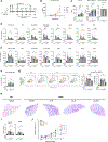
IL-9 blockade broadly ameliorates IL-33–induced airway inflammation. (A) Experimental setup of IL-33 exposed C57BL/6J wild-type, Red5+/− or IL-9R−/− mice treated with antibodies during challenge. (B and E) Cytokine levels as measured by ELISA in the supernatant of BAL fluid recovered from IL-33–challenged mice. (C) Cell counts recovered after BAL as determined by flow cytometry. TH2 cells are defined like CD4+ and ST2+. (D and F) Cell counts from enzymatically digested lungs as determined by flow cytometry. *P < .05, **P < .01, ***P < .001, ****P < .0001 vs mice treated with an irrelevant mIgG1 N297A antibody. AM, Alveolar macrophages; ns, not significant.
FIG 3.
FIG 3.
IL-9 blockade ameliorates IL-33–induced airway inflammation by suppressing ILC2s. (A) MFIs of Ki-67 and IL-13 on ILC2s as determined by flow cytometry. (B) Gating strategy shown for ILC2s in enzymatically digested lungs using Red5+/− mice. MFIs of IL-5 (tdTomato) on ILC2s and cell counts of eosinophils in the lungs as determined by flow cytometry. (C) Comparison of ILC2 numbers and MFIs (Ki-67 and IL-13) from enzymatically digested lungs between C57BL/6J WT and IL-9R−/− mice. Contour plots show frequencies representative for quantification. (D) Representative histologic comparison of hematoxylin-eosin stain from lungs after IL-33 challenge. (E) Relative lung mRNA expression levels of Muc5ac and Spdef as determined by quantitative real-time PCR. (F) BHR measured in IL-33–challenged mice in response to increasing doses of methacholine using FlexiVent (SCIREQ). Data are representative of 3 independent experiments, with at least 5 mice per group, and are presented as mean ± SEM. *P < .05, **P < .01, ***P < .001, ****P < .0001 vs mice treated with an irrelevant mIgG1 N297A antibody. MFI, Median fluorescent intensity; ns, not significant; WT, wild-type.
FIG 4.
FIG 4.
IL-21 blockade ameliorates chronic HDM-induced airway inflammation by reducing adaptive immunity. (A) Experimental setup of HDM-exposed C57BL/6J wild-type mice treated with antibodies during challenge. (B) Cytokine levels as measured by ELISA in the supernatant of BAL fluid recovered from HDM-sensitized and HDM-challenged mice. (C) Total IgE level time course as measured by ELISA in serum. (D) HDM-specific IgE and IgG1 levels at week 6 as measured by ELISA in serum. (E) Cell counts recovered after BAL as determined by flow cytometry. TH2 cells are defined as CD4+ ST2+. (F-H) Cell counts from enzymatically digested lungs as determined by flow cytometry. Quantification of TH2 cells (CD4+, ST2+, GATA3+) as determined by flow cytometry. (I) Representative histologic comparison of hematoxylin-eosin stain from lungs after challenge as measured using bright-field microscopy. Squares indicate areas shown in the panel below, depicting confocal immune stainings against B220, GL7, DAPI, and CD3e as measured by confocal microscopy. (J) Quantification of TLS structures based on hematoxylin-eosin stainings. Data are representative of 3 independent experiments, with at least 5 mice per group, and are presented as means ± SEM. *P < .05, **P < .01, ***P < .001, ****P < .0001 vs mice treated with an irrelevant mIgG1 N297A antibody. AM, Alveolar macrophages; ns, not significant.
FIG 5.
FIG 5.
Synergistic IL-9 and IL-21 inhibition ameliorates papain-induced airway inflammation. (A) Experimental setup of papain-exposed C57BL/6J wild-type mice treated with antibodies during challenge. (B and C) Total IgE time course as measured by ELISA in serum. HDM-specific IgE and IgG1 levels at week 3 as measured by ELISA in serum. (D) Cell counts recovered from BAL as determined by flow cytometry. TH2 cells are defined as CD4+ ST2+. (E-G) Cell counts from enzymatically digested lungs as determined by flow cytometry. Representative contour plots of TH2 cells (CD4+, ST2+, GATA3+) as determined by flow cytometry and quantification thereof. (H) Representative histologic comparison of hematoxylin-eosin stain from lungs after challenge. (I) Relative lung mRNA expression levels of Muc5ac and Spdef as determined by qPCR. (J) BHR measured in papain-challenged mice in response to increasing doses of methacholine using FlexiVent (SCIREQ). Data are representative of 3 independent experiments, with at least 5 mice per group, and are presented as means ± SEM. *P < .05, **P < .01, ***P < .001, ****P < .0001 vs mice treated with an irrelevant mIgG1 N297A antibody. AM, Alveolar macrophages; ns, not significant.
FIG 6.
FIG 6.
Levels of IL-21 protein and its receptor transcript are increased in human AA subjects, predicting ligand activity in multiple disease-associated cell subsets. (A) UMAP of a total of 52,152 cells recovered from 21 lower airway brushings. (B) Depiction of IL-21 receptor mRNA transcript expression (2404 cells 5 4.6% of total cells). (C) IL-21 protein levels in AA subjects and ACs before and after SAC with either diluent or allergen as determined by ELISA. (D) IL-21 receptor expression separated by group per cellular subset. (E) Predicted IL-9 and IL-21 ligand activities in the AA group. Upregulated target genes within the Treg and DC2 subsets are shown for representation. Black rectangles highlight cell subsets associated with having asthma. Target genes for these subsets are shown in Figs E8 and E9. In (C), P values were calculated using ordinary two-way ANOVA with Tukey correction for multiple comparisons. *P < .05, **P < .01, ***P < .001, ****P < .0001. ns, Not significant.

References

    1. Hammad H, Lambrecht BN. The basic immunology of asthma. Cell 2021;184:1469–85. - PubMed
    1. GBD 2019 Diseases and Injuries Collaborators. Global burden of 369 diseases and injuries in 204 countries and territories, 1990–2019: a systematic analysis for the Global Burden of Disease Study 2019. Lancet 2020;396:1204–22. - PMC - PubMed
    1. Hekking PW, Wener RR, Amelink M, Zwinderman AH, Bouvy ML, Bel EH. The prevalence of severe refractory asthma. J Allergy Clin Immunol 2015;135:896–902. - PubMed
    1. Shah PA, Brightling C. Biologics for severe asthma—which, when and why? Respirology 2023;28:709–21. - PubMed
    1. Baysoy A, Seddu K, Salloum T, Dawson CA, Lee JJ, Yang L, et al. The interweaved signatures of common-gamma-chain cytokines across immunologic lineages. J Exp Med 2023;220:e20222052. - PMC - PubMed