. 2024 Aug 15;15(1):7009.
doi: 10.1038/s41467-024-51108-z.

A broadly protective antibody targeting glycoprotein Gn inhibits severe fever with thrombocytopenia syndrome virus infection

Affiliations

A broadly protective antibody targeting glycoprotein Gn inhibits severe fever with thrombocytopenia syndrome virus infection

Xuanxiu Ren et al. Nat Commun. .

Abstract

Severe fever with thrombocytopenia syndrome virus (SFTSV) is an emerging bunyavirus that causes severe viral hemorrhagic fever and thrombocytopenia syndrome with a fatality rate of up to 30%. No licensed vaccines or therapeutics are currently available for humans. Here, we develop seven monoclonal antibodies (mAbs) against SFTSV surface glycoprotein Gn. Mechanistic studies show that three neutralizing mAbs (S2A5, S1G3, and S1H7) block multiple steps during SFTSV infection, including viral attachment and membrane fusion, whereas another neutralizing mAb (B1G11) primarily inhibits the viral attachment step. Epitope binning and X-ray crystallographic analyses reveal four distinct antigenic sites on Gn, three of which have not previously been reported, corresponding to domain I, domain II, and spanning domain I and domain II. One of the most potent neutralizing mAbs, S2A5, binds to a conserved epitope on Gn domain I and broadly neutralizes infection of six SFTSV strains corresponding to genotypes A to F. A single dose treatment of S2A5 affords both pre- and post-exposure protection of mice against lethal SFTSV challenge without apparent weight loss. Our results support the importance of glycoprotein Gn for eliciting a robust humoral response and pave a path for developing prophylactic and therapeutic antibodies against SFTSV infection.


Conflict of interest statement

The authors declare no competing interests.

Figures

Fig. 1
Fig. 1. Binding and neutralizing capabilities of Gn mAbs.
a, b The binding of mAbs to Gn head domain proteins (a) and authentic SFTSV QD02 (b) was assessed using ELISA. c-h The neutralization activity of Gn mAbs against SFTSV (genotypes A-F) pseudoviruses (psv). i, j Focus reduction neutralization tests (FRNT) against authentic SFTSV. Data represent mean ± SEM (standard error of the mean) of technical duplicates and are representative of two (dh, j), three (ac) or four (i) independent experiments. Source data are provided as a Source data file.
Fig. 2
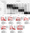
Fig. 2. Binding affinity of Gn mAbs and competition-binding relationships.
a Competition-binding assays were explored by BLI. The seven Gn mAbs were categorized into four groups (A, B, C, and D). The values in the table represent the percentage binding signal of tested antibodies in comparison to the binding signal of non-competitive antibodies. The antibodies were defined as strong competition if the percentage binding signal was less than 25%. MAb4-5 is a previously reported antibody that recognizes the SFTSV Gn protein. The results are averaged from two independent experiments. b The binding affinity of indicated mAbs to recombinant SFTSV Gn head protein was detected by BLI assay. Representative binding curves from two or three independent experiments. The values are the average of two to three independent experiments. The fitting curves are shown as red lines. Source data are provided as a Source data file.
Fig. 3
Fig. 3. Neutralizing mechanism of Gn mAbs.
a The inhibition of SFTSV attachment by mAbs at 10 or 100 μg/mL concentrations was assessed using qRT-PCR. GAPDH was used for internal control, and viral RNA fold change was compared with control cells incubated with Isotype IgG (NiV E2, anti-NiV mAb without binding to SFTSV, IsoIgG). Statistical significance was determined by a two-way ANOVA with Dunnett’s multiple comparison tests in the comparison to IsoIgG group (****p < 0.0001). The data is mean ± SEM of three independent experiments performed in triplicate. b SFTSV pre-/post-attachment inhibition was assessed through FRNT assay. The IC50 values (indicated in the upper right corner) represent mean of three independent experiments performed in duplicate, with the green line representing pre-attachment inhibition curve and the blue line representing post-attachment inhibition curve. Data represent mean ± SEM of technical duplicates and are representative of three independent experiments. c The inhibition of membrane fusion induced by the SFTSV glycoprotein (encoded by segment M) was assessed using a split-GFP/Luc system, with live-cell luciferase activity being measured. Authentic SFTSV strain QD02 (genotype D virus) was used in a and b. The statistical significance was determined by comparing with the IsoIgG group at the same mAb concentration using Dunnett’s multiple comparison tests in a two-way ANOVA analysis. The results for S1G3, S2A5, and S1H7 demonstrated a significant difference with p < 0.0001 (****), p < 0.001 (***), p < 0.01 (**), and p < 0.05 (*). Data represent mean ± SEM of three independent experiments performed in duplicate. Source data are provided as a Source data file.
Fig. 4
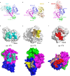
Fig. 4. Molecular determinants of three Gn mAbs.
ac Ribbon diagrams of Gn head domain complexed with antibody. For clarity, the constant domains of Fab were not displayed. The heavy chain and light chain variable domains of S2A5, B1G11, and N1D10 are shown in cyan and pale cyan, yellow and sand, red and salmon, respectively. Gn is colored by domains: DI, magenta; DII, blue; DIII, green. df Surface representation of mAb (top) and Gn head (bottom) with contacts labeled and colored as in (a). Gn residues contacted by both chains are colored gray.
Fig. 5
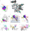
Fig. 5. Accessibility of Gn epitopes on SFTSV virion.
a Superimposition of the S2A5-Gn, B1G11-Gn, N1D10-Gn, and MAb4-5-Gn (PDB: 5Y11) complexes onto Gn head domain. MAbs S2A5, B1G11, and N1D10 are colored cyan, yellow, and red, respectively. b Epitope mapping within an asymmetric unit (ASU) of the SFTSV virion (PDB:8I4T). The epitopes of S2A5, B1G11, and N1D10 on virion are highlighted in cyan, yellow, and red, respectively. c, d Docking of the S2A5, B1G11, and N1D10 Fabs onto the hexon peplomer (hexon P) of the SFTSV virion. c Top view of the hexon peplomer. d Side view of the hexon peplomer. The Gn from reference Gn/Gc heterodimer is colored by domains (DI: magenta; DII: blue; DIII: green; domain IV: pink; linker: grey50; transmembrane domain (TM): orange). The ectodomain of Gc from reference Gn/Gc is colored sky blue, linker is colored grey50, and TM is colored olive. The other five subunits are colored white. Clashes with adjacent Gn or Gc molecules were indicated with a black circle. e Comparison of the orientations of mAbs relative to the viral membrane. f-h Close-up views of interaction/clashes between S2A5 and adjacent Gn molecule (f), B1G11 and adjacent Gc molecule (g), N1D10 and adjacent Gn/Gc heterodimer (h). The neighboring Gn molecule is colored white, and Gc is colored black.
Fig. 6
Fig. 6. S2A5 protects mice from lethal SFTSV challenge.
a, b In vivo prophylactic (a) or therapeutic (b) treatment regimen. c Mice (n = 6) were administered with indicated antibodies via an intraperitoneal route (i.p.); 24 h later, the treated mice were intraperitoneally infected with the SFTSV HBMC5 strain. Then, weight and survival rate of mice were continuously monitored for 15 days. d Mice (n = 6) were intraperitoneally infected with SFTSV HBMC5 strain first, then the infected mice were injected with indicated antibodies (i.p.) 6, 24, and 48 h post-infection. Weight data represent mean ± SEM of mice remaining at each time point. ek The experiments were performed as (b), except that mice (n = 6) were sacrificed on the third day of infection for analyses of the viral loads in different tissue samples (eg) and blood routine and biochemical indexes (h-k). Each data point represents an individual mouse within the respective groups. Isotyped IgG, IsoIgG; platelet, PLT; white blood cells, WBC; indicators for organ (liver) injury: serum aspartate aminotransferase (AST) and alanine aminotransferase (ALT). The data were subjected to one-way ANOVA with Dunnett’s test in the comparison to IsoIgG treated group. Bars show mean ± SEM, revealing a significant difference with p < 0.0001 (****), p < 0.001 (***), p < 0.01 (**), p < 0.05(*). Source data are provided as a Source data file.

References

    1. Yu, X.-J. et al. Fever with thrombocytopenia associated with a novel Bunyavirus in China. N. Engl. J. Med.364, 1523–1532 (2011). 10.1056/NEJMoa1010095 - DOI - PMC - PubMed
    1. Kuhn, J. H. et al. 2022 taxonomic update of phylum Negarnaviricota (Riboviria: Orthornavirae), including the large orders Bunyavirales and Mononegavirales. Arch. Virol.167, 2857–2906 (2022). 10.1007/s00705-022-05546-z - DOI - PMC - PubMed
    1. Casel, M. A., Park, S. J. & Choi, Y. K. Severe fever with thrombocytopenia syndrome virus: emerging novel phlebovirus and their control strategy. Exp. Mol. Med.53, 713–722 (2021). 10.1038/s12276-021-00610-1 - DOI - PMC - PubMed
    1. Yang, T., Huang, H., Jiang, L. & Li, J. Overview of the immunological mechanism underlying severe fever with thrombocytopenia syndrome (Review). Int. J. Mol. Med.50, 118 (2022). 10.3892/ijmm.2022.5174 - DOI - PMC - PubMed
    1. Zhang, X. et al. Rapid Spread of Severe Fever with Thrombocytopenia Syndrome Virus by Parthenogenetic Asian Longhorned Ticks. Emerg Infect Dis28, 363–372 (2022). 10.3201/eid2802.211532 - DOI - PMC - PubMed

MeSH terms

LinkOut - more resources