. 2024 Aug 15;15(1):7015.
doi: 10.1038/s41467-024-51458-8.

FIGNL1-FIRRM is essential for meiotic recombination and prevents DNA damage-independent RAD51 and DMC1 loading

Affiliations

FIGNL1-FIRRM is essential for meiotic recombination and prevents DNA damage-independent RAD51 and DMC1 loading

Akbar Zainu et al. Nat Commun. .

Abstract

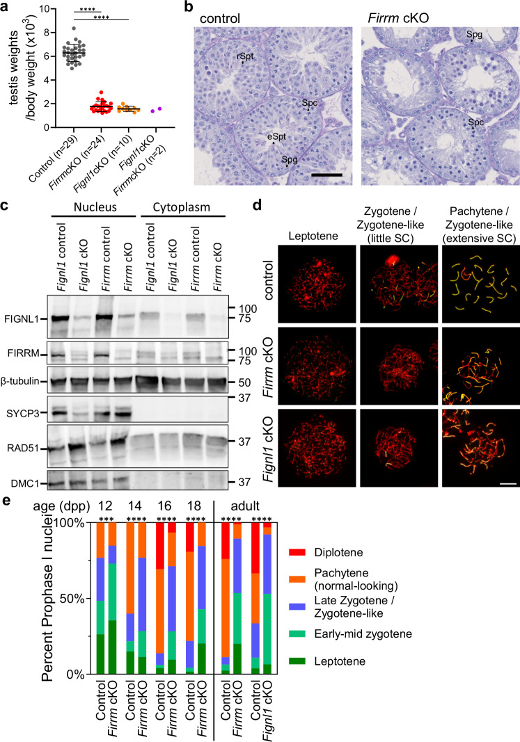
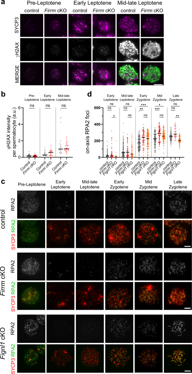
During meiosis, nucleoprotein filaments of the strand exchange proteins RAD51 and DMC1 are crucial for repairing SPO11-generated DNA double-strand breaks (DSBs) by homologous recombination (HR). A balanced activity of positive and negative RAD51/DMC1 regulators ensures proper recombination. Fidgetin-like 1 (FIGNL1) was previously shown to negatively regulate RAD51 in human cells. However, FIGNL1's role during meiotic recombination in mammals remains unknown. Here, we decipher the meiotic functions of FIGNL1 and FIGNL1 Interacting Regulator of Recombination and Mitosis (FIRRM) using male germline-specific conditional knock-out (cKO) mouse models. Both FIGNL1 and FIRRM are required for completing meiotic prophase in mouse spermatocytes. Despite efficient recruitment of DMC1 on ssDNA at meiotic DSB hotspots, the formation of late recombination intermediates is defective in Firrm cKO and Fignl1 cKO spermatocytes. Moreover, the FIGNL1-FIRRM complex limits RAD51 and DMC1 accumulation on intact chromatin, independently from the formation of SPO11-catalyzed DSBs. Purified human FIGNL1ΔN alters the RAD51/DMC1 nucleoprotein filament structure and inhibits strand invasion in vitro. Thus, this complex might regulate RAD51 and DMC1 association at sites of meiotic DSBs to promote proficient strand invasion and processing of recombination intermediates.


Conflict of interest statement

The authors declare no competing interests.

Figures

Fig. 1
Fig. 1. Spermatogenesis is defective in Firrm cKO and Fignl1 cKO mice.
a Testis weight relative to body weight in control (gray), Firrm cKO (red), Fignl1 cKO (orange) and Firrm cKO Fignl1 cKO (purple) adult mice (30 to 154 days post-partum, dpp; kinetics is shown in Supplementary Fig. 2a). The mean ± SD is shown. Unpaired t-tests, two-sided. b Periodic acid-Schiff-stained testis sections from 4-week-old control and Firrm cKO mice. Spg, spermatogonia; Spc, spermatocytes; rSpt, round spermatids; eSpt, elongated spermatids. Scale bar, 50 µm. c Western blot analysis of cytoplasmic (80 µg) and nuclear (100 µg) fractions from testes of 12 dpp mice of the indicated genotypes. d Chromosome axes (SYCP3, red) and synaptonemal complex (SYCP1, green) were detected in spread spermatocyte nuclei from control, Firrm cKO (25 dpp) and Fignl1 cKO (56 dpp) mice. Scale bar, 10 µm. e Distribution of spermatocytes at different meiotic prophase substages in juvenile Firrm cKO mice (indicated age) and in adult (8-week-old) Firrm cKO and Fignl1 cKO mice (1 mouse per condition). Two-sided chi-square tests. For all figures: ns, non-significant; *0.01 < p ≤ 0.05; **0.001 < p ≤ 0.01; ***0.0001 < p ≤ 0.001; ****p ≤ 0.0001, exact p-values are provided as Source Data. Source data are provided as a Source Data file.
Fig. 2
Fig. 2. Firrm cKO and Fignl1 cKO spermatocytes are proficient for markers of meiotic DSB formation and processing.
a Spread nuclei of spermatocytes from 12 dpp control and Firrm cKO mice stained for SYCP3 and γH2AX. Scale bar, 20 µm. b Total nuclear γH2AX signal intensity in 12 dpp control (gray) and Firrm cKO (red) spermatocytes (n = 2 mice per genotype). In cytological analyses, the black bar represents the median and two-sided Dunn’s multiple comparison tests were performed unless stated otherwise. c Spread spermatocyte nuclei from 12 dpp control and Firrm cKO mice stained for SYCP3 and RPA2. Scale bar, 10 µm. d Number of on-axis RPA2 foci in control (gray), Firrm cKO (red) and Fignl1 cKO (orange) spermatocytes. control, n = 6 (4 × 12 dpp, 1 × 16 dpp, 1 × 17 dpp), Firrm cKO, n = 4 (12 dpp) and Fignl1 cKO, n = 3 (1 × 16 dpp, 2 × 17 dpp) mice. Source data are provided as a Source Data file.
Fig. 3
Fig. 3. Firrm cKO and Fignl1 cKO spermatocytes accumulate RAD51 and DMC1, but are defective for forming foci of late-acting recombination proteins.
a Zygotene spermatocyte spreads from control and Firrm cKO mice stained for SYCP3, RAD51 and DMC1. Scale bar, 5 µm. Numbers of RAD51 (b) and DMC1 (c) foci in control (gray), Firrm cKO (red) and Fignl1 cKO (orange) spermatocytes (c). n = 2 mice per genotype. d Spreads of zygotene spermatocytes from 16 dpp control, Firrm cKO and Spo11YF/YF mice stained with SYCP3, SYCP1 and MSH4. Scale bar, 10 µm. e MSH4 focus density along SYCP1-marked synaptonemal complex fragments in control (gray), Firrm cKO (red), Spo11YF/YF Firrm cKO (purple), and Spo11YF/YF (blue) zygotene/zygotene-like spermatocytes. n = 3 mice per genotype. f Preleptotene spermatocyte spreads from control and Firrm cKO mice stained for SYCP3 (gray), DNA (DAPI, blue), RPA2 (red) and RAD51 (green). Scale bar, 10 µm. g. STED images of preleptotene spermatocyte spreads from control and Firrm cKO mice stained for RAD51 (STAR ORANGE, green) and RPA2 (STAR RED, red). Scale bar, 1 µm. Number of RAD51 foci that colocalized with RPA2 foci (h) and of RPA2 foci that colocalized with RAD51 (i) in spreads of preleptotene control (gray) and Fignl1 cKO (orange) spermatocyte nuclei. n = 2 (control) or n = 3 mice (Fignl1 cKO). The observed (obs) and expected by chance (random) numbers of colocalized foci are shown. Wilcoxon two-tailed test. Source data are provided as a Source Data file.
Fig. 4
Fig. 4. RAD51 and DMC1 patterns in mouse meiotic chromosomes.
a STED images of spreads of leptotene spermatocyte nuclei stained for SYCP3 (STAR GREEN, gray), RAD51 (STAR ORANGE, green), and DMC1 (guinea-pig primary antibody, STAR RED, red). b STED images of spreads of zygotene/zygotene-like spermatocyte nuclei with extensive synaptonemal complexes, stained for SYCP3 (STAR 460 L, white), RAD51 (STAR RED, red) and DMC1 (mouse primary antibody, STAR ORANGE, green). Scale bars, 1 µm (main panels) or 200 nm (insets). c Relative intensity of SYCP3 (black), RAD51 (red) cd and DMC1 (green) signal across the synaptonemal complex in control (across RAD51-DMC1 mixed foci) and Firrm cKO (outside regions of stronger focus-like RAD51-DMC1 staining). The mean of 12 sections from STED images of 3 different nuclei.
Fig. 5
Fig. 5. FIRRM prevents DSB-independent accumulation of RAD51 and DMC1 in mouse spermatocyte chromosomes.
a Spreads of representative control, Firrm cKO, Spo11YF/YF Firrm cKO, and Spo11YF/YF early zygotene spermatocytes stained for SYCP3 (blue), DMC1 (red) and RAD51 (green). Scale bar, 10 µm. Counts of on-axis RAD51 (b) and DMC1 (c) foci in spreads from control (gray), Firrm cKO (red), Spo11YF/YF Firrm cKO (purple), and Spo11YF/YF (blue) spermatocytes from 12 dpp mice. n = 2 mice per genotype. Percentage (corrected for random colocalization, see Methods) of on-axis RAD51 foci colocalized with on-axis DMC1 foci (d) and vice-versa (e), in spreads from control and Firrm cKO spermatocytes from 12 dpp mice. Source data are provided as a Source Data file.
Fig. 6
Fig. 6. DMC1 is recruited at meiotic DSB hotspots in Firrm cKO spermatocytes.
Percentages of on-axis RPA2 foci colocalized with on-axis DMC1 foci (a), and vice-versa (b) in spreads from mid-late leptotene to mid-zygotene/zygotene-like spermatocyte nuclei from 12-dpp control (gray), Firrm cKO (red) and Fignl1 cKO (orange) mice. n = 2 mice per genotype. c Numbers of and shared hotspots identified by DMC1-SSDS in spermatocytes from 12 dpp control and Firrm cKO mice. d DMC1-SSDS signal correlation between control and Firrm cKO mice at hotspots identified in both genotypes. The Spearman rho and associated p-value (two-sided) are shown. Red and green dots indicate hotspots that were significantly over- and under-represented in Firrm cKO compared with control spermatocytes (DESeq2, p-value < 0.1, log2FC > 0 and log2FC < 0, respectively). Unchanged autosomal hotspots are represented in gray and chromosome X hotspots by black circled diamonds. e Average plots (top) and corresponding heatmaps (bottom) of DMC1-SSDS intensity (fragments per million, FPM) in control (left) and Firrm cKO mice (right) for hotspots that overlap with SPO11-oligo hotspots detected in both genotypes (common peaks), in control only (control-specific), or in Firrm cKO only (Firrm cKO-specific)(see Supplementary Fig. 7a–c). The center of intervals is defined as the center of SPO11-oligo peaks detected in B6 mice, as defined in ref. . f Normalized average distribution of ssDNA type 1 fragments (see Methods) originating from forward (fwd) and reverse strands (rev) at common peaks, defined in (e), for control (dark red, orange) and Firrm cKO (dark blue, light blue). The SSDS signal was normalized to have the same cumulated amount of normalized average signal over common peaks (on a 5-kb window) for both genotypes. Source data are provided as a Source Data file.
Fig. 7
Fig. 7. Firrm and Fignl1 deletions restore DMC1 loading in Swsap1−/− spermatocytes.
a Spreads of control, Firrm cKO, Swsap1−/− Firrm cKO and Swsap1−/− early zygotene spermatocytes stained for SYCP3 (gray), RAD51 (yellow) and RPA2 (magenta). Scale bar, 10 µm. Total numbers of on-axis RPA2 (b) or DMC1 (c) foci, or number of on-axis RPA2 foci colocalized with on-axis DMC1 foci (corrected for the numbers expected by chance) (d) in spreads from control (gray, n = 3 mice), Fignl1 cKO (orange, n = 1), Swsap1−/− Fignl1 cKO (pink, n = 1), Swsap1−/− Firrm cKO (purple, n = 1) and Swsap1−/− (blue, n = 2) spermatocytes from 17 dpp mice. Source data are provided as a Source Data file.
Fig. 8
Fig. 8. FIGLN1 alters the architecture and the activity of RAD51 and DMC1 nucleoprotein filaments.
Electrophoretic Mobility Shift Assay (EMSA). Human RAD51 or DMC1 was incubated with a Cy5-labeled 400-nt ssDNA (a) or 200-bp dsDNA (b) fragment with or without human FIGNL1ΔN. RAD51 or DMC1 was incubated with DNA for 5 min before (pre-formed nucleofilament) or concomitantly (no pre-formed filament) to FIGNL1ΔN addition. c Quantification of free dsDNA in the EMSA in (b) (n = 2). Representative TEM images in positive (d) and negative staining (e) and length distribution (f) of RAD51 filaments assembled on 400-nt ssDNA fragments without (ss400-RAD51) or with human FIGNL1ΔN (ss400-RAD51 + FIGNL1ΔN). Scale bar, 200 nm (main) or 50 nm (detail). Median, quartiles, minima, and maxima are represented. Sidak’s multiple comparison tests. Two replicates were made with 35-40 filaments each. >450 nm-long filaments formed with FIGNL1ΔN (d) were not included in the quantification in (f) (see Supplementary Fig. 10b). FIGNL1ΔN inhibits the formation of a D-loop by RAD51 and DMC1 in vitro. Representative gel (RAD51 with 0.0 to 1.6 µM of FIGNL1ΔN) (g) and titration of FIGNL1ΔN (h) in the D-loop assay. Average of 2 replicates ±SD. i Model for possible roles of the FIGNL1-FIRRM complex in regulating RAD51 and DMC1 in mouse spermatocytes. i FIGNL1-FIRRM might limit the nuclear RAD51 level by sequestering a cytoplasmic RAD51 pool. (ii) FIGNL1-FIRRM might prevent the stabilization of transient dsDNA-RAD51 associations (and ensuing DMC1 recruitment) at replication forks during premeiotic replication. (iii) During meiotic recombination, FIGNL1-FIRRM might prevent stable RAD51 loading on the 3′ region of DSB ssDNA tails, promoting indirectly the 5’ to 3′ polymerization of an uninterrupted DMC1 filament (arrows) up to the 3′ ends. A factor (e.g., the SWSAP1-SWS1-SPIDR complex, SWS) might protect the RAD51 filament located in dsDNA-proximal regions from FIGNL1-FIRRM-dependent dissociation. RAD51/DMC1 filaments formed in the absence of FIGL1-FIRRM might not be fully functional for homology search, strand invasion and D-loop stabilization. Post-strand invasion, FIGNL1-FIRRM might contribute to remove RAD51/DMC1 from invading ends involved in intersister and/or interhomolog interactions. Source data are provided as a Source Data file.

References

    1. Gerton, J. L. & Hawley, R. S. Homologous chromosome interactions in meiosis: diversity amidst conservation. Nat. Rev. Genet.6, 477–487 (2005). 10.1038/nrg1614 - DOI - PubMed
    1. Hunter, N. Meiotic recombination: the essence of heredity. Cold Spring Harb. Perspect. Biol.7, a016618 (2015). 10.1101/cshperspect.a016618 - DOI - PMC - PubMed
    1. Baudat, F., Imai, Y. & de Massy, B. Meiotic recombination in mammals: localization and regulation. Nat. Rev. Genet.14, 794–806 (2013). 10.1038/nrg3573 - DOI - PubMed
    1. Robert, T., Vrielynck, N., Mézard, C., de Massy, B. & Grelon, M. A new light on the meiotic DSB catalytic complex. Semin. Cell Dev. Biol.54, 165–176 (2016). 10.1016/j.semcdb.2016.02.025 - DOI - PubMed
    1. Paiano, J. et al. ATM and PRDM9 regulate SPO11-bound recombination intermediates during meiosis. Nat. Commun.11, 857 (2020). 10.1038/s41467-020-14654-w - DOI - PMC - PubMed

MeSH terms

LinkOut - more resources