. 2024 Aug 15;16(1):185.
doi: 10.1186/s13195-024-01536-2.

Effects of strategic white matter hyperintensities of cholinergic pathways on basal forebrain volume in patients with amyloid-negative neurocognitive disorders

Affiliations

Effects of strategic white matter hyperintensities of cholinergic pathways on basal forebrain volume in patients with amyloid-negative neurocognitive disorders

Ye Eun Kim et al. Alzheimers Res Ther. .

Abstract

Background: The cholinergic neurotransmitter system is crucial to cognitive function, with the basal forebrain (BF) being particularly susceptible to Alzheimer's disease (AD) pathology. However, the interaction of white matter hyperintensities (WMH) in cholinergic pathways and BF atrophy without amyloid pathology remains poorly understood.

Methods: We enrolled patients who underwent neuropsychological tests, magnetic resonance imaging, and 18F-florbetaben positron emission tomography due to cognitive impairment at the teaching university hospital from 2015 to 2022. Among these, we selected patients with negative amyloid scans and additionally excluded those with Parkinson's dementia that may be accompanied by BF atrophy. The WMH burden of cholinergic pathways was quantified by the Cholinergic Pathways Hyperintensities Scale (CHIPS) score, and categorized into tertile groups because the CHIPS score did not meet normal distribution. Segmentation of the BF on volumetric T1-weighted MRI was performed using FreeSurfer, then was normalized for total intracranial volume. Multivariable regression analysis was performed to investigate the association between BF volumes and CHIPS scores.

Results: A total of 187 patients were enrolled. The median CHIPS score was 12 [IQR 5.0; 24.0]. The BF volume of the highest CHIPS tertile group (mean ± SD, 3.51 ± 0.49, CHIPSt3) was significantly decreased than those of the lower CHIPS tertile groups (3.75 ± 0.53, CHIPSt2; 3.83 ± 0.53, CHIPSt1; P = 0.02). In the univariable regression analysis, factors showing significant associations with the BF volume were the CHIPSt3 group, age, female, education, diabetes mellitus, smoking, previous stroke history, periventricular WMH, and cerebral microbleeds. In multivariable regression analysis, the CHIPSt3 group (standardized beta [βstd] = -0.25, P = 0.01), female (βstd = 0.20, P = 0.04), and diabetes mellitus (βstd = -0.22, P < 0.01) showed a significant association with the BF volume. Sensitivity analyses showed a negative correlation between CHIPS score and normalized BF volume, regardless of WMH severity.

Conclusions: We identified a significant correlation between strategic WMH burden in the cholinergic pathway and BF atrophy independently of amyloid positivity and WMH severity. These results suggest a mechanism of cholinergic neuronal loss through the dying-back phenomenon and provide a rationale that strategic WMH assessment may help identify target groups that may benefit from acetylcholinesterase inhibitor treatment.

Keywords: Amyloid-negative, vascular cognitive impairment; Basal forebrain; Cholinergic pathway; Neurodegeneration; White matter hyperintensities.


Conflict of interest statement

The authors declare no competing interests.

Figures

Fig. 1
Fig. 1
Enrollment and flow chart of the study participants
Fig. 2
Fig. 2
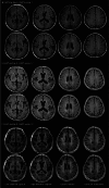
Representative fluid-attenuated inversion recovery (FLAIR) images of patients in each CHIPS tertile group. In Panels A through C, the top row of each panel is the original MRI image, and the bottom row is the image with the region of interest added. Colored lines represent the medial (blue) and lateral (red) cholinergic pathways. The Montreal Neurological Institute (MNI) z-coordinates of the associated index images are about − 1, 10, 25, and 37. Panels A through C shows representative FLAIR images of each CHIPS tertile group at the low external capsule, high external capsule, corona radiata, and centrum semiovale levels, from left to right. A represents CHIPSt1 with a CHIPS score of 7, C represents CHIPSt2 with a CHIPS score of 17, and D represents CHIPSt3 with a CHIPS score 72
Fig. 3
Fig. 3
Segmentation of the basal forebrain using FreeSurfer ScLimbic toolbox. Segmentation of limbic structures including BF was performed using the ScLimbic toolbox from the FreeSurfer toolbox (v.7.2.0, FMRIB, Oxford, UK) (https://surfer.nmr.mgh.harvard.edu/fswiki/ScLimbic). The basal forebrain is marked in brown color in the axial, sagittal, and coronal images of the 3D T1-weighted MRI (A-C) and the 3-dimensional reconstruction image (D). Total basal forebrain volume was calculated by adding the volumes of left and right basal forebrains. For subsequent analyses, basal forebrain volume was normalized to head size by dividing it by total intracranial volume
Fig. 4
Fig. 4
Differences in BF volume between tertile groups. Box plots show a significant difference in BF volume between tertile groups. The difference in BF volume was significant between the CHIPSt1 and CHIPSt3 groups (P < 0.01) and the CHIPSt2 and CHIPSt3 groups (P < 0.01), but not between the CHIPSt1 and CHIPSt2 groups (P = 0.99). The top and bottom of each box indicate the 75th and 25th percentiles, and the horizontal line inside each box indicates the 50th percentile. P values were obtained using a Tukey test, with an asterisk indicating P < 0.05. Abbreviations: BF, basal forebrain; CHIPS, Cholinergic Pathways Hyperintensities Scale; ns, not significant
Fig. 5
Fig. 5
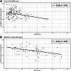
Sensitivity analysis for the correlations between CHIPS scores and BF volume. The sensitivity analyses examined the relationship between the CHIPS score and the BF volume in subgroups based on WMH severity. The results showed a negative correlation between CHIPS score and normalized BF volume in the subgroup with none to mild WMH (A) and with moderate to severe WMH (B)

References

    1. Chen Z-R, Huang J-B, Yang S-L, Hong F-F. Role of Cholinergic Signaling in Alzheimer’s Disease. Molecules. 2022;27:1816. 10.3390/molecules27061816 - DOI - PMC - PubMed
    1. Geula C, Dunlop SR, Ayala I, Kawles AS, Flanagan ME, Gefen T, et al. Basal forebrain cholinergic system in the dementias: vulnerability, resilience, and resistance. J Neurochem. 2021;158:1394–411. 10.1111/jnc.15471 - DOI - PMC - PubMed
    1. Giacobini E, Cuello AC, Fisher A. Reimagining cholinergic therapy for Alzheimer’s disease. Brain. 2022;145(7):2250–75. 10.1093/brain/awac096 - DOI - PubMed
    1. Nemy M, Dyrba M, Brosseron F, Buerger K, Dechent P, Dobisch L, et al. Cholinergic white matter pathways along the Alzheimer’s disease continuum. Brain. 2023;146(5):2075–88. 10.1093/brain/awac385 - DOI - PMC - PubMed
    1. Ray NJ, Bradburn S, Murgatroyd C, Toseeb U, Mir P, Kountouriotis GK, et al. In vivo cholinergic basal forebrain atrophy predicts cognitive decline in de novo Parkinson’s disease. Brain. 2018;141:165–76. 10.1093/brain/awx310 - DOI - PMC - PubMed