. 2024 Aug 15;19(1):65.
doi: 10.1186/s13062-024-00518-6.

Disulfidptosis signature predicts immune microenvironment and prognosis of gastric cancer

Affiliations

Disulfidptosis signature predicts immune microenvironment and prognosis of gastric cancer

Zitao Liu et al. Biol Direct. .

Abstract

Background: Disulfidptosis is a newly identified mechanism of cell death triggered by disulfide stress. Thus, gaining a comprehensive understanding of the disulfidptosis signature present in gastric cancer (GC) could greatly enhance the development of personalized treatment strategies for this disease.

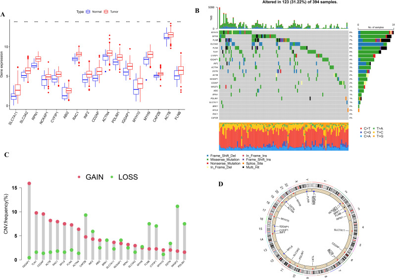
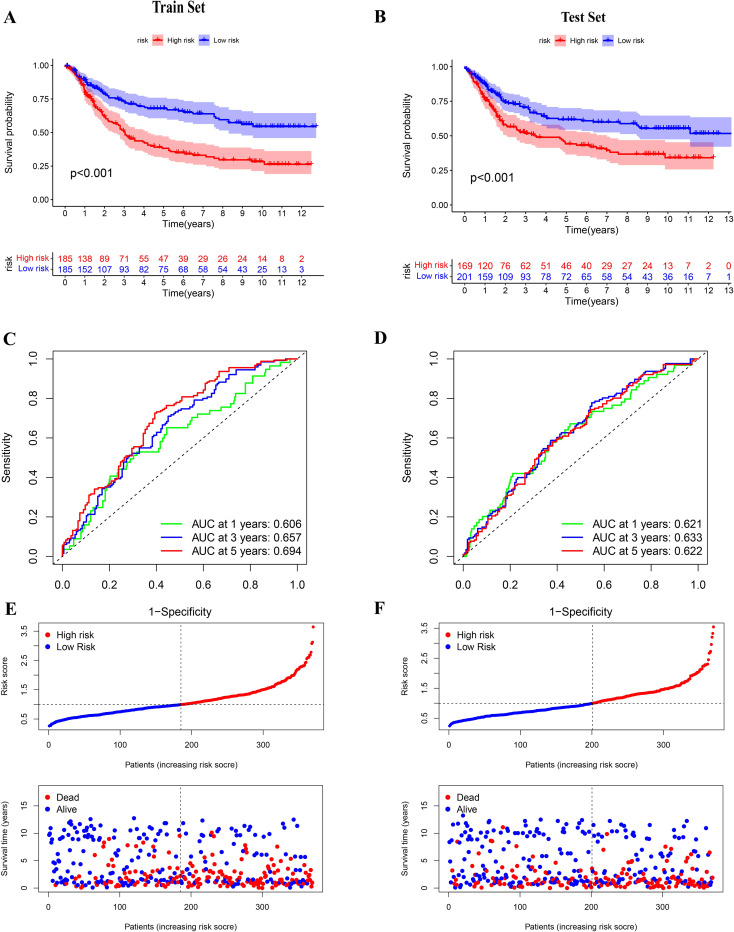
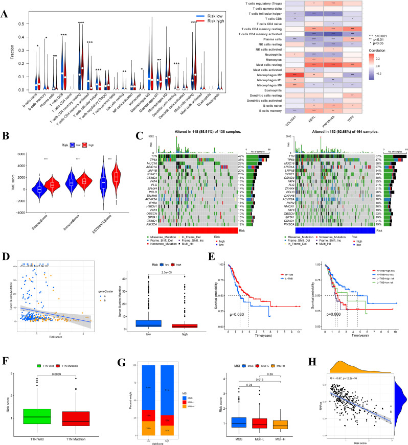
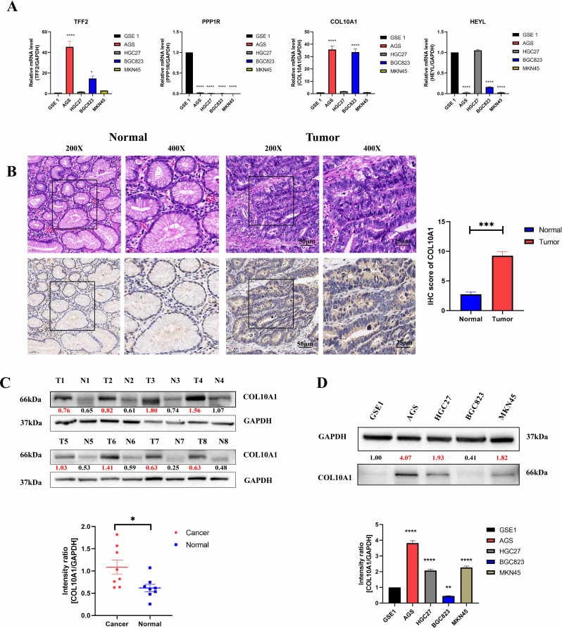
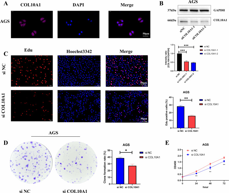
Methods: We employed consensus clustering to identify various subtypes of disulfidptosis and examined the distinct tumor microenvironment (TME) associated with each subtype. The Disulfidptosis (Dis) score was used to quantify the subtype of disulfidptosis in each patient. Subsequently, we assessed the predictive value of Dis score in terms of GC prognosis and immune efficacy. Finally, we conducted in vitro experiments to explore the impact of Collagen X (COL10A1) on the progression of GC.

Results: Two disulfidptosis-associated molecular subtypes (Discluster A and B) were identified, each with distinct prognosis, tumor microenvironment (TME), immune cell infiltration, and biological pathways. Discluster A, characterized by high expression of disulfidptosis genes, exhibited a high immune score but poor prognosis. Furthermore, the Dis score proved useful in predicting the prognosis and immune response in GC patients. Those in the low Dis score group showed better prognosis and increased sensitivity to immunotherapy. Finally, our experimental findings validated that downregulation of COL10A1 expression attenuates the proliferation and migration capabilities of GC cells while promoting apoptosis.

Conclusions: This study demonstrates that the disulfidptosis signature can assist in risk stratification and personalized treatment for patients with GC. The results offer valuable theoretical support for anti-tumor strategies.

Keywords: Disulfidptosis; Gastric cancer; Immunotherapy; Prognosis; Tumor microenvironment.


Conflict of interest statement

The authors declare no competing interests.

Figures

Fig. 1
Fig. 1
Genetic mutational landscape of disulfidptosis genes in GC. (A) Expression distributions of DEGs between GC and normal tissues. (B) Genetic alterations in the disulfidptosis gene. (C) Alteration frequency of CNV in disulfidptosis gene. (D) Location of CNV alterations in disulfidptosis genes on chromosomes
Fig. 2
Fig. 2
2 Discluster identified by consensus clustering. (A) A consensus matrix heatmap defining three clusters (k = 2) and their associated regions. (B) Differences in clinical features and disulfidptosis gene expression levels among different Discluster. (C) PCA analysis showed significant transcriptome differences between the two subgroups. (C) KM curve analysis of OS differences among different Discluster
Fig. 3
Fig. 3
Biological pathways and TME characteristics associated with Discluster. (A) GSVA analysis between subtypes. (B) Enrichment scores of 2 Discluster in the Hallmark pathway. (C) Infiltration levels of immune cells in 2 Discluster. (D) Correlation of Discluster with TME scores
Fig. 4
Fig. 4
Constructing Dis score. (A, B) KM curve analysis of differences in OS between two subgroups. (C, D) ROC curves of Dis score predicting 1-year, 3-year and 5-year OS. (E, F) Risk curve of Dis score
Fig. 5
Fig. 5
Expression level of COL10A1. (A) Expression of four characteristic genes in gastric cancer cell lines (GSE 1, AGS, HGC27, BGC823 and MKN45). The experiment was repeated three times and analyzed by one-way ANOVA, P < 0.05. (B) Immunohistochemical results of 4 pairs of gastric cancer tissues. (C) Expression of COL10A1 protein in 8 pairs of gastric cancer. Using t-test analysis, P < 0.05. (D) Expression level of COL10A1 protein in gastric cancer cell line. The experiment was repeated three times and analyzed by one-way ANOVA, P < 0.05. (*P < 0.05, **P < 0.01, ***P < 0.001, ****P < 0.0001)
Fig. 6
Fig. 6
Effect of COL10A1 on the progression of gastric cancer. (A) The expression of COL10A1 was analyzed by immunofluorescence. (B) The expression of COL10A1 in AGS was knocked down by siRNA, and the efficiency was verified by WB. (C, D, E) EDU, colony formation and CCK8 assay showed that the proliferative ability of AGS cells decreased after knockout of COL10A1. The results of CCK8 are shown as a line chart, and the other experimental results are shown as a bar chart. The above experiments were repeated three times, and the expression of COL10A1 was analyzed by one-way ANOVA. Other experiments were analyzed by t test. (*P < 0.05, **P < 0.01, ***P < 0.001)
Fig. 7
Fig. 7
Effect of COL10A1 on the progression of gastric cancer. (A, B) Transwell and wound healing experiments showed that the migration ability of AGS cells decreased after knocking down COL10A1. (C, D) After knocking down COL10A1, the apoptosis rate of AGS cells increased. (E) After knocking down COL10A1, the expression of F-actin in AGS cells decreased. The above experiments were analyzed by t test. (*P < 0.05, **P < 0.01, ***P < 0.001)
Fig. 8
Fig. 8
Construction and validation of a nomogram. (A) Dis score was positively correlated with T and N stages. The later the T and N stages, the higher the DIS scores. (B) Results of univariate and multivariate COX regression. (C) A nomogram predicting 1-, 3-, and 5-year OS in STAD patients in the entire cohort. (D) Calibration curve of the nomogram. (E, F) ROC curve and DCA curve of nomogram predicting 3-year OS of patients
Fig. 9
Fig. 9
Levels of immune cell infiltration and TME characteristics between the two ICDRS groups. (A) Correlation between Dis score and immune cell types. In addition, correlation of signature genes and immune cell infiltration in the Dis score model. (B) Correlation between Dis score and TME score. (C, D) Correlation between Dis score and TMB. (E) KM curve analysis of OS between high and low TMB groups. The prognosis of patients with high TMB and low risk group is obviously better than other groups. (F) Differences in the distribution of Dis score in TNN wild and mutant types. (G) Correlation of Dis score with MSI. (H) The relationship between Dis score and tumor stemness index
Fig. 10
Fig. 10
Relationship between Dis score and immunotherapy and chemotherapy drug sensitivity. (A) Differences in risk scores between immunotherapy responders and non-responders. (B) TIDE scores between different Dis score subgroups. (C) Expression levels of ICPs in high and low risk groups. (D) IPS scores between different ICDRS subgroups. (E) Correlation between Dis score and chemotherapeutic drug sensitivity

References

    1. Bray F, Ferlay J, Soerjomataram I, Siegel RL, Torre LA, Jemal A. Global Cancer statistics 2018: Globocan estimates of incidence and Mortality Worldwide for 36 cancers in 185 countries. Cancer J Clin. 2018;68(6):394–424. 10.3322/caac.21492. Epub 2018/09/13.10.3322/caac.21492 - DOI - PubMed
    1. Gambardella V, Castillo J, Tarazona N, Gimeno-Valiente F, Martínez-Ciarpaglini C, Cabeza-Segura M, et al. The role of Tumor-Associated macrophages in gastric Cancer Development and their potential as a therapeutic target. Cancer Treat Rev. 2020;86:102015. 10.1016/j.ctrv.2020.102015. Epub 2020/04/06. 10.1016/j.ctrv.2020.102015 - DOI - PubMed
    1. Russo AE, Strong VE. Gastric Cancer Etiology and Management in Asia and the West. Annual review of medicine (2019) 70:353 – 67. Epub 2018/10/26. 10.1146/annurev-med-081117-043436 - PubMed
    1. Chalabi M, Fanchi LF, Dijkstra KK, Van den Berg JG, Aalbers AG, Sikorska K, et al. Neoadjuvant Immunotherapy leads to pathological responses in Mmr-Proficient and Mmr-deficient early-stage Colon cancers. Nat Med. 2020;26(4):566–76. 10.1038/s41591-020-0805-8. Epub 2020/04/07. 10.1038/s41591-020-0805-8 - DOI - PubMed
    1. Sheih A, Voillet V, Hanafi LA, DeBerg HA, Yajima M, Hawkins R, et al. Clonal kinetics and single-cell transcriptional profiling of Car-T cells in patients undergoing Cd19 Car-T immunotherapy. Nat Commun. 2020;11(1):219. 10.1038/s41467-019-13880-1. Epub 2020/01/12. 10.1038/s41467-019-13880-1 - DOI - PMC - PubMed

Publication types

LinkOut - more resources