. 2024 Jul 15;15(32):12957-12963.
doi: 10.1039/d4sc01721h. eCollection 2024 Aug 14.

Structural modulation of aggregation-induced emission luminogens for NIR-II fluorescence imaging/photoacoustic imaging of tumors

Affiliations

Structural modulation of aggregation-induced emission luminogens for NIR-II fluorescence imaging/photoacoustic imaging of tumors

Xue Liu et al. Chem Sci. .

Abstract

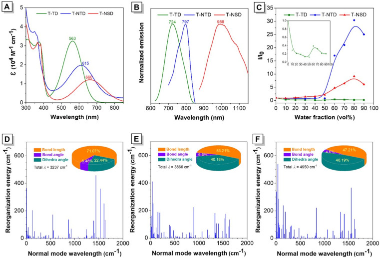
Concurrent near-infrared-II (NIR-II) fluorescence imaging (FLI) and photoacoustic imaging (PAI) holds tremendous potential for effective disease diagnosis owing to their combined benefits and complementary features, in particular on the basis of a single molecule. However, the simultaneous guarantee of high-quality NIR-II FLI and PAI is recognized to be challenging impeded by the competitive photophysical processes at the molecular level. Herein, a simple organic fluorophore, namely T-NSD, is finely engineered with facile synthetic procedures through delicately modulating the rigidity and electron-withdrawing ability of the molecular acceptor. The notable advantages of fabricated T-NSD nanoparticles include a large Stokes shift, intense fluorescence emission in the NIR-II region, and anti-quenching properties in the aggregated states, which eventually enable the implementation of dual-modal NIR-II FLI/PAI in a 4T1 tumor-xenografted mouse model with reliable performance and good biocompatibility. Overall, these findings present a simple strategy for the construction of NIR-II optical agents to allow multimodal disease diagnosis.


Conflict of interest statement

There are no conflicts to declare.

Figures

Scheme 1
Scheme 1. Preparation of NIR-II AIE nanoparticles for tumor diagnosis. (A) Molecular design philosophy by modulating the rigidity and electron-withdrawing ability of the molecular acceptor. (B) Illustration of the fabrication of T-NSD nanoparticles and their application in dual-modal NIR-II FLI/PAI of tumors.
Fig. 1
Fig. 1. Structure details of T-BTD, T-NTD and T-NSD. (A) Chemical structures, (B) optimized S0 geometries and (C) illustration of the frontier molecular orbitals (LUMOs and HOMOs) determined at the M06-2X/6-31G(d,p) level of theory, using the Gaussian 16 program.
Fig. 2
Fig. 2. Photophysical property study of T-BTD, T-NTD and T-NTD. (A) The absorption and (B) emission spectra of the obtained compounds dissolved in THF solution (10 μM, excited at 563 nm, 615 nm and 660 nm, respectively). (C) Plots of relative PL intensity (I/I0) of the obtained compounds versus different water fractions. Inset: enlarged plot of T-BTD. I0 and I are the values of PL intensities at maximum peaks in THF and THF/water mixtures, respectively. Calculated reorganization energy versus the normal mode wavenumbers of (D) T-BTD, (E) T-NTD, and (F) T-NSD, respectively.
Fig. 3
Fig. 3. Characterization of the T-NSD NPs. (A) The diagrammatic drawing of the fabrication process of T-NSD NPs. (B) DLS analysis of T-NSD NPs. Inset: corresponding TEM image of T-NSD NPs. (C) Normalized absorption and fluorescence spectra of T-NSD NPs in aqueous solution (10 μM).
Fig. 4
Fig. 4. The cytotoxicity and in vitro bioimaging study of T-NSD NPs. (A) HeLa cell and (B) 4T1 cell viabilities after staining with different concentrations of T-NSD NPs. (C) Co-localization test of FITC-NPs with Nile Red, Mito-Tracker, and Lyso-Tracker, respectively in 4T1 cells.
Fig. 5
Fig. 5. In vivo NIR-II FLI and PAI. (A) NIR-II FLI on 4T1 tumor-bearing mice after injection of T-NSD NPs. (B) Corresponding average fluorescence intensity of tumor sites at different time points based on the images in (A) (n = 3). (C) PAI on 4T1 tumor-bearing mice after injection of T-NSD NPs (PA 700 nm). (D) The photoacoustic signal intensity within the solid coil at the tumor site based on the images in (C) (n = 3).
Fig. 6
Fig. 6. The biosafety evaluation of T-NSD NPs (n = 3). (A) In vitro hemolysis experiments. (B) Liver function and kidney function indicators (n = 3). (C) H&E-stained photomicrographs of major organs.

References

    1. Zhang P. Li W. Liu C. Qin F. Lu Y. Qin M. Hou Y. Exploration. 2023;3:20230070. - PMC - PubMed
    1. Cheng P. Pu K. Nat. Rev. Mater. 2021;6:1095–1113. doi: 10.1038/s41578-021-00328-6. - DOI
    1. Huang X. Song J. Yung B. C. Huang X. Xiong Y. Chen X. Chem. Soc. Rev. 2018;47:2873–2920. doi: 10.1039/C7CS00612H. - DOI - PMC - PubMed
    1. Chen Y. Wang S. Zhang F. Nat. Rev. Bioeng. 2023;1:60–78.
    1. Li C. Chen G. Zhang Y. Wu F. Wang Q. J. Am. Chem. Soc. 2020;142:14789–14804. doi: 10.1021/jacs.0c07022. - DOI - PubMed

LinkOut - more resources