. 2024 Jul 9;15(32):12710-12720.
doi: 10.1039/d4sc02787f. eCollection 2024 Aug 14.

Exploring the influence of H-bonding and ligand constraints on thiolate ligated non-heme iron mediated dioxygen activation

Affiliations

Exploring the influence of H-bonding and ligand constraints on thiolate ligated non-heme iron mediated dioxygen activation

Maike N Lundahl et al. Chem Sci. .

Abstract

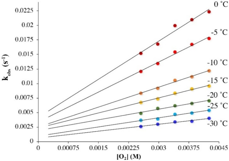
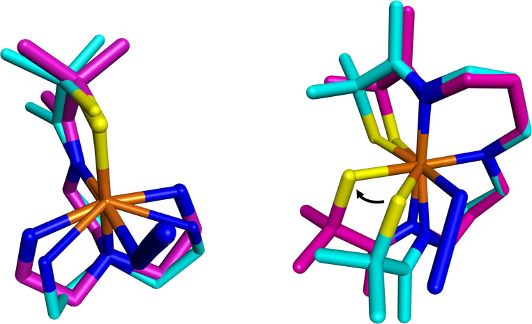
Converting triplet dioxygen into a powerful oxidant is fundamentally important to life. The study reported herein quantitatively examines the formation of a well-characterized, reactive, O2-derived thiolate ligated FeIII-superoxo using low-temperature stopped-flow kinetics. Comparison of the kinetic barriers to the formation of this species via two routes, involving either the addition of (a) O2 to [FeII(S2 Me2N3(Pr,Pr))] (1) or (b) superoxide to [FeIII(S2 Me2N3(Pr,Pr))]+ (3) is shown to provide insight into the mechanism of O2 activation. Route (b) was shown to be significantly slower, and the kinetic barrier 14.9 kJ mol-1 higher than route (a), implying that dioxygen activation involves inner-sphere, as opposed to outer sphere, electron transfer from Fe(ii). H-bond donors and ligand constraints are shown to dramatically influence O2 binding kinetics and reversibility. Dioxygen binds irreversibly to [FeII(S2 Me2N3(Pr,Pr))] (1) in tetrahydrofuran, but reversibly in methanol. Hydrogen bonding decreases the ability of the thiolate sulfur to stabilize the transition state and the FeIII-superoxo, as shown by the 10 kJ mol-1 increase in the kinetic barrier to O2 binding in methanol vs. tetrahydrofuran. Dioxygen release from [FeIII(S2 Me2N3(Pr,Pr))O2] (2) is shown to be 24 kJ mol-1 higher relative to previously reported [FeIII(SMe2N4(tren))(O2)]+ (5), the latter of which contains a more flexible ligand. These kinetic results afford an experimentally determined reaction coordinate that illustrates the influence of H-bonding and ligand constraints on the kinetic barrier to dioxygen activation an essential step in biosynthetic pathways critical to life.


Conflict of interest statement

There are no conflicts to declare.

Figures

Scheme 1
Scheme 1. Pathways for the formation of [FeIII(S2Me2N3(Pr,Pr))(O2)] (2) (top), and reversible dioxygen binding to [FeII(SMe2N4(tren))]+ (4) to form a putative superoxo intermediate [FeIII(SMe2N4(tren))(O2)]+ (5) (bottom).
Fig. 1
Fig. 1. Time-resolved spectral changes obtained upon mixing THF solutions of five – coordinate [FeII(S2Me2N3(Pr,Pr))] (1, 0.25 mM) and O2 (3.95 mM) at −40 °C. Inset: kinetic trace (λ = 523 nm) showing the formation of the Fe–O2 intermediate 2. All reported concentrations are after mixing in the stopped-flow cell.
Fig. 2
Fig. 2. Temperature-dependent rate constants kobs for the formation of superoxo 2 in the reaction between 1 (0.25 mM) and O2 in THF plotted against [O2]. The intercept of approximately 0.0 would be consistent with irreversible O2 binding.
Fig. 3
Fig. 3. Temperature-dependent rate constants kobs for the formation of superoxo 2 in the reaction between 1 (0.1 mM) and O2 in MeOH, plotted against [O2]. The non-zero intercepts would be consistent with reversible O2 binding in MeOH.
Fig. 4
Fig. 4. Eyring plots from which the activation parameters reported were obtained for the following reactions. (A) O2 binding to 1 in THF and (B) O2 binding to 1 in MeOH, which use the second order rate constants, kon, obtained from the slope of kobsvs. [O2] plots (Fig. 2 and 3) with [FeII] = 0.25 mM and 0.1 mM respectively. (C) The reaction of 3 and KO2 (solubilized with KryptoFix) in THF plotted with second order rate constants, kon, obtained from the slope of kobsvs. [KO2] plots. [FeIII] = 0.1 mM, after mixing. (D) The release of O2 from 2 in MeOH which uses first order rate constants, koff, obtained from the intercept of kobsversus [O2] plots (Fig. 3).
Fig. 5
Fig. 5. Time-resolved spectral changes observed in the reaction between 3 (0.1 mM) and KO2 (5.0 mM; solubilized with 222-Kryptofix) in THF at −40 °C. Inset: kinetic trace (λ = 523 nm) showing the formation of 2. All reported concentrations are after mixing in the stopped-flow cell.
Fig. 6
Fig. 6. Temperature-dependent rate constants, kobs, for the formation of superoxo 2 in the reaction between 3 and KO2 (solubilized with KryptoFix) in THF, plotted against [KO2]. The ∼ zero intercepts would be consistent with irreversible O2˙ binding. [Fe(iii)] = 0.1 mM. Concentrations listed correspond to after mixing in the stopped-flow cell.
Fig. 7
Fig. 7. Possible mechanisms for the reaction between 1 and O2 involving either outer-sphere electron transfer (ET) followed by superoxide (O2˙) binding to oxidized 3, O2 binding followed by inner-sphere ET, or a concerted mechanism (diagonal).
Fig. 8
Fig. 8. H-bonding to MeOH causes the RS → Fe band of reduced 1 (0.238 mM) to blue-shift relative to its energy in THF.
Fig. 9
Fig. 9. ORTEP diagram of 1 crystallized from MeOH showing the MeOH that is H-bonded to one of the thiolate sulfurs, S(1). Shown with 50% probability ellipsoids and numbering scheme. All hydrogen atoms excluding H(2) and H(1) have been deleted for clarity.
Fig. 10
Fig. 10. Comparison of the experimentally determined barrier to O2 binding to 1 in THF (green) and MeOH (red) and 4 in MeOH (black), as well as the release of O2 from 2 and 5 in MeOH.
Fig. 11
Fig. 11. Space-filling models generated from the crystallographically-determined structure of 1 (left) and 3 (left), and the DFT-optimized geometry of 2calc (middle) displaying differences in the helical wrapping angle, Φ, and the larger amount of structural rearrangement required for O2˙ binding to 3 than O2 binding to 1. The least squared planes of N(1)FeN(2)N(3) and C(6)N(2)C(7)Fe shown in orange and purple respectively.
Fig. 12
Fig. 12. Visualization of experimentally obtained structural changes that occur upon oxidation and binding of a sixth ligand, azide. Left: [FeII(SMe2N4(tren))]+ (4, cyan) to [FeIII(SMe2N4(tren))N3] (magenta). Right: [FeII(S2Me2N3(Pr,Pr))] (1, cyan) and [FeIII(S2Me2N3(Pr,Pr))(N3)] (magenta). Depiction utilized previously reported crystallographically determined structures.

References

    1. Solomon E. I. DeWeese D. E. Babicz Jr J. T. Mechanisms of O2 activation by mononuclear non-heme iron enzymes. Biochemistry. 2021;60:3497–3506. doi: 10.1021/acs.biochem.1c00370. - DOI - PMC - PubMed
    1. Gennari M. Duboc C. Bio-inspired, multifunctional metal–thiolate motif: from electron transfer to sulfur reactivity and small-molecule activation. Acc. Chem. Res. 2020;53:2753–2761. doi: 10.1021/acs.accounts.0c00555. - DOI - PubMed
    1. Jeong D. Selverstone Valentine J. Cho J. Bio-inspired mononuclear nonheme metal peroxo complexes: synthesis, structures and mechanistic studies toward understanding enzymatic reactions. Coordin. Chem. Rev. 2023;480:215021. doi: 10.1016/j.ccr.2023.215021. - DOI
    1. Nastri F. Chino M. Maglio O. Bhagi-Damodaran A. Lu Y. Lombardi A. Design and engineering of artificial oxygen-activating metalloenzymes. Chem. Soc. Rev. 2016;45:5020–5054. doi: 10.1039/C5CS00923E. - DOI - PMC - PubMed
    1. Kennepohl P. Neese F. Schweitzer D. Jackson H. L. Kovacs J. A. Solomon E. I. Spectroscopy of non-heme iron thiolate complexes: insight into the electronic structure of the low-spin active site of nitrile hydratase. Inorg. Chem. 2005;44:1826–1836. doi: 10.1021/ic0487068. - DOI - PMC - PubMed

LinkOut - more resources