Case Reports
. 2024 Jul 31:14:1362090.
doi: 10.3389/fonc.2024.1362090. eCollection 2024.

Intranodal palisaded myofibroblastoma in the submandibular gland region: a case report

Affiliations
Case Reports

Intranodal palisaded myofibroblastoma in the submandibular gland region: a case report

Han Zhang et al. Front Oncol. .

Abstract

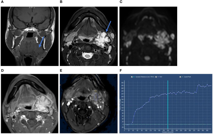
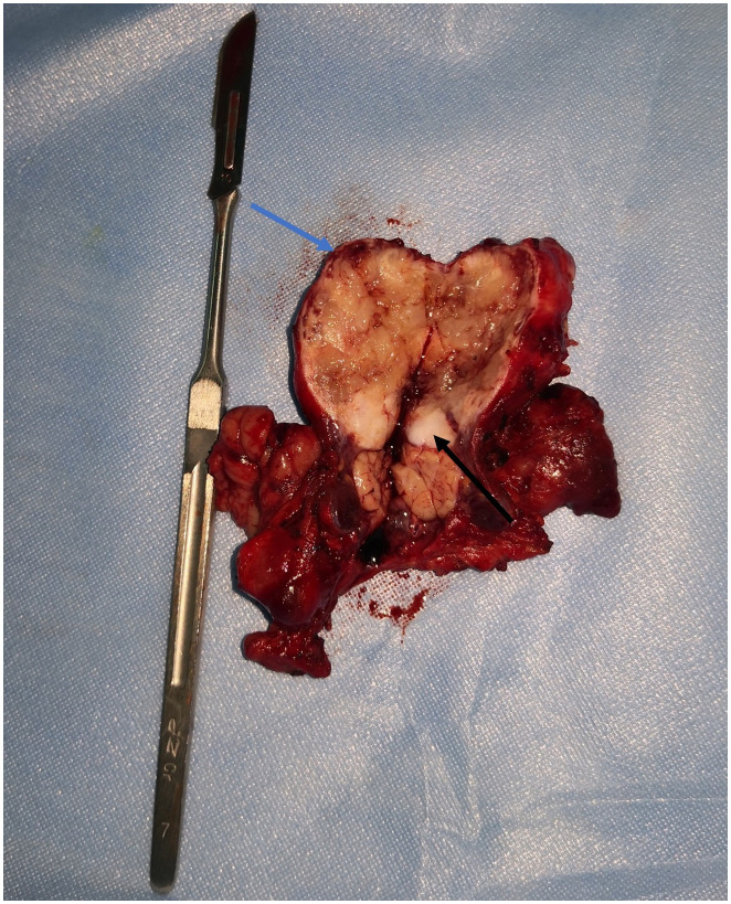
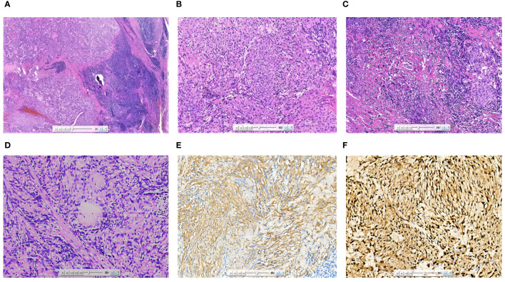
Intranodal palisaded myofibroblastoma (IPM) is a rare benign tumor of the lymph nodes, particularly in inguinal lymph nodes. IPM originating from the submandibular gland lymph nodes is rarely encountered in clinical practice. Herein, we report the case of a 31-year-old male patient with IPM of the submandibular gland region and describe in detail magnetic resonance imaging findings and pathology. Magnetic resonance imaging detected a heterogeneous lesion with a hypointense rim on T2-weighted imaging with specificity in the left submandibular gland region. This case report will contribute to the accumulation of experience in the diagnosis of this disease.

Keywords: case report; intranodal palisaded myofibroblastoma; lymph node; magnetic resonance imaging; pathology; submandibular gland.


Conflict of interest statement

The authors declare that the research was conducted in the absence of any commercial or financial relationships that could be construed as a potential conflict of interest.

Figures

Figure 1
Figure 1
Coronal T1WI scan (A) showed the tumor mass on the upper pole of the left submandibular gland (blue arow). Axial T2WI-FS (B) showed heterogeneous hyperintense with a hypointense lobulated rim (blue arow). The tumor showed heterogeneous high signal intensity on DWI (C). Axial gadolinium-enhanced T1-weighted image (D) showed heterogeneous enhancement with a hypointense rim. Time-intensity curve (TIC) was progressive ascending type (E, F). Its x-axis represents the time of the enhanced scan, from 0 seconds to 380 seconds, with the extension of time, the curve is on the rise.
Figure 2
Figure 2
Lesion surgically excised. The submandibular mass had a grey and white appearance with multifocal hemorrhagic areas on the cut surface. It was surrounded by tissues similar to capsule (blue arrow) and the attached salivary gland tissue (black arrow) was observed.
Figure 3
Figure 3
Microscopic image (A) of the excised specimen showing the tumor was located in the lymph nodes, and residual lymphoid tissue could be seen at the edge (HE×20). The tumor cells were arranged in a fence-like and woven pattern (B) (HE×100). The surrounding area of the tumor was accompanied by significant hyaline degeneration (C) (HE×100). The characteristic morphological asbestos-like fibrous nodules (D) (HE×200). SMA (E) was partially expressed in tumor cells (IHC×200). Cyclin-D1 (F) was diffusely expressed in tumor cells (IHC×200).

Similar articles

References

    1. Bhullar JS, Herschman BR, Dubay L. Intranodal palisaded myofibroblastoma: A new entity of axillary tumors. Am Surgeon. (2013) 79:19–21. doi: 10.1177/000313481307900111 - DOI - PubMed
    1. Nguyen T, Eltorky MA. Intranodal palisaded myoflbroblastoma. Arch Pathol Lab Med. (2007) 131:306–10. doi: 10.5858/2007-131-306-IPM - DOI - PubMed
    1. Pérez-Serrano C, Bargalló X, Rodríguez-Carunchio L, Ganau S, Úbeda B. Intranodal palisaded myofibroblastoma of the axilla. Breast J. (2019) 25:1288–9. doi: 10.1111/tbj.13447 - DOI - PubMed
    1. Karabulut YY, Kara T, Berkeşoğlu M. Intranodal palisaded myofibroblastoma – a rare case report and literature review. APMIS. (2016) 124:905–10. doi: 10.1111/apm.12580 - DOI - PubMed
    1. Hisaoka M, Hashiomoto H, Daimaru Y. Intranodal palisaded myofibroblastoma with so-called amianthoid fibers: A report of two cases with a review of the literature. Pathol Int. (1998) 48:307–12. doi: 10.1111/j.1440-1827.1998.tb03911.x - DOI - PubMed

Publication types

LinkOut - more resources