

Pulsed Vibro-Acoustic Analysis Technique for Monitoring Bone Health in Preterm Infants: A Pilot Study
- PMID: 39148928
- PMCID: PMC11324250
- DOI: 10.1109/access.2024.3437375
Pulsed Vibro-Acoustic Analysis Technique for Monitoring Bone Health in Preterm Infants: A Pilot Study
Abstract
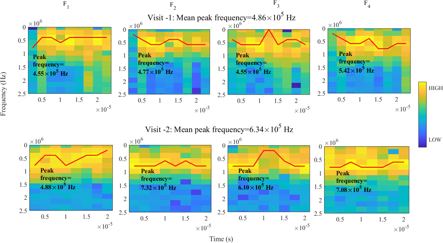
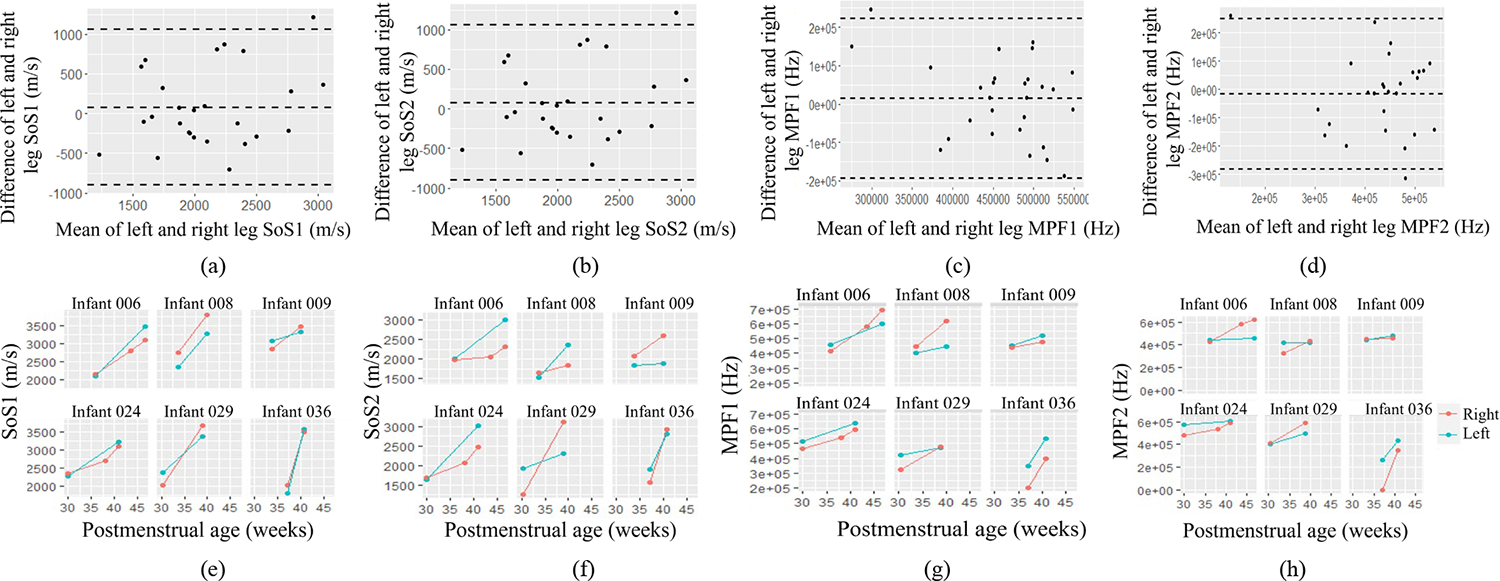
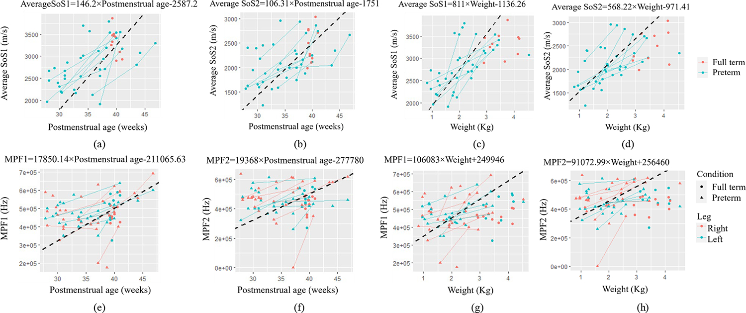
Despite advances in neonatal care, metabolic bone disease of prematurity (MBDP) remains a common problem in preterm infants. The development of non-invasive and affordable diagnostic approaches can be highly beneficial in the diagnosis and management of preterm infants at risk of MBDP. In this study, we present an ultrasound method called pulsed vibro-acoustic analysis to investigate the progression of bone mineralization in infants over time versus weight and postmenstrual age. The proposed pulsed vibro-acoustic analysis method is used to evaluate the vibrational characteristics of the bone. This method uses the acoustic radiation force of ultrasound to vibrate the bone. The generated acoustic waves are detected using a hydrophone placed on the skin over the tibia. The frequency of vibration and the speeds of received acoustic waves have information regarding the material property of the bone. We examined the feasibility of this method through an in vivo study consisting of 25 preterm and 10 full term infants. The pulsed vibro-acoustic data were acquired longitudinally in preterm infants with multiple visits and at a single visit in full term infants. Speed of sound and mean peak frequency of slow and fast sound waves recorded by hydrophone were used to analyze bone mineralization progress. Linear mixed model was used for statistical analysis in characterizing the mineralization progress in preterm infants compared to data from full term subjects. Significance changes in wave parameters (speed of sound and mean peak frequency) with respect to the postmenstrual age and weight in preterm infants were observed with p-values less than 0.05. Statistical significances in speed of sound measurement for both fast and slow waves were observed between preterm and full term infants, with p-values of <0.01 and 0.02, respectively. The results of this pilot study indicate the potential use of vibro-acoustic analysis for monitoring the progression of bone mineralization in preterm infants.
Keywords: Bone mineralization; Premature infant; Quantitative ultrasound; Speed of Sound; Vibro-acoustic analysis.
Figures


Similar articles
-
Noise or sound management in the neonatal intensive care unit for preterm or very low birth weight infants.Cochrane Database Syst Rev. 2024 May 30;5(5):CD010333. doi: 10.1002/14651858.CD010333.pub4. Cochrane Database Syst Rev. 2024. PMID: 38813836 Free PMC article.
-
Cup feeding versus other forms of supplemental enteral feeding for newborn infants unable to fully breastfeed.Cochrane Database Syst Rev. 2016 Aug 31;2016(8):CD005092. doi: 10.1002/14651858.CD005092.pub3. Cochrane Database Syst Rev. 2016. PMID: 27577968 Free PMC article.
-
Effects of targeting lower versus higher arterial oxygen saturations on death or disability in preterm infants.Cochrane Database Syst Rev. 2017 Apr 11;4(4):CD011190. doi: 10.1002/14651858.CD011190.pub2. Cochrane Database Syst Rev. 2017. PMID: 28398697 Free PMC article.
-
Late (>7 days) postnatal corticosteroids for chronic lung disease in preterm infants.Cochrane Database Syst Rev. 2009 Jan 21;(1):CD001145. doi: 10.1002/14651858.CD001145.pub2. Cochrane Database Syst Rev. 2009. Update in: Cochrane Database Syst Rev. 2014 May 13;(5):CD001145. doi: 10.1002/14651858.CD001145.pub3. PMID: 19160189 Updated.
-
Sound reduction management in the neonatal intensive care unit for preterm or very low birth weight infants.Cochrane Database Syst Rev. 2015 Jan 30;1:CD010333. doi: 10.1002/14651858.CD010333.pub2. Cochrane Database Syst Rev. 2015. Update in: Cochrane Database Syst Rev. 2020 Jan 27;1:CD010333. doi: 10.1002/14651858.CD010333.pub3. PMID: 25633155 Updated.
References
-
- Moreira A, Jacob R, Lavender L, and Escaname E, “Metabolic Bone Disease of Prematurity,” NeoReviews, vol. 16, no. 11, pp. e631–e641, Nov. 2015, doi: 10.1542/neo.16-11-e631. - DOI
Grants and funding
LinkOut - more resources
Full Text Sources