. 2024 Aug 1:12:1412811.
doi: 10.3389/fbioe.2024.1412811. eCollection 2024.

Key quality parameter comparison of mesenchymal stem cell product cryopreserved in different cryopreservation solutions for clinical applications

Affiliations

Key quality parameter comparison of mesenchymal stem cell product cryopreserved in different cryopreservation solutions for clinical applications

Yuan Tan et al. Front Bioeng Biotechnol. .

Abstract

Introduction: Cryopreservation is a critical process of cell products for achieving a commercial viability through wide scale adoption. By preserving cells in a lower temperature, cryopreservation enables a product to be off-the-shelf and ready for infusion. An optimized cryopreservation strategy can maintain the viability, phenotype, and potency of thawed mesenchymal stromal/stem cells (MSCs) while being regulatory compliant. We compared three clinical-ready formulations with one research cryopreservation solutions and evaluated key quality parameters of post thawed MSCs.

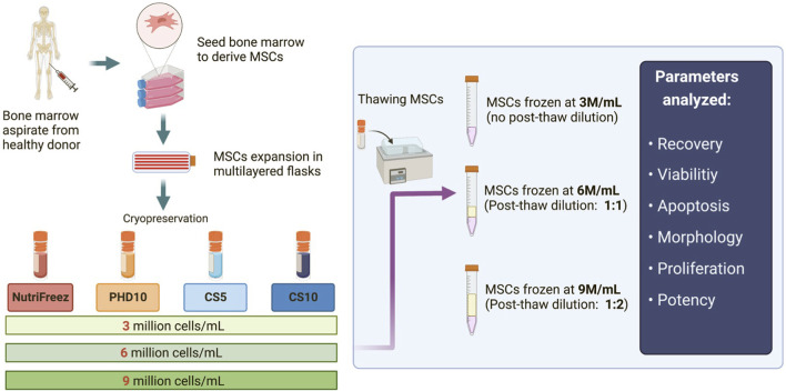
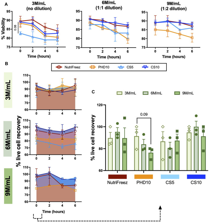
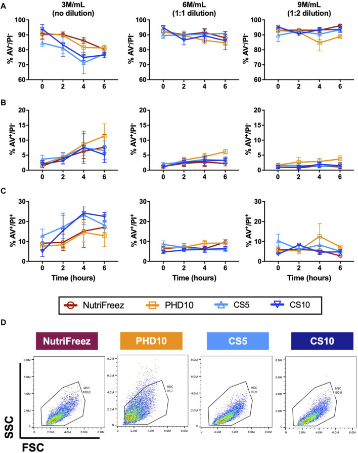
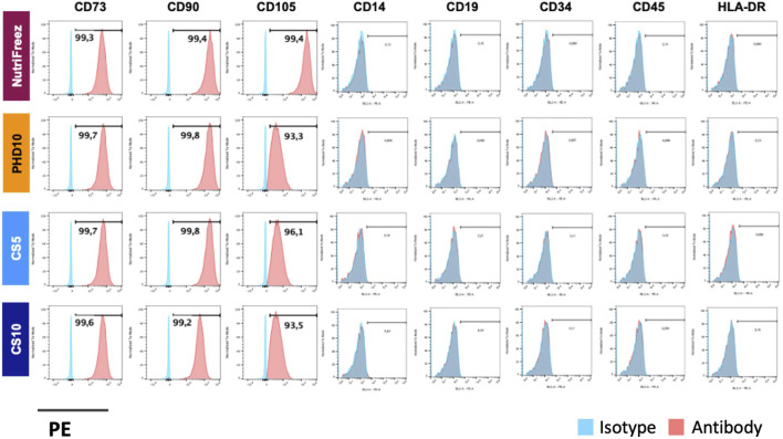
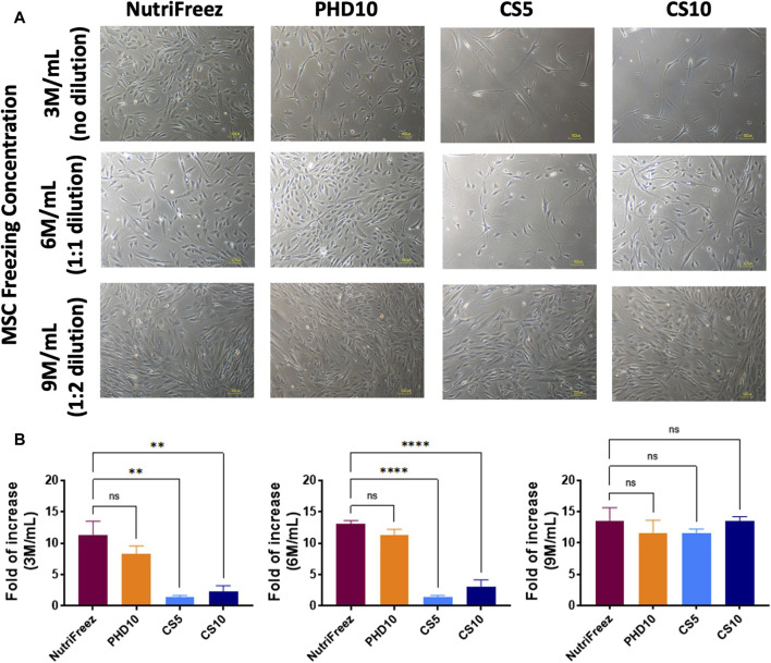
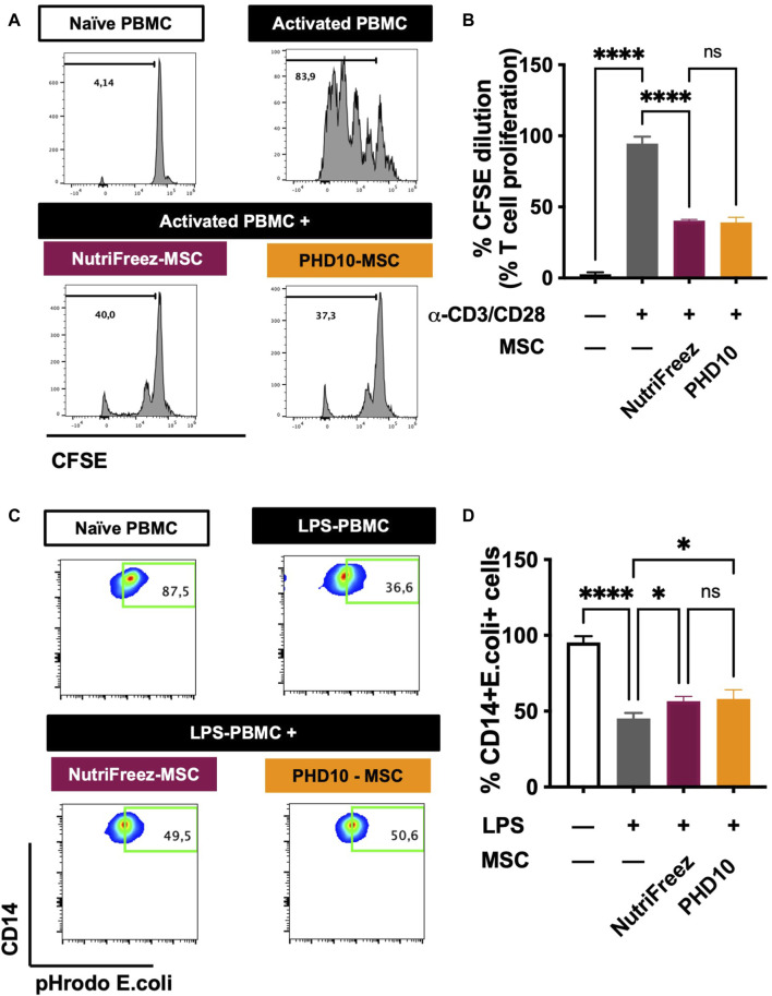
Method and result: MSCs were cryopreserved at 3, 6, and 9 million cells/mL (M/mL) in four different cryopreservation solutions: NutriFreez (10% dimethyl sulfoxide [DMSO]), Plasmalyte A (PLA)/5% human albumin (HA)/10% DMSO (PHD10), CryoStor CS5 (5% DMSO), and CryoStor CS10 (10% DMSO). To establish post thaw viability, cells were evaluated with no dilution of DMSO (from 3 M/mL), 1:1 dilution (from 6 M/mL), or 1:2 dilution (from 9 M/mL) with PLA/5% HA, to achieve uniform concentration at 3 M/mL. Cell viability was measured at 0-, 2-, 4-, and 6-h post thaw with Trypan blue exclusion and Annexin V/PI staining. Dilution (1:2) of final cell products from 9M/mL resulted in an improvement of cell viability over 6 h but showed a trend of decreased recovery. MSCs cryopreserved in solutions with 10% DMSO displayed comparable viabilities and recoveries up to 6 h after thawing, whereas a decreasing trend was noted in cell viability and recovery with CS5. Cells from all groups exhibited surface marker characteristics of MSCs. We further evaluated cell proliferation after 6-day recovery in culture. While cells cryopreserved in NutriFreez and PHD10 presented similar cell growth post thaw, MSCs cryopreserved in CS5 and CS10 at 3 M/mL and 6M/mL showed 10-fold less proliferative capacity. No significant differences were observed between MSCs cryopreserved in NutriFreez and PHD10 in their potency to inhibit T cell proliferation and improve monocytic phagocytosis.

Conclusion: MSCs can be cryopreserved up to 9 M/mL without losing notable viability and recovery, while exhibiting comparable post thaw potency with NutriFreez and PHD10. These results highlight the importance of key parameter testing for selecting the optimal cryopreservation solution for MSC-based therapy.

Keywords: cell therapy; cryopreservation; final cell products; mesenchymal stem cells; off-the-shelf; potency; quality; stability.


Conflict of interest statement

The authors declare that the research was conducted in the absence of any commercial or financial relationships that could be construed as a potential conflict of interest.

Figures

FIGURE 1
FIGURE 1
Schematic of the experimental design for MSC isolation, culture expansion, and cryopreservation. Bone marrow aspirates were first obtained from healthy donors, and MSCs were derived, expanded, and cryopreserved in four different cryopreservation solutions: NutriFreez, PHD10 (Plasmalyte-A, 5% human albumin, 10% DMSO), Cryostor CS5, and Cryostor CS10. For each cryopreservation solution, cells were frozen at concentrations of 3 million cells/mL (M/mL), 6 M/mL, or 9 M/mL. Cryopreserved MSCs were thawed, with or without dilution (pending on freezing cell concentrations), for downstream analysis.
FIGURE 2
FIGURE 2
Assessment and comparison of MSC viability and recovery post thaw over 6 h. Trypan blue exclusion was used to assess (A) post thaw viabilities and (B) viable cell recoveries, and (C) viable cell recoveries at 0h post thaw. Aliquot of cells were collected and used for measurements at 0, 2, 4, and 6 h n = 3 independent experiments with MSCs from one donor, data graphed as mean ± SEM. Group comparisons were analyzed by one-way ANOVA with Dunnett’s post hoc test. For A, p = 0.08 comparing CS5 vs. Nutrifreez D10.
FIGURE 3
FIGURE 3
Assessment and comparison of apoptosis levels in cryopreserved MSC over 6 h post thaw. Annexin V (AV) and PI staining was performed on the MSCs that had been cryopreserved in NutriFreez, PH10, CS5, and CS10. Flow cytometry analysis was carried out to assess levels of cellular apoptosis at 0, 2, 4, and 6 h. Data represents (A) live cells from AV-/PI- population, (B) early apoptotic cells from AV+/PI- population, and (C) dead cells from AV+/PI + population. (D) Representative forward scatter (FSC) and side scatter (SSC) from each cryopreservation conditions. n = 3 independent experiments with MSCs from one donor, data plotted as mean ± SEM.
FIGURE 4
FIGURE 4
Surface marker characterization of cryopreserved MSCs. Representative flow cytometric plots indicate positive markers (CD73, CD90, CD105) and negative markers (CD14, CD19, CD34, CD45, and HLA-DR) for MSC characterization profile. n = 3 independent experiments with MSCs from one donor.
FIGURE 5
FIGURE 5
Post thaw expansion of cryopreserved MSCs. Post thaw, MSCs were seeded and cultured for 6 days to examine (A) cell morphology, and (B) proliferation potential (fold of cell number increases, calculated by dividing the number of cells harvested at day 6 to the number of cells used at initial seeding). Scale bar = 100 μm n = 3 independent experiments with MSCs from one donor, data plotted as mean ± SEM. Group comparisons were analyzed by one-way ANOVA with Dunnett’s post hoc test, *P < 0.05, **P < 0.01, and ****P < 0.0001.
FIGURE 6
FIGURE 6
Effect of MSCs on the inhibition of T-cell proliferation and improvement of monocytic phagocytic capacity post LPS injury. (A) Representative flow cytometric plots of CFSE dilution of naïve PBMCs, CD3/CD28 activated PMBCs without and with MSCs co-culture (previously cryopreserved in NutriFreez or PHD10), showing the ability of MSC to inhibit T-cell proliferation. (B) Quantification and summary data is plotted as a bar graph, compared to negative control of naïve PBMCs and activated PBMCs. (C) Representative flow cytometric plots of naïve PBMCs, LPS-treated PBMCs without and with MSCs (previously cryopreserved in NutriFreez or PHD10) demonstrating the PBMC’s ability to phagocytose bacteria as indicated by the percentage of CD14+ cells positive for green, fluorescent signal. (D) Quantification and summary data is plotted as a bar graph, compared to negative control of naïve PBMCs and activated PBMCs. n = 3 independent experiments using three different donors derived MSCs, data represent mean ± SEM. Group comparisons were analyzed by one-way ANOVA with Tukey’s post hoc test, *P < 0.05 while ns = non-.significant.

Similar articles

Cited by

References

    1. Abramson J. S., Palomba M. L., Gordon L. I., Lunning M. A., Wang M., Arnason J., et al. (2020). Lisocabtagene maraleucel for patients with relapsed or refractory large B-cell lymphomas (TRANSCEND NHL 001): a multicentre seamless design study. Lancet 396 (10254), 839–852. 10.1016/s0140-6736(20)31366-0 - DOI - PubMed
    1. Acker J. P. (2005). “Biopreservation of cells and engineered tissues,” in Tissue engineering II, Editors Lee K., Kaplan D. (Berlin Heidelberg: Springer; ), 157–187. - PubMed
    1. Alessandrino E. P., Bernasconi P., Caldera D., Colombo A., Bonfichi M., Malcovati L., et al. (1999). Adverse events occurring during bone marrow or peripheral blood progenitor cell infusion: analysis of 126 cases. Bone Marrow Transplant. 23 (6), 533–537. 10.1038/sj.bmt.1701609 - DOI - PubMed
    1. Bahsoun S., Coopman K., Akam E. C. (2019). The impact of cryopreservation on bone marrow-derived mesenchymal stem cells: a systematic review. J. Transl. Med. 17 (1), 397–429. 10.1186/s12967-019-02136-7 - DOI - PMC - PubMed
    1. Barnes D. W., Loutit J. F. (1955). The radiation recovery factor: preservation by the Polge-Smith-Parkes technique. J. Natl. Cancer Inst. 15 (4), 901–905. 10.1093/jnci/15.4.901 - DOI - PubMed

LinkOut - more resources