

Pathological and Immunohistochemical Assessment of Aging of the Abdominal Skin Treated with Carboxytherapy: A Randomized, Split-body Trial
- PMID: 39148961
- PMCID: PMC11324193
Pathological and Immunohistochemical Assessment of Aging of the Abdominal Skin Treated with Carboxytherapy: A Randomized, Split-body Trial
Abstract
Background: Skin aging as a continuous and irreversible process is mainly the result of alterations of function and structure of the dermis. Among the modalities used for treating skin aging, carboxytherapy has been introduced as a safe minimally-invasive method for rejuvenation, reparation, and reconditioning of the skin.
Objective: We assessed the efficacy of carboxytherapy for the treatment of intrinsic skin aging through pathological and immunohistochemical (IHC) investigations.
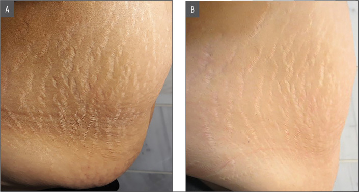
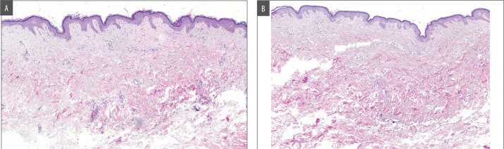
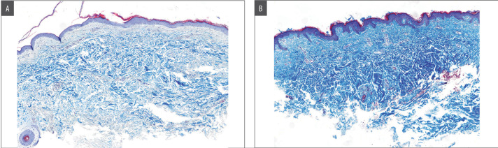
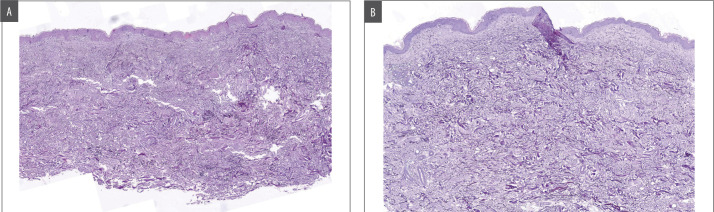
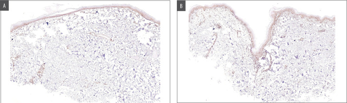
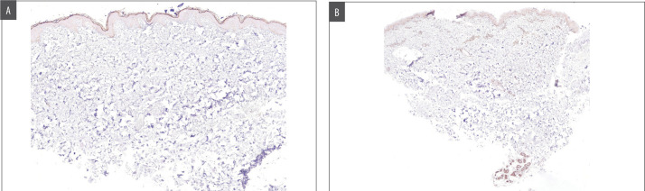
Methods: Our study was a split-body, randomized clinical trial on 15 female patients with intrinsic skin aging of the abdomen. Carboxytherapy was performed on one side of the abdomen, weekly for 10 sessions, while the other side was left untreated. Two weeks after the last session, skin biopsies were taken from both sides of the abdomen. Staining with hematoxylin-eosin, Masson-trichrome, and Orcein Giemsa was performed for the assessment of epidermal and dermal thickness, collagen, and elastin organization, respectively. IHC examination was performed for investigation of TGF-β1 and VEGF.
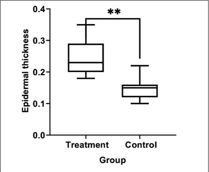
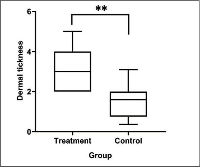
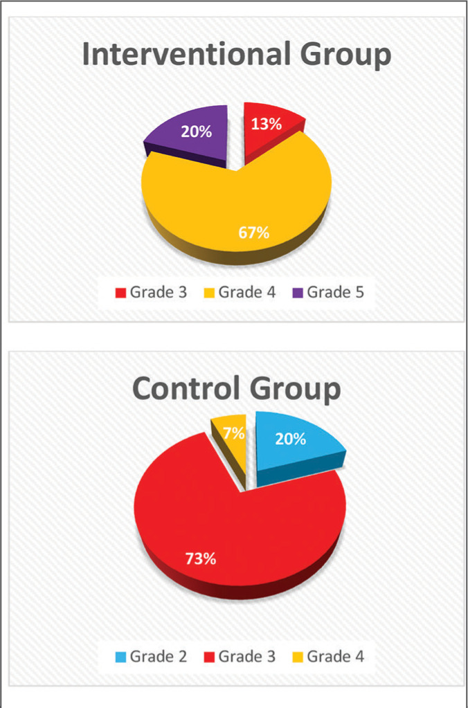
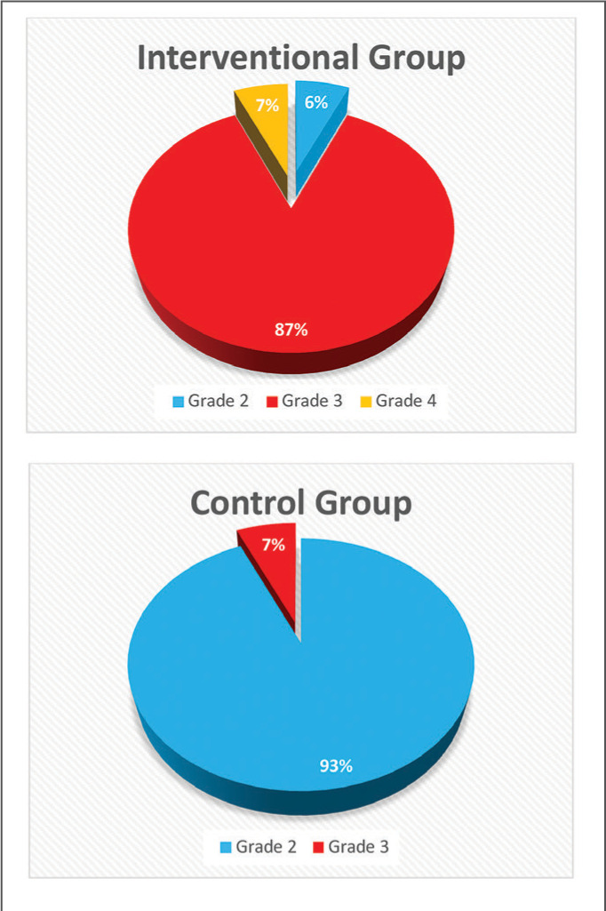
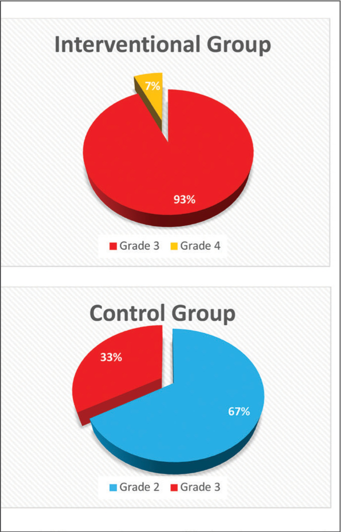
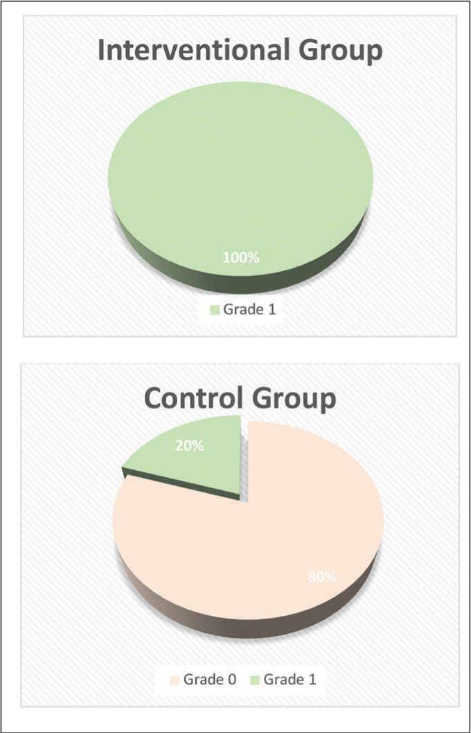
Results: Pathological examination showed a significant increase in epidermal and dermal thickness and re-organization of collagens and elastic fibers with carboxytherapy. IHC examinations revealed a significantly increased expression of TGF-β1 and VEGF with carboxytherapy.
Conclusion: Our study demonstrated the effectiveness of carboxytherapy in treating and reversing intrinsic aging skin through pathological and IHC studies.
Keywords: carboxytherapy; immunohistochemistry; pathology; skin aging.
Copyright © 2024. Matrix Medical Communications. All rights reserved.
Conflict of interest statement
DISCLOSURES: This study was supported by Tehran University of Medical Sciences in partial fulfillment of the requirement of PhD of molecular medicine. Nik Fannavaran Plasma Co., supported through renting the carboxytherapy machine and accepting machine service-related fees.
Figures

References
-
- Damle M, Mallya R. Development and evaluation of a novel delivery system containing phytophospholipid complex for skin aging. AAPS PharmSciTech. 2016;17(3):607–617. - PubMed
-
- Eklouh-Molinier C, Gaydou V, Froigneux E et al. In vivo confocal Raman microspectroscopy of the human skin: highlighting of spectral markers associated to aging via a research of correlation between Raman and biometric mechanical measurements. Anal Bioanal Chem. 2015;407(27):8363–8372. - PubMed
-
- Fenske NA, Lober CW. Structural and functional changes of normal aging skin. J Am Acad Dermatol. 1986;15(4 Pt 1):571–585. - PubMed
LinkOut - more resources
Full Text Sources