[Preprint]. 2024 May 20:2024.05.15.594393.
doi: 10.1101/2024.05.15.594393.

ISGylation of the SARS-CoV-2 N protein by HERC5 impedes N oligomerization and thereby viral RNA synthesis

Affiliations

ISGylation of the SARS-CoV-2 N protein by HERC5 impedes N oligomerization and thereby viral RNA synthesis

Junji Zhu et al. bioRxiv. .

Update in

Abstract

Interferon (IFN)-stimulated gene 15 (ISG15), a ubiquitin-like protein, is covalently conjugated to host (immune) proteins such as MDA5 and IRF3 in a process called ISGylation, thereby limiting the replication of Severe acute respiratory syndrome coronavirus 2 (SARS-CoV-2). However, whether SARS-CoV-2 proteins can be directly targeted for ISGylation remains elusive. In this study, we identified the nucleocapsid (N) protein of SARS-CoV-2 as a major substrate of ISGylation catalyzed by the host E3 ligase HERC5; however, N ISGylation is readily removed through de-ISGylation by the papain-like protease (PLpro) activity of NSP3. Mass spectrometry analysis identified that the N protein undergoes ISGylation at four lysine residues (K266, K355, K387 and K388), and mutational analysis of these sites in the context of a SARS-CoV-2 replicon (N-4KR) abolished N ISGylation and alleviated ISGylation-mediated inhibition of viral RNA synthesis. Furthermore, our results indicated that HERC5 targets preferentially phosphorylated N protein for ISGylation to regulate its oligomeric assembly. These findings reveal a novel mechanism by which the host ISGylation machinery directly targets SARS-CoV-2 proteins to restrict viral replication and illuminate how an intricate interplay of host (HERC5) and viral (PLpro) enzymes coordinates viral protein ISGylation and thereby regulates virus replication.

Keywords: HERC5; ISG15; ISGylation; N protein; SARS-CoV-2; papain-like protease.


Conflict of interest statement

Competing interests The authors declare that they have no competing interests.

Figures
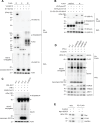
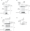
Fig 1.
Fig 1.. The SARS-CoV-2 N protein is ISGylated by HERC5.
(A) ISGylation of FLAG-tagged N, E, and M of SARS-CoV-2 in HEK293T cells that were transfected for 24 h with either empty vector or V5-tagged ISG15 along with plasmids encoding E1 (UBE1L), E2 (UBCH8), and E3 (HERC5), determined by FLAG pull-down (PD:FLAG) and immunoblotting (IB) with anti-V5 and anti-FLAG. Whole cell lysates (WCLs) were immunoblotted with the indicated antibodies. (B) ISGylation of FLAG-tagged NSP16 and N in HEK293T cells that were co-transfected for 24 h with empty vector or V5-tagged PLpro WT or C111A together with E1, E2, E3, and V5-ISG15, determined by PD:FLAG and IB with anti-V5 and anti-FLAG. (C) ISGylation of N protein in A549-hACE2 cells that were transfected for 24 h with either control non-targeting siRNA (si.C) or ISG15-specific siRNA (si.ISG15), followed by mock treatment or stimulation with 1000 U/mL IFN-α for 24 h and subsequent infection with SARS-CoV-2 (MOI 1) for 24 h in the absence or presence of the PLpro inhibitor GRL-0617 (25 μM), determined in the WCLs by IB with anti-N (SARS-CoV-2). WCLs were further immunoblotted with the indicated antibodies. (D) ISGylation of FLAG-tagged N in A549 cells transfected with the indicated siRNAs for 24 h, followed by mock treatment or stimulation with 1000 U/mL IFN-α for 24 h and subsequent transfection with FLAG-N for 24 h, determined by PD:FLAG and IB with the indicated antibodies. (E) Binding of Myc-tagged HERC5 to FLAG-tagged N in transiently transfected HEK293T cells, determined by PD:FLAG and IB with anti-Myc and anti-FLAG. si.C, nontargeting control siRNA. Asterisks (*) in (A), (C) and (D) indicate non-specific bands. Arrowheads in (A) and (B) indicate non-ISGylated form of transfected proteins. Data are representative of at least two independent experiments.
Fig 2.
Fig 2.. Identification of the ISGylation sites in the SARS-CoV-2 N protein by mass spectrometry.
(A) Schematic diagram of the domain organization of the SARS-CoV-2 N protein with the ISGylated lysines (Ks) identified by MS analysis illustrated in red. The three intrinsically disordered regions, including the N-IDR, the linker (LKR) and C-IDR, as well as the N-terminal domain (NTD) and C-terminal domain (CTD) are illustrated. (B) Amino acid sequence spanning the region of the N protein that contains K266, K355, K387 and K388 for the indicated SARS-CoV-2 variants. The four lysine residues found to be conjugated with ISG15 are highlighted in red. (C) Mass spectra of the tryptic peptides of affinity-purified FLAG-tagged N that identified K-GlyGly modifications at K266, K355, K387, and K388. b- and y-ion designations are shown. See also Supplementary Fig. 2A. (D) Location of K266, K355, K387, and K388 in the monomeric Cryo-EM structure of full-length N protein (PDB: 8FD5). (E) ISGylation of FLAG-tagged N WT or mutants in transiently transfected HEK293T cells that were co-transfected for 24 h with either empty vector or V5-tagged ISG15 together with E1, E2 and E3, determined by PD:FLAG and IB with anti-V5 and anti-FLAG. WCLs were immunoblotted with anti-V5. Data in (A and C) are from one unbiased MS screen. Data in (E) are representative of at least two independent experiments.
Fig 3.
Fig 3.. A SARS-CoV-2 replicon encoding an ISGylation-defective mutant N protein escapes HERC5-mediated virus restriction.
(A and B) Replication of a SARS-CoV-2 replicon encoding either N WT or a mutant in which the four ISGylation sites were mutated to arginine (SARS-CoV-2Rep WT or 4KR), determined by fluorescence microscopy analysis of mScarlet signals (A) and qPCR analysis of viral subgenomic and genomic RNA (B) in HEK293T cells that were transfected for 48 h with the WT or mutant replicon and empty vector or E1, E2 and E3/HERC5 with or without FLAG-tagged ISG15-WT or ISG15-AA. Scale bar, 100 μm. (C) ISGylation of N protein in HEK293T cells that were transfected as in (B), determined in the WCLs by IB with anti-N (SARS-CoV-2). WCLs were further immunoblotted with anti-FLAG (to confirm the expression of FLAG-tagged ISG15 WT or AA) and anti-Actin (loading control). Data are representative of at least two independent experiments (mean ± SD of n = 3 biological replicates in (B)). ****P < 0.0001 (two-tailed, unpaired Student’s t-test).
Fig 4.
Fig 4.. ISGylation of the SARS-CoV-2 N protein impedes its oligomerization.
(A) Schematic diagram of the co-IP approach to determine the binding of ISGylated vs. non-ISGylated HA-tagged N with FLAG-tagged N. (B) Left: FLAG-tagged N (or empty vector as a control) was transfected into HEK293T cells, followed by affinity purification and immobilization using PD:FLAG. Right: In parallel, HA-tagged N was co-transfected with either empty vector or V5-tagged ISG15, together with E1, E2, and E3/HERC5, into HEK293T cells. WCLs were prepared and incubated in vitro with immobilized FLAG-N. Binding was determined by IB with anti-HA and anti-FLAG. (C) Oligomerization of FLAG-tagged N WT or 4KR in HEK293T cells that were co-transfected for 24 h with either empty vector or V5-tagged ISG15 together with E1, E2, and E3/HERC5, determined by SDD-AGE and IB with anti-FLAG. WCLs were analyzed by SDS-PAGE and probed by IB with anti-FLAG, anti-V5 and anti-Actin (loading control). Data in (B) and (C) are representative of at least two independent experiments.
Fig 5.
Fig 5.. Phosphorylation of the SARS-CoV-2 N protein promotes its ISGylation.
(A) ISGylation of FLAG-tagged N WT or S188,206A in HEK293T cells that were co-transfected for 24 h with either empty vector or V5-ISG15 along with E1, E2, and E3/HERC5, determined by PD:FLAG and IB with anti-V5 and anti-FLAG. (B) ISGylation of FLAG-tagged N in HEK293T cells that were co-transfected for 24 h with either empty vector or V5-ISG15 along with E1, E2, and E3/HERC5 in the absence or presence of the GSK3 inhibitor kenpaullone (5 μM, 10 μM, or 50 μM), determined by PD:FLAG and IB with anti-V5 and anti-FLAG. (C) ISGylation of FLAG-tagged N WT or the indicated mutants in HEK293T cells that were co-transfected for 24 h with either empty vector or V5-ISG15 along with E1, E2, and E3/HERC5, determined by PD:FLAG and IB with anti-V5 and anti-FLAG. (D) Binding of Myc-tagged HERC5 to FLAG-tagged N S188,206A or 10D in transiently transfected HEK293T cells, determined by PD:FLAG and IB with anti-Myc and anti-FLAG. Asterisks (*) in (A) and (B) indicate non-specific bands. Data are representative of at least two independent experiments.

Similar articles

References

    1. Yuki K, Fujiogi M, Koutsogiannaki S. 2020. COVID-19 pathophysiology: A review. Clin Immunol 215:108427. - PMC - PubMed
    1. V’Kovski P, Kratzel A, Steiner S, Stalder H, Thiel V. 2021. Coronavirus biology and replication: implications for SARS-CoV-2. Nat Rev Microbiol 19:155–170. - PMC - PubMed
    1. Merad M, Blish CA, Sallusto F, Iwasaki A. 2022. The immunology and immunopathology of COVID-19. Science 375:1122–1127. - PubMed
    1. Harrison AG, Lin T, Wang P. 2020. Mechanisms of SARS-CoV-2 Transmission and Pathogenesis. Trends Immunol 41:1100–1115. - PMC - PubMed
    1. Baggen J, Vanstreels E, Jansen S, Daelemans D. 2021. Cellular host factors for SARS-CoV-2 infection. Nat Microbiol 6:1219–1232. - PubMed

Publication types

LinkOut - more resources