[Preprint]. 2024 Aug 8:2024.08.08.607154.
doi: 10.1101/2024.08.08.607154.

SKAP binding to microtubules reduces friction at the kinetochore-microtubule interface and increases attachment stability under force

Affiliations

SKAP binding to microtubules reduces friction at the kinetochore-microtubule interface and increases attachment stability under force

Miquel Rosas-Salvans et al. bioRxiv. .

Update in

Abstract

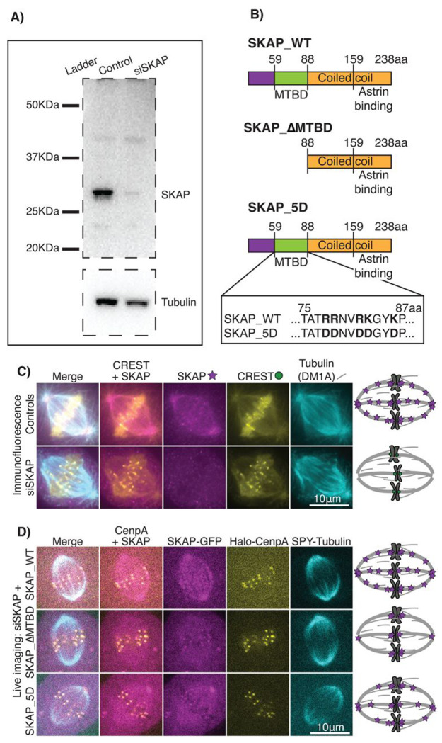
The kinetochore links chromosomes to spindle microtubules to drive chromosome segregation at cell division. We recently uncovered that the kinetochore complex Astrin-SKAP, which binds microtubules, reduces rather than increases friction at the mammalian kinetochore-microtubule interface. How it does so is not known. Astrin-SKAP could affect how other kinetochore complexes bind microtubules, reducing their friction along microtubules, or it could itself bind microtubules with similar affinity but lower friction than other attachment factors. Using SKAP mutants unable to bind microtubules, live imaging and laser ablation, we show that SKAP's microtubule binding is essential for sister kinetochore coordination, force dissipation at the interface and attachment responsiveness to force changes. Further, we show that SKAP's microtubule binding is essential to prevent chromosome detachment under both spindle forces and microneedle-generated forces. Together, our findings indicate that SKAP's microtubule binding reduces kinetochore friction and increases attachment responsiveness and stability under force. We propose that having complexes with both high and low sliding friction on microtubules, making a mechanically heterogeneous interface, is key to maintaining robust attachments under force and thus accurate segregation.


Conflict of interest statement

DECLARATION OF INTEREST The authors declare no conflict of interest.

Figures

Figure 1.
Figure 1.. SKAP_ΔMTBD and SKAP_5D display graded disruption of spindle microtubule binding in vivo.
(A) Representative western blot showing SKAP siRNA efficiency (90% depletion average). (B) Schematic representation of SKAP_WT and two mutants expected to disrupt microtubule binding. SKAP_ΔMTBD lacks the first 87 amino acids, including the microtubule-binding domain (MTBD), and SKAP_5D has five point mutations making SKAP’s microtubule-binding domain more negatively charged thus weakening microtubule binding. Coiled coil domains (yellow), including the Astrin-binding domain, microtubule-binding domain (green) and the rest of SKAP’s N-terminus (purple). (C) Representative control and siSKAP immunofluorescence images of metaphase RPE1 cells stained for kinetochores (CREST), SKAP and tubulin. Schematic representation of SKAP (magenta) localization at kinetochores (green) and spindle microtubules (grey) (right). (D) Representative live images of three above SKAP-GFP constructs in metaphase RPE1 cells under SKAP RNAi. SKAP localizes to kinetochores for all constructs but SKAP_ΔMTBD has much weaker and SKAP_5D weaker spindle microtubule localization than control. Schematic representation of SKAP mutant (magenta) localization at kinetochores and spindle microtubules (grey) (right).
Figure 2.
Figure 2.. SKAP binding to microtubules increases sister kinetochore coordination and reduces tension at the kinetochore-microtubule interface.
(A) Cartoon of parameters measured to probe the role of SKAP’s microtubule binding in metaphase kinetochore movement. (B) Representative kymographs of sister kinetochore metaphase oscillations timelapse imaging in RPE1 Halo-CenpA (white, JF549) control, siSKAP, and SKAP RNAi cells with either SKAP_WT-GFP, SKAP_ΔMTBD-GFP or SKAP_5D-GFP expression with SPY-tubulin (not shown). Comparison across these five conditions (ANNOVA test) of (C) standard deviation of kinetochore position and (D) average movement speed of individual kinetochores over time, (E) velocity correlation between sister kinetochores, (F) fraction of time that individual sister kinetochores move in opposite directions (percentage time) and (G) average inter-kinetochore distance for each sister pair. Number of kinetochores (KT) and cells marked for each condition.
Figure 3.
Figure 3.. SKAP’s microtubule binding increases the kinetochore-microtubule interface’s responsiveness to force.
(A) Cartoon of experiment probing the role of SKAP’s microtubule binding in the kinetochore-microtubule interface responding to force: metaphase kinetochore ablation (yellow) reduces tension at the sister kinetochore (green), inducing k-fiber depolymerization (purple) that pulls the sister kinetochore towards the pole, and increasing polar ejection forces (red) approaching the pole induce kinetochore directional switch and k-fiber rescue (orange). (B) Representative timelapse images of the above experiment in RPE1 Halo-CenpA (green, Halo tag ligand Oregon Green) cells with SPY-tubulin (magenta) under control, siSKAP, and SKAP RNAi with either SKAP_WT-GFP or SKAP_ΔMTBD-GFP. Timelapse shows kinetochore ablation (yellow bolt, 0 s first frame post-ablation), sister poleward movement (white arrowhead) and subsequent reversal and anti-poleward movement (orange arrowhead) under increasing force. (C) Examples of kinetochore movement trajectories defining directional switch and plateau events. For the above cellular conditions, (D) kinetochore distance from the pole at the time of directional switch from poleward to antipoleward movement after sister ablation (Mann-Whitney test) and (E) percentage of kinetochores with a clear directional switch, no directional switch (plateau) and inconsistent switch near the pole (Fisher exact test). Number of ablation events (N) and cells (also N) marked for each condition.
Figure 4.
Figure 4.. SKAP preserves attachment integrity under force through its microtubule-binding activity.
(A) Cartoon of experiment probing the role of SKAP’s microtubule binding in attachment robustness under force. A glass microneedle (red) pulls on the k-fiber (grey), increasing tension and inter-kinetochore (green) distance, which leads to either detachment (left) or attachment maintenance (right). (B) Representative timelapse images of a kinetochore detachment from its k-fiber in a microneedle (green/black, BSA-Alexa-488) manipulated RPE1 Halo-CenpA control (green, JF549nm, white arrows marking kientochores) cells with SPY-tubulin 650nm (magenta, white arrow marking k-fiber plus-end) expressing SKAP_ΔMTBD-GFP. Whole cell (left) and cropped region (right) timelapse showing increased inter-kinetochore distance (0-10 s) before detachment, the detachment event (between 10 and 15s) and a k-fiber disconnected from the kinetochore and the inter-kinetochore distance decrease after detachment (15-30 s). (C) Kinetochore-microtubule attachment outcomes after microneedle pulling in RPE1 Halo-CenpA control, siSKAP, and control cells expressing SKAP_WT-GFP or SKAP_ΔMTBD-GFP (Fisher exact test). Inter-kinetochore distance outcomes for dataset in (C): (D) Inter-kinetochore distance relaxation during the first 15 s after maximum inter-kinetochore distance with linear regression fits (dashed lines) for microneedle pulling in control (green) and siSKAP (gold) cells on the left, and control cells expressing SKAP_WT-GFP (blue) and SKAP_ΔMTBD-GFP (green) on the right. (E) Average inter-kinetochore distance relaxation over time per kinetochore pair with color indicating detachment (red) vs no detachment (blue) (Mann-Whitney test). (F) Inter-kinetochore distance relative relaxation (to final relaxed inter-kinetochore distance) for individual manipulated kinetochore pairs (Mann-Whitney test). (G) Maximum inter-kinetochore distance achieved per manipulated pair before relaxation starts (Mann-Whitney test).
Figure 5.
Figure 5.. Model for how SKAP’s microtubule binding reduces friction at the kinetochore-microtubule interface, making the attachment more responsive and robust under force.
With SKAP (left, 1): 2) Under tension at the kinetochore, SKAP slides before other components due to its lower friction for sliding on microtubules, rearranging the interface and making it responsive to force. 3) This increases tension the other microtubule binders (Ndc80 and SKA) experience, inducing their subsequent sliding. The whole system slides without detachment. Without SKAP or its microtubule-binding activity (right, 1): 2) Under a similar tension as above, there is no rearrangement of the attachment interface since only high friction binders are present. Higher tension is needed to slide components due to their higher friction. 3) Under this higher tension, either the whole system slides and kinetochore and microtubules remain attached or the attachment breaks due to its low rearrangement capacity. SKAP (yellow), SKA (purple), Ndc80 (black), the rest of the kinetochore (green), and microtubules (grey) are depicted.

Similar articles

Cited by

References

    1. Akiyoshi B, Sarangapani KK, Powers AF, Nelson CR, Reichow SL, Arellano-Santoyo H, Gonen T, Ranish JA, Asbury CL & Biggins S (2010) Tension directly stabilizes reconstituted kinetochore-microtubule attachments. Nature 468: 576–579 - PMC - PubMed
    1. Auckland P, Clarke NI, Royle SJ & McAinsh AD (2017) Congressing kinetochores progressively load Ska complexes to prevent force-dependent detachment. Journal of Cell Biology 216: 1623–1639 - PMC - PubMed
    1. Bayart E, Svetlizky I & Fineberg J (2016) Slippery but Tough: The Rapid Fracture of Lubricated Frictional Interfaces. Phys Rev Lett 116: 194301. - PubMed
    1. Cheerambathur DK, Prevo B, Hattersley N, Lewellyn L, Corbett KD, Oegema K & Desa A (2017) Dephosphorylation of the Ndc80 Tail Stabilizes Kinetochore-Microtubule Attachments via the Ska Complex. Developmental Cell 41: 424–437 - PMC - PubMed
    1. Cheeseman IM, Chappie JS, Wilson-Kubalek EM & Desai A (2006) The Conserved KMN Network Constitutes the Core Microtubule-Binding Site of the Kinetochore. Cell 127: 983–997 - PubMed

Publication types

LinkOut - more resources