[Preprint]. 2024 Aug 8:2024.08.06.606693.
doi: 10.1101/2024.08.06.606693.

TBK1 and IKKε protect target cells from IFNγ-mediated T cell killing via an inflammatory apoptotic mechanism

Affiliations

TBK1 and IKKε protect target cells from IFNγ-mediated T cell killing via an inflammatory apoptotic mechanism

Nicholas D Sun et al. bioRxiv. .

Abstract

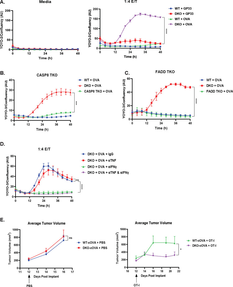
Cytotoxic T cells produce interferon gamma (IFNγ), which plays a critical role in anti-microbial and anti-tumor responses. However, it is not clear whether T cell-derived IFNγ directly kills infected and tumor target cells, and how this may be regulated. Here, we report that target cell expression of the kinases TBK1 and IKKε regulate IFNγ cytotoxicity by suppressing the ability of T cell-derived IFNγ to kill target cells. In tumor targets lacking TBK1 and IKKε, IFNγ induces expression of TNFR1 and the Z-nucleic acid sensor, ZBP1, to trigger RIPK1-dependent apoptosis, largely in a target cell-autonomous manner. Unexpectedly, IFNγ, which is not known to signal to NFκB, induces hyperactivation of NFκB in TBK1 and IKKε double-deficient cells. TBK1 and IKKε suppress IKKα/β activity and in their absence, IFNγ induces elevated NFκB-dependent expression of inflammatory chemokines and cytokines. Apoptosis is thought to be non-inflammatory, but our observations demonstrate that IFNγ can induce an inflammatory form of apoptosis, and this is suppressed by TBK1 and IKKε. The two kinases provide a critical connection between innate and adaptive immunological responses by regulating three key responses: (1) phosphorylation of IRF3/7 to induce type I IFN; (2) inhibition of RIPK1-dependent death; and (3) inhibition of NFκB-dependent inflammation. We propose that these kinases evolved these functions such that their inhibition by pathogens attempting to block type I IFN expression would enable IFNγ to trigger apoptosis accompanied by an alternative inflammatory response. Our findings show that loss of TBK1 and IKKε in target cells sensitizes them to inflammatory apoptosis induced by T cell-derived IFNγ.


Conflict of interest statement

Conflict of Interest We declare that there are no financial conflicts of interest.

Figures

Figure 1.
Figure 1.. TBK1/IKKε-deficient cells are sensitive to IFNγ-induced RIPK1-mediated apoptosis.
(A) Western blot analysis of the indicated proteins in wildtype (WT), IKKε single knockout, TBK1 single knockout, and double knockout (DKO) B16-F1 melanoma cells generated by CRISPR. (B) Control sgRNA (WT), IKKε and TBK1 single KO, and DKO B16 cell lines were stimulated in triplicates with media, 100 ng/mL TNF, or 100 ng/mL IFNγ and imaged for 48 h in an IncuCyte S3 in the presence of YOYO-3 to measure cell death. The confluency of the cells in each well were also quantified. Data is presented as YOYO-3 counts normalized to confluency in each well. Values are triplicate mean ± SD. ****P<0.0001 by unpaired t test comparing last data point. (C) WT and DKO B16 cells were treated with IFNγ (100 ng/mL) in the presence of DMSO or Nec-1s (10 μM) and analyzed in the IncuCyte. Values are triplicate mean ± SD. ****P<0.0001 by unpaired t test comparing last data point. (D) Western blot analysis of the indicated proteins in WT and DKO B16 cells pretreated with zVAD-fmk (20 μM) for 30 min, followed by IFNγ (100 ng/mL) for 0, 24, and 48 h. (E) Western blot analysis of the indicated proteins in WT and DKO B16 cells treated with 0, 10, 100 ng/mL IFNγ for 24 h. (F-G) WT, DKO, TBK1/IKKε/FADD TKO (F), and TBK1/IKKε/CASP8 TKO (G) B16 cells were treated with IFNγ (100 ng/mL) and analyzed by IncuCyte. Values are triplicate mean ± SD. ****P<0.0001 by unpaired t test comparing last data point.
Figure 2.
Figure 2.. TBK1/IKKε-deficient targets are killed by T cells in an IFNγ-dependent manner.
(A) WT and DKO B16 cells were pulsed with control LCMV GP33 peptide or OVA SIINFEKL peptide followed by co-culture with either control media or OT-I T cells at 1:4 effector to target (E/T) ratio. Target cell death was analyzed by IncuCyte. Values are triplicate mean ± SD. ****P<0.0001 by unpaired t test comparing last data point. (B-C) WT, DKO, TBK1/IKKε/CASP8 TKO (B), and TBK1/IKKε/FADD TKO (C) B16 cells were pulsed with OVA peptide followed by co-culture with OT-I T cells at 1:4 E/T ratio. Target cell death was analyzed by IncuCyte. Values are triplicate mean ± SD. ***P<0.001, ****P<0.0001 by unpaired t test comparing last data point. (D) DKO B16 cells were pulsed with OVA peptide followed by co-culture with OT-I T cells at 1:4 E/T ratio and treated with control IgG (50 μg/mL), anti-TNF (50 μg/mL), anti-IFNγ (50 μg/mL) or both mAbs (50 μg/mL). Values are triplicate mean ± SD. ****P<0.0001, not significant (ns) by unpaired t test comparing last data point. (E) Tumor volume analysis of NSG mice bearing B16 WT- or DKO-cOVA tumors treated once at day 12 post tumor implant with PBS or OT-I T cells (10 million/100 μL) i.v.; WT (PBS) n = 13, DKO (PBS) n = 11, WT (OT-I) n = 7, DKO (OT-I) n = 8. Values are mean ± SEM. Not significant (ns), *P<0.05 by paired t test comparing all combined data points.
Figure 3.
Figure 3.. TNF/TNFR1 signaling is required for IFNγ-mediated killing of TBK1/IKKε-deficient cells.
(A) WT, DKO, STAT1 single KO, and TBK1/IKKε/STAT1 TKO B16 cell were treated with IFNγ (100 ng/mL) and analyzed by IncuCyte. Values are triplicate mean ± SD. ****P<0.0001 by unpaired t test comparing last data point. (B) WT and DKO B16 cells were treated with IFNγ (100 ng/mL) in the presence of DMSO or ruxolitinib (1 μM) and analyzed by IncuCyte. Values are triplicate mean ± SD. ****P<0.0001 by unpaired t test comparing last data point. (C) WT and DKO B16 cells were stimulated with IFNγ (100 ng/mL) for 0, 16, 24 h and RNA isolated for sequencing. Values displayed as log2 CPM comparing Tnfrsf1a from three independent experiments. Statistical analysis was performed using one-way ANOVA with Sidak’s multiple-comparison test. ns = not significant. (D) Western blot analysis of the indicated proteins in WT and DKO B16 cells treated with IFNγ (100 ng/mL) for 0, 4, 8 h. (E) WT, DKO, and TBK1/IKKε/TNFR1 TKO B16 cells were treated with IFNγ (100 ng/mL) (left) or pulsed with OVA peptide followed by co-culture with OT-I T cells at 1:4 E/T ratio (right) and analyzed by IncuCyte. Values are triplicate mean ± SD. ****P<0.0001 by unpaired t test comparing last data point. (F) WT, DKO, and TBK1/IKKε/TNF TKO B16 cells were treated with media alone or IFNγ (100 ng/mL) and analyzed by IncuCyte. Values are triplicate mean ± SD. ***P<0.001 by unpaired t test comparing last data point. (G) WT, DKO, TBK1/IKKε/TNFR1 TKO, TBK1/IKKε/TNF TKO, and DKO with anti-TNF (50 μg/mL) B16 cells were treated with IFNγ (100 ng/mL) and analyzed by IncuCyte. Values are triplicate mean ± SD. *P<0.05, **P<0.01, ***P<0.001 by unpaired t test comparing last data point.
Figure 4.
Figure 4.. ZBP1 in tandem with TNFR1 mediate IFNγ killing of TBK1/IKKε-deficient cells.
(A) Western blot analysis of the indicated proteins in WT and DKO B16 cells pretreated with zVAD-fmk (20 μM) for 30 min, followed by IFNγ (100 ng/mL) for 0, 24, and 48 h. Equal concentration of lysates were then immunoprecipitated with antibody against ZBP1. (B) WT, DKO, TBK1/IKKε/TNFR1 TKO, TBK1/IKKε/ZBP1 TKO, and TBK1/IKKε/TNFR1/ZBP1 QKO B16 cells were treated with IFNγ (100 ng/mL) and analyzed by IncuCyte. Values are triplicate mean ± SD. ****P<0.0001 by unpaired t test comparing last data point. (C) Western blot analysis of the indicated proteins in WT, DKO, TNFR1 TKO, ZBP1 TKO, and QKO B16 cells pretreated with zVAD-fmk (20 μM) for 30 min, followed by IFNγ (100 ng/mL) for 0 and 24 h. (D) Western blot analysis of the indicated proteins in WT, DKO, TNFR1 TKO, ZBP1 TKO, and QKO B16 cells treated with 0, 10, or 100 ng/mL IFNγ for 24 h. (E) WT, DKO, TNFR1 TKO, ZBP1 TKO, and QKO B16 cells were pulsed with OVA peptide followed by co-culture with OT-I T cells at 1:4 E/T ratio. Target cell death was analyzed by IncuCyte. Values are triplicate mean ± SD. ****P<0.0001 by unpaired t test comparing last data point.
Figure 5.
Figure 5.. IFNγ induces inflammatory gene expression in TBK1/IKKε-deficient cells.
(A) Global transcriptomics analysis of enriched gene pathways measured as log2 fold change between IFNγ-stimulated vs unstimulated control (Ctrl) WT cells (left), IFNγ-stimulated vs unstimulated control (Ctrl) DKO cells (middle), and a ratio of the IFNγ-stimulated change in DKO versus IFNγ-stimulated change in WT cells (right). (B) Heatmap expression profile of inflammatory genes obtained from RNAseq analysis of WT and DKO B16 cells stimulated with IFNγ (100 ng/mL) for 0, 16, 24 h. (C) Global proteomics analysis of enriched proteins in pathways measured as log2 fold change between IFNγ-stimulated vs unstimulated control (Ctrl) WT cells (left), IFNγ-stimulated vs unstimulated control (Ctrl) DKO cells (middle), and a ratio of the IFNγ-stimulated change in DKO versus IFNγ-stimulated change in WT cells (right). (D) Western blot analysis of the indicated proteins in nuclear extracts obtained from WT, DKO, TNFR1 TKO, ZBP1 TKO, and QKO B16 cells treated with IFNγ (100 ng/mL) for 0 or 24 h. (E) ELISA analysis of mouse CCL2 and CXCL9 in WT, DKO, TNFR1 TKO, ZBP1 TKO, and QKO B16 cells treated with IFNγ (100 ng/mL) for 0 or 24 h. ns = not significant, **P<0.01, ****P<0.0001, statistical analysis was performed using unpaired t test. (F) Western blot analysis of the indicated proteins in WT and DKO B16 cells treated with TNF (10 ng/mL) for 0, 15, 30, and 60 min.
Figure 6.
Figure 6.. Model for the regulation of IFNγ-mediated death and inflammation by TBK1 and IKKε.
Schematic of how multiple responses downstream of IFNγ stimulation are regulated by TBK1 and IKKε. (Left) In addition to their function in inducing type I IFN expression, TBK1 and IKKε also suppress RIPK1-dependent death and NFκB-driven inflammation. (Right) In their absence, IFNγ induces RIPK1-dependent apoptosis and NFκB-dependent inflammatory gene expression driven by autocrine activation of TNFR1. ZBP1 also plays a secondary role in these processes that is redundant to TNFR1. The figure was created using BioRender.

References

    1. Maraskovsky E., Chen W.F. & Shortman K. IL-2 and IFN-gamma are two necessary lymphokines in the development of cytolytic T cells. J Immunol 143, 1210–1214 (1989). - PubMed
    1. Curtsinger J.M., Agarwal P., Lins D.C. & Mescher M.F. Autocrine IFN-gamma promotes naive CD8 T cell differentiation and synergizes with IFN-alpha to stimulate strong function. J Immunol 189, 659–668 (2012). - PMC - PubMed
    1. Yang Y., Waters J.B., Fruh K. & Peterson P.A. Proteasomes are regulated by interferon gamma: implications for antigen processing. Proc Natl Acad Sci U S A 89, 4928–4932 (1992). - PMC - PubMed
    1. Alspach E., Lussier D.M. & Schreiber R.D. Interferon gamma and Its Important Roles in Promoting and Inhibiting Spontaneous and Therapeutic Cancer Immunity. Cold Spring Harb Perspect Biol 11 (2019). - PMC - PubMed
    1. Perry A.K., Chow E.K., Goodnough J.B., Yeh W.C. & Cheng G. Differential requirement for TANK-binding kinase-1 in type I interferon responses to toll-like receptor activation and viral infection. J Exp Med 199, 1651–1658 (2004). - PMC - PubMed

Publication types

LinkOut - more resources