[Preprint]. 2024 Aug 5:2024.08.05.606604.
doi: 10.1101/2024.08.05.606604.

The molecular basis of Human FN3K mediated phosphorylation of glycated substrate

Affiliations

The molecular basis of Human FN3K mediated phosphorylation of glycated substrate

Ankur Garg et al. bioRxiv. .

Update in

Abstract

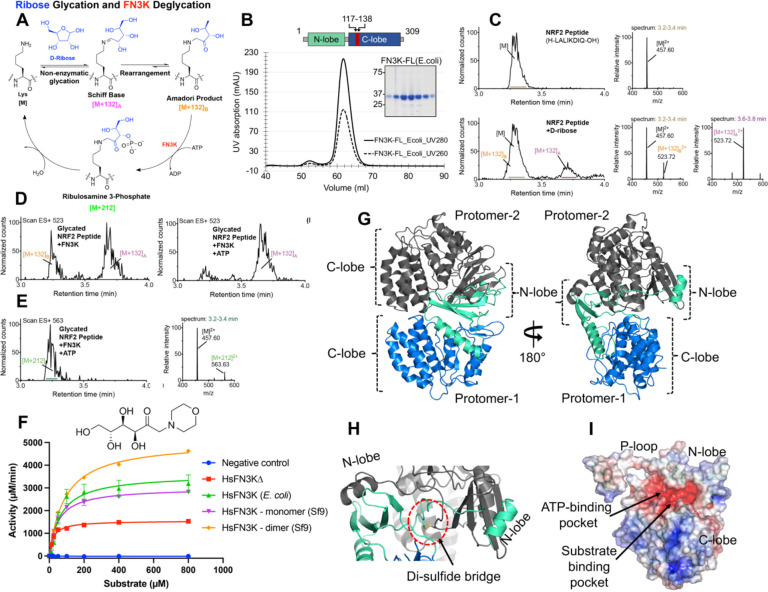
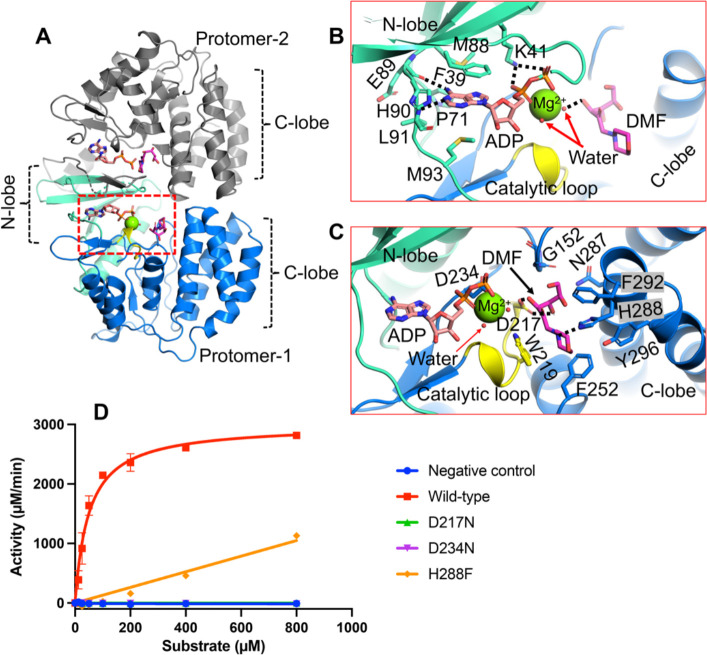
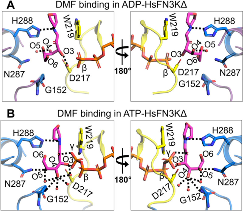
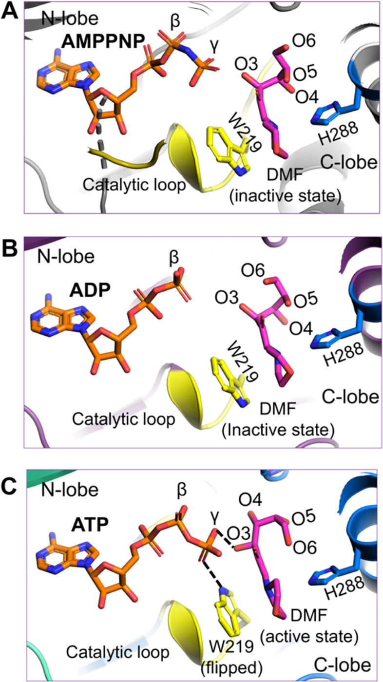
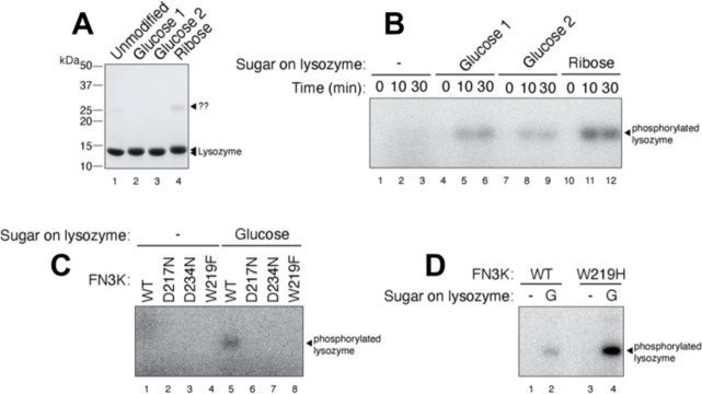
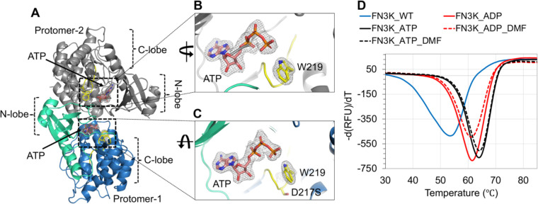
Glycation, a non-enzymatic post-translational modification occurring on proteins, can be actively reversed via site-specific phosphorylation of the fructose-lysine moiety by FN3K kinase, to impact the cellular function of target protein. A regulatory axis between FN3K and glycated protein targets has been associated with conditions like diabetes and cancer. However the molecular basis of this relationship has not been explored so far. Here, we determined a series of crystal structures of HsFN3K in apo-state, and in complex with different nucleotide analogs together with a sugar substrate mimic to reveal the features important for its kinase activity and substrate recognition. Additionally, the dynamics in sugar substrate binding during the kinase catalytic cycle provide important mechanistic insights into HsFN3K function. Our structural work provides the molecular basis for rationale small molecule design targeting FN3K.


Conflict of interest statement

Declaration of interest Authors declare no competing financial interest.

Figures

Figure 1-
Figure 1-
HsFN3K is an active kinase. (A) Mechanism of D-ribose glycation and FN3K-mediated deglycation of a lysine substrate. (B) Size-exclusion chromatogram (SEC) and SDS-PAGE showing the homogeneously purified full-length (FL) HsFN3K protein from E. coli. The UV280 and UV260 traces are shown in solid and dotted lines respectively. Domain architecture showing the N-lobe and C-lobe of HsFN3K is also shown. The truncated loop (aa 117–138) from the C-lobe to generate HsFN3KΔ is colored red (C) Synthetic NRF2 peptide (H-LALIKDIQ-OH, M.W.=912.56 g/mol) was incubated with excess D-ribose in PBS for 24 h at 37°C and analyzed by UPLC-MS. Representative chromatograms and combined mass spectra for the unglycated (top) and glycated (bottom) peptides were shown. (D) Extracted ion chromatograms of m/z 523 ([M+132]2+) reveal that FN3K deglycates the Amadori product ([M+132]B2+) in an ATP-dependent manner. (E) Extracted ion chromatograms of m/z 563 and the combined mass spectrum confirm the presence of the phosphorylated intermediate, ribulosamines 3-phosphate ([M+212]), following FN3K treatment with ATP. (F) HsFN3K in vitro kinase assays on DMF substrate. The HsFN3KΔ used for crystallization also showed kinase activity. (C) Cartoon representation of the crystal structure of apo-HsFN3K showing two molecules arranged as a domain-swapped dimer via the N-lobe. (D) Shown is the Cys24 mediated di-sulfide bridge, supporting the domain-swapped dimeric arrangement. (E) Electrostatic surface potential of the apo-HsFN3K showing the deep negatively charged pockets for ATP and substrate binding. The electrostatic surface potential is displayed in a range from −5 (red) to +5 kT/e (blue).
Figure 2-
Figure 2-
ADP and DMF binding to HsFN3K. (A) The crystal structure of HsFN3KΔ in complex with ADP and the substrate DMF. Both protomers in the asymmetric unit contain ADP-DMF, while only protomer-1 shows a coordinated Mg2+ ion (green sphere). The molecular interactions of the (B) ADP and the substrate (C) DMF observed in the crystal structure. The ADP base shows specific interactions and is surrounded by bulky hydrophobic amino acids. The conserved Lys41 binds both phosphates. The substrate binding pocket is lined with several aromatic residues including His288 (blue sticks) and Trp219 from the catalytic loop (yellow). The fructose sugar moiety interacts with the catalytic Asp217 (yellow sticks). The coordinating water molecules are shown as red nd-spheres. Direct interactions are shown as black dotted lines (D) In vitro kinase assays showing the phosphorylation of DMF by WT and several mutants of HsFN3K. D217N and D234N show no kinase activity and the H288F mutant shows significantly reduced kinase activity on DMF compared to the WT-HsFN3K.
Figure 3-
Figure 3-
The pre-catalytic state of HsFN3K mediated phosphorylation. (A) The crystal structure of HsFN3K in complex with ATP and substrate DMF is shown in cartoon representation. Protomer-1 shows only ADP in the catalytic site, while ATP and DMF are both observed bound to protomer-2. (B) A close-up view of the molecular interaction of ATP and DMF bound to HsFN3K protomer-2. Direct interactions are shown as black dotted lines. The adenosine base and K41 interactions are conserved. Several interactions with the ATP γ-phosphate, including a direct interaction with the sugar moiety, are shown. W219 (yellow sticks) is flipped compared to the ADP-bond structure and establishes direct interaction with ATP, representing the true pre-catalytic state. The side-by-side comparison of ADP (C) and ATP (D) nucleotide binding in HsFN3K shows Trp219 in opposite orientations. The 2Fo-Fc electron density corresponding to W219 is shown as a mesh at 1.0 σ level with a carved radius of 1.6 Å. The protomer lacking the Mg2+ ion in the active site is shown for the ADP structure. The predominant alternative conformation of W219 is shown in ADP bound form. (E) In vitro kinase assays show the phosphorylation of DMF by WT and W219 mutant HsFN3K. W219H shows substantially elevated kinase activity on DMF compared to the WT, acting as a FN3K super kinase.
Figure 4-
Figure 4-
Structural reorientation of the sugar moiety in the HsFN3K kinase cycle. Close-up views of the fructose sugar moiety coordination in the (A) ADP and (B) ATP-bound structures. The sugar O3’ interactions are conserved in the two FN3K states. In the ATP bound pre-catalytic state, the geometry of the sugar moiety is stabilized by several water molecules and an interaction between O4 and D217. The γ-phosphate interaction with sugar O2’ and O3’ atoms is also shown. Direct interactions are shown as black dotted lines.
Figure 5-
Figure 5-
The AMPPNP-bound pre-catalytic state of HsFN3K. Close-up view of DMF substrate binding to HsFN3K in (A) AMPPNP, (B) ADP, and (C) ATP-bound structures. The β-phosphate in ADP is positioned far from the sugar moiety of DMF (inactive conformation). The AMPPNP γ-phosphate is positioned close to the DMF sugar moiety, but with W219 (yellow sticks) in a predominant unflipped alternative conformation, is unable to induce the sugar conformational changes. The flipped W219 interaction with the ATP γ-phosphate allows it to stably interact with DMF (magenta sticks) sugar O3’ and triggers the active conformation. The H288 (blue sticks) interactions with the DMF morpholino group are conserved in all structures.
Figure 6-
Figure 6-
In vitro phosphorylation of a glycated protein substrate. (A) SDS-PAGE showing the purified glycated lysozyme substrate following in vitro glycation with glucose or ribose sugar. The protein ladder is also marked. (B) HsFN3K phosphorylates the glycated lysozyme only. No phosphorylation signal is observed for unmodified lysozyme within the 30 min time course. (C) The phosphorylation of glucose glycated lysozyme with different HsFN3K proteins (WT and mutant). D217N, D234N, and W219F mutants showed no phosphorylation signal compared to WT HsFN3K. (D) The HsFN3K W219H mutation exhibits substantially elevated kinase activity on the glycated lysozyme, without affecting specificity.
Figure 7-
Figure 7-
Structure of HsFN3K-D217S. (A) The crystal structure of the HsFN3KΔ_D217S mutant showing the bound ATP nucleotide in both protomers in the asymmetric unit. The electron density of both ATP molecules and the flipped W219 is shown for (B) protomer-1 and (C) protomer-2 in the ASU. No substrate was observed in the substrate binding site. (D) The thermal melting spectra for HsFN3K-FL (WT) (blue solid lines) in the presence of DMF and different nucleotides. The analysis shows that the Tm change is significantly higher with ATP (red solid lines) compared to ADP (black solid lines). DMF binding shows no further Tm increase.

Similar articles

References

    1. Zhang Q., Ames J.M., Smith R.D., Baynes J.W. and Metz T.O. (2009) A perspective on the Maillard reaction and the analysis of protein glycation by mass spectrometry: probing the pathogenesis of chronic disease. J Proteome Res, 8, 754–769. - PMC - PubMed
    1. Hodge J.E. (1955) The Amadori rearrangement. Adv Carbohydr Chem, 10, 169–205. - PubMed
    1. Baynes J.W., Watkins N.G., Fisher C.I., Hull C.J., Patrick J.S., Ahmed M.U., Dunn J.A. and Thorpe S.R. (1989) The Amadori product on protein: structure and reactions. Prog Clin Biol Res, 304, 43–67. - PubMed
    1. Brownlee M. (1991) Glycosylation products as toxic mediators of diabetic complications. Annu Rev Med, 42, 159–166. - PubMed
    1. Ahmed M.U., Thorpe S.R. and Baynes J.W. (1986) Identification of N epsilon-carboxymethyllysine as a degradation product of fructoselysine in glycated protein. J Biol Chem, 261, 4889–4894. - PubMed

Publication types

LinkOut - more resources