[Preprint]. 2024 Aug 5:2024.08.01.606165.
doi: 10.1101/2024.08.01.606165.

Drug Metabolism and Transport Capacity of Endothelial Cells, Pericytes, and Astrocytes: Implications for CNS Drug Disposition

Affiliations

Drug Metabolism and Transport Capacity of Endothelial Cells, Pericytes, and Astrocytes: Implications for CNS Drug Disposition

Hannah N Wilkins et al. bioRxiv. .

Abstract

Therapeutically targeting the brain requires interactions with endothelial cells, pericytes, and astrocytes at the blood brain barrier (BBB). We evaluated regional and cell-type specific drug metabolism and transport mechanisms using rhesus macaques and in vitro treatment of primary human cells. Here, we report heterogenous distribution of representative drugs, tenofovir (TFV), emtricitabine (FTC), and their active metabolites, which cerebrospinal fluid measures could not reflect. We found that all BBB cell types possessed functional drug metabolizing enzymes and transporters that promoted TFV and FTC uptake and pharmacologic activation. Pericytes and astrocytes emerged as pharmacologically dynamic cells that rivaled hepatocytes and were uniquely susceptible to modulation by disease and treatment. Together, our findings demonstrate the importance of considering the BBB as a unique pharmacologic entity, rather than viewing it as an extension of the liver, as each cell type possesses distinct drug metabolism and transport capacities that contribute to differential brain drug disposition.

Keywords: Antiretroviral therapy; Blood brain barrier; Central nervous system; Drug disposition; Drug metabolism; Drug transporter; HIV.


Conflict of interest statement

Declaration of interests The authors declare no competing interests.

Figures

Figure 1.
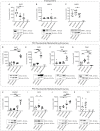
Figure 1.. TFV and FTC parent and metabolite quantification in BBB cells and rhesus macaque tissues.
(A and B) The thalamus, frontal cortex (denoted as F. Cortex), and cerebellum were sectioned from rhesus macaque brains collected 200 days post inoculation with SIVmac251 followed by a subcutaneous dose of 20 mg/kg TFV, 40 g/kg FTC, and 2.5 g/kg DTG administered once daily. Within these brain tissues, (A) Parent TFV and FTC as well as (B) TFV-DP and FTC-TP metabolites were quantified by LC-MS/MS from cerebellum, frontal cortex, and thalamus (n=2 macaques, represented as individual dots). (C) Astrocytes were obtained from healthy rhesus macaques that received six months of a once daily intramuscular ART administration (5.1 mg/kg TFV, 50 mg/kg FTC, and 2.5 mg/kg DTG). Astrocytes were isolated from five macaque brains (represented as individual dots) and TFV-DP and FTC-TP quantified by LC-MS/MS. Below limit of quantification (BLQ) samples reported (represented as a singular dot) had concentrations less than 5 fmol/sample (TFV-DP), 50 fmol/sample (FTC-TP), 0.05 ng/sample (TFV), and 0.25 ng/sample (FTC). (D) Primary human BBB cells were treated with 10 μM TFV, 2 μM FTC, and 6 μM DTG for 24h at 37°C, 5% CO2. After this time, TFV-DP and FTC-TP were quantified by LC-MS/MS. Three to four independent experiments were performed (represented as individual dots). Data represented as mean ± standard deviation. Statistical analysis was performed using Brown-Forsythe and Welch ANOVA. *p < 0.05, **p < 0.01, ****p < 0.0001.
Figure 2.
Figure 2.. BBB cells differentially express TFV and FTC transporters and nucleotide metabolizing kinases.
Primary human BBB cell monoculture and hepatocyte lysates were processed for LC-MS/MS-based proteomics, and protein concentrations were reported by Proteomic Ruler for (A) ENT1, (B) MRP4, and (C) MRP1 TFV/FTC transporters, (D) AK2, (E) CKB, (F) PKLR, and (G) PKM TFV nucleotide metabolizing kinases, as well as (H) CMPK1, (I) DCK, (J) PGK1, and (K) TK1 FTC nucleotide metabolizing kinases, each with respective protein western blots to confirm protein abundance in BBB cells. Four to five independent experiments with two LC-MS/MS injection replicates each were performed (represented as individual dots). Replicates with missing LC-MS/MS values were omitted from plot. >2 independent experiments with missing values were reported as not reliably detected (ND) by proteomics analyses. Data represented as mean ± standard deviation. Statistical analysis was performed using Brown-Forsythe and Welch ANOVA or Kruskal-Wallis ANOVA. *p < 0.05, **p < 0.01, ***p<0.001, ****p < 0.0001.
Figure 3.
Figure 3.. TFV/FTC efflux transporters and nucleotide metabolizing kinases are differentially active in BBB cells.
Brain microvascular endothelial cell (BMVEC), pericyte, and astrocyte monocultures were loaded with dyes for (A) P-gp (rhodamine 123, 10 μM), (B) MRP4 (monobromobimane, 10 μM), and (C) BCRP (Hoechst 33342, 5 μg/mL) for 15 minutes at 37°C, 5% CO2. The dyes were allowed to efflux out for 2 hours at 37°C, 5% CO2, after which flow cytometry was performed to quantify intracellular fluorescence (mean fluorescence intensity, MFI) as an indicator of efflux capacity. Statistical analysis was performed using one-way ANOVA. (D) P-gp and (E) BCRP expression were confirmed by western blot, as these proteins were not reliably measurable by proteomics analyses. (F) Endogenous pyruvate kinase and (H) thymidine kinase activities were measured in primary human BBB cell monocultures by colorimetric activity assay and DiviTum activity assay, respectively. Four to eight independent experiments (represented by individual dots) were performed. Statistical analysis was performed using a Brown-Forsythe and Welch ANOVA test. (G) TFV-DP formation in human primary BBB cell lysates was measured by a CKB-mediated tenofovir metabolism assay using LC-MS/MS. Cell lysates were incubated with tenofovir-monophosphate (TFV-MP) and phosphocreatine (+CKB Substrates) or a mixture of phosphocreatine, phosphoenolpyruvate, and ATP (+Phosphate Donors) for 30 minutes at 37°C. The reaction was quenched by LC-MS grade methanol, and peak area of TFV-DP was measured by LC-MS/MS. Three to five independent experiments were performed (represented by individual dots. Statistical analysis was performed using by one-way ANOVA. *p < 0.05, **p < 0.01, ***p<0.001, ****p < 0.0001.
Figure 4.
Figure 4.. BBB TFV/FTC nucleotide metabolizing kinases and transporters are impacted in a cell-dependent manner by ART and HIV.
Primary human pericyte and astrocyte monocultures were exposed to ART (10 μM TFV, 10 μM FTC, and 10 μM DTG), HIV (5 ng/mL), or HIV+ART (10 μM TFV, 10 μM FTC, 10 μM DTG, and 5 ng/mL HIV) for 24 h at 37°C, 5% CO2. Treatment with vehicle was used as a control. Pericytes and astrocytes were lysed and processed for proteomics analyses. Concentrations of proteins of interest were measured by Proteomic Ruler. After exposure, changes in protein concentration were assessed in (A-G) astrocytes (Astro.) for (A) AK2, (B) CMPK1, (C) ENT1, (D) P-gp, (E) PGK1, (F) MRP4, and (G) MRP1. Similarly, changes in protein concentration were assessed in (H-L) pericytes (Per.) for (H) CKB, (I) MRP4, (J) MRP1, (K) PGK1, (L) TK1. Four independent experiments with two LC-MS/MS injection replicates each were performed per condition (represented as individual dots). Replicates with missing LC-MS/MS values were omitted from plot. >2 independent experiments with missing values were reported as not reliably detected (ND) by proteomics analyses. Data represented as mean ± standard deviation. Statistical analysis was performed using Brown-Forsythe and Welch ANOVA or Kruskal-Wallis ANOVA. *p < 0.05, **p < 0.01, ***p<0.001, ****p < 0.0001.
Figure 5.
Figure 5.. HIV and ART differentially impact astrocyte and pericyte cell pathways involved in cell transport and regulation, metabolism, immune response and signaling, and cell-cell communication.
Primary human pericyte and astrocyte monocultures were exposed to ART (10 μM TFV, 10 μM FTC, and 10 μM DTG), HIV (5 ng/mL), or HIV+ART (10 μM TFV, 10 μM FTC, 10 μM DTG, and 5 ng/mL HIV) for 24 h at 37 °C, 5% CO2. Treatment with vehicle was used as a control. Pericytes and astrocytes were lysed and processed for proteomics analyses. Pathways significantly altered in astrocytes after (A) HIV, (B) ART, and (C) HIV+ART exposure as well as in pericytes after (D) HIV, (E) ART, and (F) HIV+ART (relative to vehicle) were analyzed by SimpliFi proteomics software. Pathways of interest were filtered by pathway changes greater than one log fold and hypergeometric p-value < 0.05. Pathway hits of interest were ranked by −log10(hypergeometric p-value). Pathway analyses reflect four independent experiments per condition with two LC-MS/MS injection replicates each.

References

    1. Cogill S.A., Lee J.-H., Jeon M.-T., Kim D.-G., and Chang Y. (2024). Hopping the Hurdle: Strategies to Enhance the Molecular Delivery to the Brain through the Blood–Brain Barrier. Cells 13, 789. 10.3390/cells13100789. - DOI - PMC - PubMed
    1. Schiera G., Liegro C.M.D., Schirò G., Sorbello G., and Liegro I.D. (2024). Involvement of Astrocytes in the Formation, Maintenance, and Function of the Blood–Brain Barrier. Cells 13, 150. 10.3390/cells13020150. - DOI - PMC - PubMed
    1. Procter T.V., Williams A., and Montagne A. (2021). Interplay between Brain Pericytes and Endothelial Cells in Dementia. Am. J. Pathol. 191, 1917–1931. 10.1016/j.ajpath.2021.07.003. - DOI - PubMed
    1. Wilhelm I., Nyúl-Tóth Á., Suciu M., Hermenean A., and Krizbai I.A. (2016). Heterogeneity of the blood-brain barrier. Tissue Barriers 4, e1143544. 10.1080/21688370.2016.1143544. - DOI - PMC - PubMed
    1. Chaillon A., Gianella S., Dellicour S., Rawlings S.A., Schlub T.E., Oliveira M.F.D., Ignacio C., Porrachia M., Vrancken B., and Smith D.M. (2020). HIV persists throughout deep tissues with repopulation from multiple anatomical sources. J. Clin. Investig. 130, 1699–1712. 10.1172/jci134815. - DOI - PMC - PubMed

Publication types

LinkOut - more resources