[Preprint]. 2024 Aug 9:2024.08.09.607272.
doi: 10.1101/2024.08.09.607272.

Outcome of H5N1 clade 2.3.4.4b virus infection in calves and lactating cows

Affiliations

Outcome of H5N1 clade 2.3.4.4b virus infection in calves and lactating cows

Nico Joel Halwe et al. bioRxiv. .

Abstract

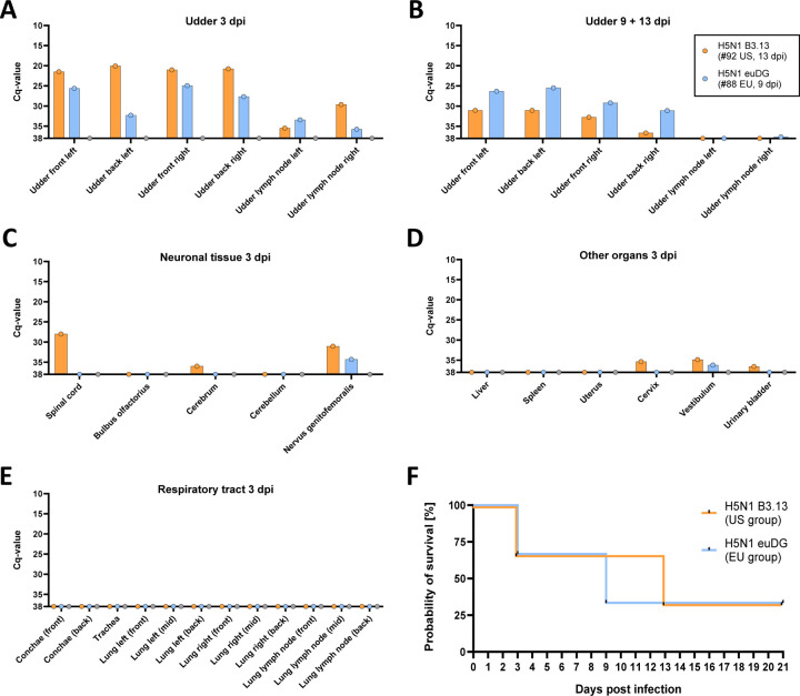
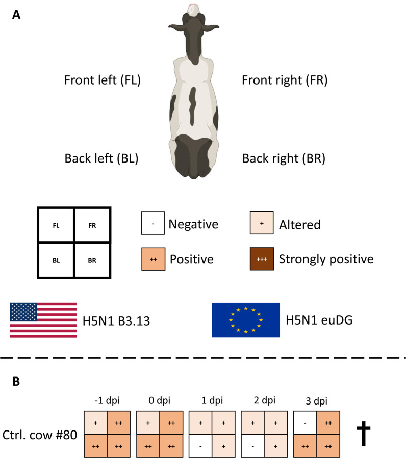
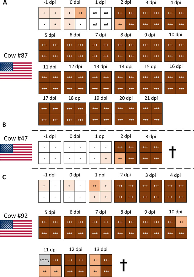
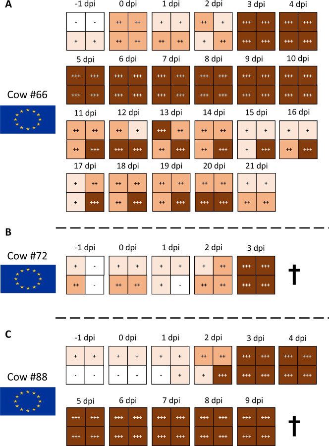
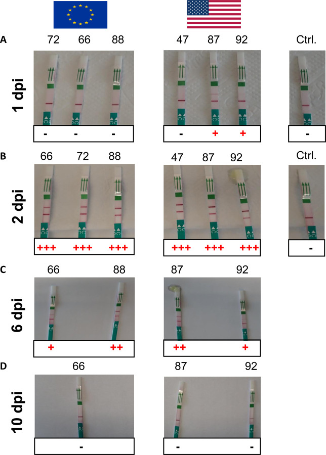
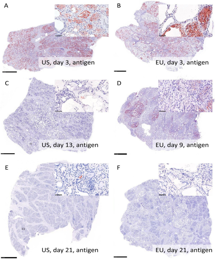
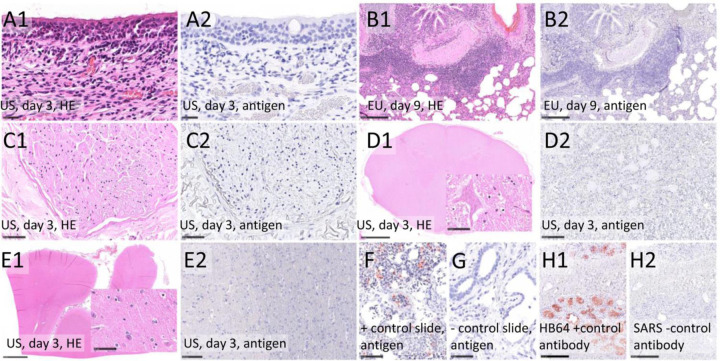
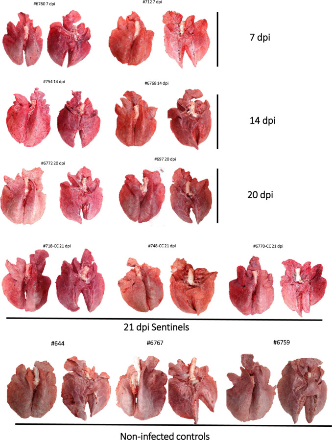
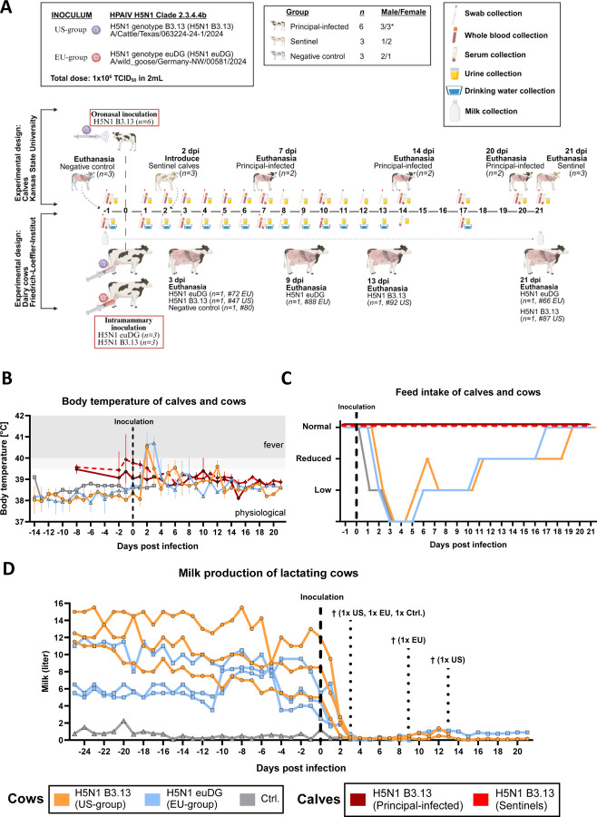
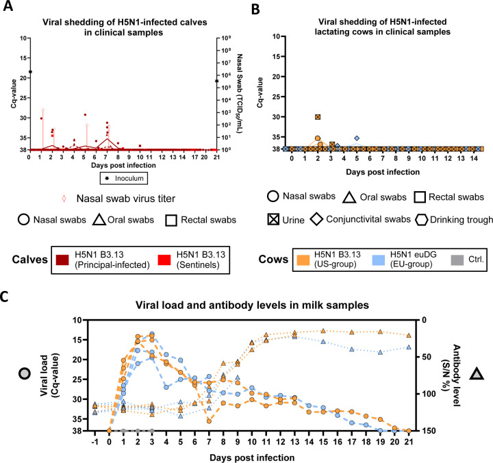
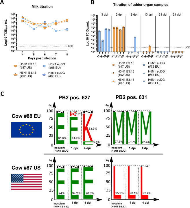
In March 2024, highly pathogenic avian influenza virus (HPAIV) clade 2.3.4.4b H5N1 infections in dairy cows were first reported from Texas, USA. Rapid dissemination to more than 190 farms in 13 states followed. Here, we provide results of two independent clade 2.3.4.4b experimental infection studies evaluating (i) oronasal susceptibility and transmission in calves to a US H5N1 bovine isolate genotype B3.13 (H5N1 B3.13) and (ii) susceptibility of lactating cows following direct mammary gland inoculation of either H5N1 B3.13 or a current EU H5N1 wild bird isolate genotype euDG (H5N1 euDG). Inoculation of the calves resulted in moderate nasal replication and shedding with no severe clinical signs or transmission to sentinel calves. In dairy cows, infection resulted in no nasal shedding, but severe acute mammary gland infection with necrotizing mastitis and high fever was observed for both H5N1 genotypes/strains. Milk production was rapidly and drastically reduced and the physical condition of the cows was severely compromised. Virus titers in milk rapidly peaked at 108 TCID50/mL, but systemic infection did not ensue. Notably, adaptive mutation PB2 E627K emerged after intramammary replication of H5N1 euDG. Our data suggest that in addition to H5N1 B3.13, other HPAIV H5N1 strains have the potential to replicate in the udder of cows and that milk and milking procedures, rather than respiratory spread, are likely the primary routes of H5N1 transmission between cattle.


Conflict of interest statement

Competing interests The J.A.R. laboratory received support from Tonix Pharmaceuticals, Genus plc, Xing Technologies, and Zoetis, outside of the reported work. J.A.R. is inventor on patents and patent applications on the use of antivirals and vaccines for the treatment and prevention of virus infections, owned by Kansas State University. The other authors declare no competing interests.

Figures

Extended Data Fig. 1 |
Extended Data Fig. 1 |. Viral genome load in organ samples and survival data of lactating cows
Orange: lactating cows infected with H5N1 B3.13. Blue: Lactating cows infected with H5N1 euDG. Grey: Uninfected negative control cow. A Viral genome load in udder organ samples of lactating cows euthanized at 3 dpi (#72 EU, #47 US, #80 Ctrl.) B Viral RNA load in udder organ samples of lactating cows euthanized at 9 dpi (#88 EU) or 13 dpi (#92 US). C Viral genome load in organ samples from neuronal tissues. D Viral RNA load in other internal organs of lactating cows. E Viral genome load in organ samples of the respiratory tract. F Survival curve of lactating cows over the course of the experiment.
Extended Data Fig. 2 |
Extended Data Fig. 2 |. Exemplary milk consistency and appropriate CMT of H5N1-infected lactating dairy cows during the animal trial
A CMT-picture of an H5N1 euDG-infected lactating dairy cow at 2 dpi B Milk consistency of H5N1 B3.13 and euDG infected dairy cows during the experiment (4 dpi)
Extended Data Fig. 3 |
Extended Data Fig. 3 |. California Mastitis Test (CMT)
A Legend for semi-quantification. Milk samples from individual quarters (front left/right and back left/right were gained and collected on appropriate CMT-plates. CMT-reagent was applied ~ 1:1 to the milk samples and was graded by eye with the help of a defined template. B CMT of milk samples from the uninfected control animal (#80) during the course of the experiment until its euthanasia timepoint.
Extended Data Fig. 4 |
Extended Data Fig. 4 |. California Mastitis Test (CMT) of lactating cows infected with H5N1 B3.13 (US-group)
A – C CMT of milk samples from cattle infected with H5N1 B3.13 during the course of the experiment until their respective euthanasia timepoint.
Extended Data Fig. 5 |
Extended Data Fig. 5 |. California Mastitis Test (CMT) of lactating cows infected with H5N1 euDG (EU-group)
A – C CMT of milk samples from cattle infected with H5N1 euDG during the course of the experiment until their respective euthanasia timepoint.
Extended Data Fig. 6 |
Extended Data Fig. 6 |. Megacor-RAT from milk samples of H5N1 experimentally infected dairy cattle
A H5-specific Megacor-RAT used for milk samples of H5N1-infected cattle at 1 dpi. Positive samples are depicted with a red cross. B H5-specific Megacor-RAT used for milk samples of H5N1-infected cattle at 2 dpi. All H5N1-infected lactating dairy cattle have become positive via the H5-specific RAT from Megacor already at 2 dpi, irrespective of the H5N1-virus isolate used. C H5-specific Megacor-RAT used for milk samples of H5N1-infected cattle at 6 dpi. D H5-specific Megacor-RAT used for milk samples of H5N1-infected cattle at 10 dpi. All cows have become already negative via the RAT at 10 dpi.
Extended Data Fig. 7 |
Extended Data Fig. 7 |. Influenza A virus nucleoprotein detection using immunohistochemistry in the mammary gland of cattle after intramammary infection with H5N1 B3.13 and H5N1 euDG.
The distribution was graded on an ordinal scale with scores 0 = no antigen, 1 = focal, affected cells/tissue <5% or up to 3 foci per tissue; 2 = multifocal, 6%–40% affected; 3 = coalescing, 41%–80% affected; 4 = diffuse, >80% affected. Representative pictures were taken from the most severely affected quarter from each cow. A Score 4, H5N1 B3.13, 3 dpi. B Score 3, H5N1 euDG, 3 dpi. C Score 1, H5N1 B3.13, 13 dpi. D Score 3, H5N1 euDG, 9 dpi. E Score 1, H5N1 B3.13, 21 dpi. F Score 0, H5N1 euDG, 21 dpi. Scale bar 2.5 mm and 50 µm (inlay).
Extended Data Fig. 8 |
Extended Data Fig. 8 |. Histopathology and Influenza A virus nucleoprotein detection (antigen) of cattle after intramammary infection with H5N1 B3.13 and H5N1 euDG including tissue controls.
A Nasal concha: Chronic-active rhinitis (A1) lacking IAV antigen (A2). B: Lung: Chronic bronchointerstitial pneumonia in convalescence phase (B1), lacking IAV NP (B2). C Genitofemoral nerve: No findings (C1), no IAV antigen (C2). D Spinal cord: No findings (D1), no IAV antigen (D2). E Brain, cortex: No findings (E1), no IAV antigen (E2). F Positive control slide, HPAIV infected chicken, lung: abundant IAV antigen. G Negative control slide, uninfected cow, mammary gland: no IAV antigen. H1 Mammary gland: cow infected with H5N1 B3.13, 3 dpi, abundant IAV antigen. H2 Consecutive slide of H1: an irrelevant antibody (anti Sars clone 4F3C4) yielded no immunopositive reaction. Hematoxylin and eosin (HE) stain (A1, B1, C1, E1) and immunohistochemistry (antigen) on consecutive (A2, B2, C2, E2, H1, H2) or independent (F, G) slides. Scale bar 25 µm (A1–2), 50 µm (C1–2, inlay D1, E1, F, G), 100 µm (D2, E2, H1–2), 250 µm (B1–2), 2.5 mm (D1, E1).
Extended Data Fig. 9 |
Extended Data Fig. 9 |. Tissue controls used for immunohistochemistry.
The anti-NP antibody used strongly labels influenza virus A H3N2 and H5N1-infected epithelial cells lining bronchioles.
Extended Data Fig. 10 |
Extended Data Fig. 10 |. Gross lung pathology of calves
(A) At 7 dpi, multiple well-defined pulmonary lobules were red and slightly depressed on the right cranial lobe (congestion and partial atelectasis) affecting approximately 20% of the cranial and caudal portions of the right cranial lobe extending into the right middle and caudal lobe of one of the two principal-infected calves (#712). There was a focal area of mild subpleural hemorrhage on the ventral surface of the left caudal lung lobe of animal #6760. (B) At 14 dpi, one of the two principal-infected calves (#754) had multifocal to coalescing red and depressed foci of congestion and atelectasis on the left and right cranial lobes. Approximately 60% of the caudal portion of the left cranial lobe, 55–60% of both the cranial and caudal portions of the right cranial lobe and <5% of the accessory lobe were affected. There were also multiple pleural adhesions to the thoracic wall. (C) At 20 dpi, the two principal-infected calves #6772 and #697) had either few small red and slightly depressed foci of congestion and atelectasis on the left cranial lobes (#6772), or a focal, similar area on the apical portion of the right middle lobe (#697). (D) Postmortem examinations of the three sentinel animals were performed at 21 dpi and revealed scattered red foci of pulmonary congestion/atelectasis. In animal #748, there were multiple, small foci of mild consolidation in the left and right cranial lobes (5% of lung affected) and few pleural adhesions to the thoracic cavity. For animal #6770, congestion and atelectasis were accompanied by mild to moderate edema affecting predominately the right lung lobes. (E) One of the three negative control calves (#6767) had a small isolated focus of consolidation of the pulmonary parenchyma at the apical margin of the right middle lobe. Gross lesions were not appreciated in the remaining negative control animals.
Fig. 1 |
Fig. 1 |. Experimental design and clinical outcomes following infection with HPAIV H5N1 clade 2.3.4.4b isolates
A Experimental study timeline. (Top) Twelve Holstein calves of mixed sex (* indicates one calf was hermaphroditic) were allocated to three experimental groups: 1 – principal-infected (n=6); 2 – sentinel (n=3); 3 – negative control (n=3). Negative control calves were euthanized prior to experimental infection and tissues were collected for baseline comparison. Principal-infected calves were oronasally inoculated with 1x10 TCID50/calf of H5N1 B3.13. Sentinel calves were introduced 48 hours post-infection. Rectal temperatures and clinical samples, including whole blood, urine, nasal-, oral-, and rectal swabs, were collected daily for 14 dpi and every 3 days thereafter. Serum was collected at 0, 7, 10, 14, 17, and 20/21 dpi. Post-mortem examinations and extensive tissue collections were performed on days 7 (n=2, principal-infected), 14 (n=2, principal-infected), and 20/21 (n=2/3, principal-infected/sentinel) post infection. (Bottom) Seven Holstein-Friesian multiparous lactating dairy cattle were used in this experiment. Three animals were inoculated intramammary with 10. TCID50/cattle of H5N1 B3.13 (A/Cattle/Texas/063224–24-1/2024, US-group, n=3) and three animals were inoculated intramammary with 10. TCID50/cattle of H5N1 euDG (A/wild_goose/Germany-NW/00581/2024, EU-group, n=3). One cow served as a negative control. Swab samples (nasal, conjunctival, and rectal) were taken daily until 9 dpi. EDTA blood samples were taken from individual cattle at 1, 3, 7 and 10 dpi. Urine was taken regularly until 14 dpi. Serum samples were obtained from 7, 14 dpi and the day of euthanasia. One cow of each group (#47 US and #72 EU) reached the humane endpoint at 3 dpi, one further cow at 9 dpi (#88 EU) and one additional cow at 13 dpi (#92 US) and were subsequently subjected to necropsy. Created with BioRender under agreement number YH275PUF4T. B The mean and standard deviation of rectal temperatures are shown for both principal-infected and sentinel calves and each group of lactating cattle prior to and following inoculation. C The average feed intake of each group of calves and cows following H5N1-infection D Individual milk production of lactating cows prior to and following experimental infection. Milk production of individual cows was tracked daily from −25 dpi through until the end of the experiment at 21 dpi. The control animal never produced high amounts of milk, which is why this cow was picked as control. Dark red: Principal-infected calves (n=6). Bright red: Sentinel calves (n=3). Orange: H5N1 B3.13 infected lactating cows (US-group, n=3, #47, #87, #92) Blue: H5N1 euDG infected lactating cows (EU-group, n=3, #66, #72, #88) Grey: Uninfected negative control cow (#80).
Fig. 2 |
Fig. 2 |. Viral shedding of influenza A/H5N1 clade 2.3.4.4b virus isolates in experimentally infected calves and lactating cows
A RT-qPCR was used for the detection of influenza A M gene (left y-axis) in nasal, oral and rectal swabs collected from H5N1 B3.13 oronasally infected calves post-inoculation an sentinel calves (dark red: principal-infected; bright red: sentinel). Viral titers of nasal swabs (right y-axis) are represented as the mean and standard deviation of nasal swabs with 46.4 TCID50/mL on each day. The Cq-value and titer of inoculum are also shown (*) on the respective y-axis. B RT-qPCR of nasal, oral, rectal and conjunctivital swabs of lactating cows intramammary infected with H5N1 B3.13 or H5N1 euDG. Orange: Cattle infected with the H5N1 B3.13. Blue: Cattle infected with H5N1 euDG. Grey: Uninfected negative control C H5N1 viral genome load (left y-axis) and corresponding H5-specific antibody titers (right y-axis) in milk samples over time. All cattle were milked daily and individual pooled milk samples were analyzed via RT-qPCR for the detection of H5N1 viral RNA. Detection of H5-specific antibodies in selected milk samples was achieved using an H5-specific ELISA and reported as sample OD450 / negative control OD450 percentage (S/N%).
Fig. 3 |
Fig. 3 |. Infectious virus yields in milk and udder tissue of H5N1 infected lactating cows and genetic adaptations over time
A H5N1 viral titers recovered from milk samples of H5N1 B3.13 (orange) and H5N1 euDG (blue) infected lactating cows throughout the experiment. B Experimental titration of H5N1 infectious viral particles from individual udder quarters (FL = front left; BL = back left; FR = front right; BR= back right) of each H5N1-infected lactating cow harvested at their respective euthanasia timepoints. C Genetic adaptation at position 627 and 631 in the Polymerase Basic Protein 2 (PB2) of H5N1 B3.13 and H5N1 euDG. A sequence logo plot displays the relative proportion of amino acids (E - ; K- ; N - ; M – ; L - ) present at positions 627 and 631 of polymerase basic protein 2 (PB2) for H5N1 B3.13 and H5N1 euDG present in milk sampled from cows #88 EU and #87 US over time.
Fig. 4 |
Fig. 4 |. Histological changes observed in respiratory tissues of calves.
Histological changes in calves oronasally infected with H5N1 B3.13. Calves at 7 (A-D) and 14 dpi (E-F) are depicted in the figure. (A and B) H&E staining showing there was a segmental region of suppurative tracheitis at 7 dpi (#6760). Degenerate neutrophils filled the tracheal lumina (arrows). No viral antigen was detected by IHC (B). (C and D) In animal #712 (7 dpi), there were multiple small and discrete foci of interstitial pneumonia (C) with fibrin filling regional alveolar spaces (asterisks) and mild numbers of neutrophils, macrophages and lymphocytes expanding alveolar septa. No viral antigen was detected (D). (E and F) In animal #754 (14 dpi), bronchioles were frequently lined by hyperplastic epithelium, filled with degenerate neutrophils, and partially occluded by papillary projections composed of a core of fibrous connective tissue with few inflammatory cells and lined by bronchiolar epithelium (bronchiolitis obliterans, asterisk). Bronchioles were also frequently delimited by prominent lymphoid aggregates (BALT hyperplasia). No viral antigen was detected (F).
Fig. 5 |
Fig. 5 |. Histopathology and Influenza A virus (IAV) nucleoprotein (NP) detection in the mammary gland and teat of multiparous cattle after intramammary infection with H5N1 B3.13 and H5N1 euDG.
(A) Abundant IAV NP detection 3 days post infection (dpi), inlay showing juxtaposition of intact, lactating alveoli lacking antigen (black asterisk) and affected areas (green asterisk) in a lobular pattern. Immunohistochemistry, IHC, using AEC chromogen and Mayer’s hematoxylin counterstain. H5N1 B3.13. Scale bar 2.5 mm and 100 µm (inlay). (B1) Full necrosis of the alveolar epithelium with cellular debris filling the lumen and (B2) intralesional detection of IAV antigen (green asterisk) on a consecutive slide. Some adjacent alveoli remain unaffected (black asterisk), 3 dpi, H5N1 B3.13. HE (B1) and IHC (B2). Scale bar 50µm. (C) Alveoli affected by necrosis with mostly intact basal lamina lined by basal/myoepithelial cells (blue arrow), indicative for regenerative capacity, 3 dpi, H5N1 euDG. HE. Scale bar 25µm. (D) Target cells identified based on morphology following IHC included alveolar secretory epithelium, 3 dpi, H5N1 B3.13. IHC. Sale bar 50µm. (E) Teat with diffuse degeneration and necrosis of the lining epithelium, subepithelial edema and mainly neutrophilic infiltrates (inlay), 3 dpi, H5N1 B3.13. HE. Scale bar 100 µm. (F) Target cells identified based on morphology following IHC included teat canal epithelium (inlay), 3 dpi, H5N1 B3.13. IHC. Scale bar 100 µm. (G) Necrotic alveoli filled with cellular debris admixed with degenerate neutrophils (blue arrow) in acute lesions and many lymphocytes, fewer macrophages, neutrophils and plasma cells in the interstitium (green arrow), H5N1 euDG, 9 dpi. HE. Scale bar 50 µm. (H) Abundant intralesional IAV NP detection in secretory alveoli, mainly within cellular debris (inlay), 9 dpi, H5N1 euDG. IHC. Scale bar 50 µm. (I) Simultaneous occurrence of either intact, lactating alveoli (black asterisk), disruption of alveolar epithelium by necrosis (green asterisk) and beginning regeneration (blue asterisk), 13 dpi H5N1 B3.13. HE. Scale bar 50 µm. (J) Late stage IAV NP detection restricted to cellular debris, found scattered at 13 dpi, H5N1 B3.13. IHC. Scale bar 25 µm. (K1) Mammary alveolus with intraluminal sloughed epithelium and cellular debris (green asterisk), and (K2) intralesional detection of IAV antigen on a consecutive slide (green asterisk), 21 dpi, H5N1 B3.13. HE (K1) and IHC (K2). Scale bar 50µm. (L) Regenerating alveoli (blue asterisk) with lack of IAV antigen labeling (not shown). Interstitial immune cell infiltrates constitute many lymphocytes and plasma cells (inlay), 21 dpi, H5N1 euDG. HE. Scale bar 50 µm.

Similar articles

Cited by

References

    1. (CDC), C. f. D. C. a. P. Technical Report: June 2023 Highly Pathogenic Avian Influenza A(H5N1) Viruses, <https://www.cdc.gov/bird-flu/php/technical-report/h5n1-070723.html?CDC_A...> (2023).
    1. (WHO), W. H. O. The panzootic spread of highly pathogenic avian influenza H5N1 sublineage 2.3.4.4b: a critical appraisal of One Health preparedness and prevention, <https://www.who.int/publications/m/item/the-panzootic-spread-of-highly-p...> (2023). - PubMed
    1. Bevins S. N. et al. Intercontinental Movement of Highly Pathogenic Avian Influenza A(H5N1) Clade 2.3.4.4 Virus to the United States, 2021. Emerg Infect Dis 28, 1006–1011 (2022). https://doi.org:10.3201/eid2805.220318 - DOI - PMC - PubMed
    1. Baechlein C. et al. Neurotropic Highly Pathogenic Avian Influenza A(H5N1) Virus in Red Foxes, Northern Germany. Emerg Infect Dis 29, 2509–2512 (2023). https://doi.org:10.3201/eid2912.230938 - DOI - PMC - PubMed
    1. Plaza P. I., Gamarra-Toledo V., Rodriguez Eugui J., Rosciano N. & Lambertucci S. A. Pacific and Atlantic sea lion mortality caused by highly pathogenic Avian Influenza A(H5N1) in South America. Travel Med Infect Dis 59, 102712 (2024). https://doi.org:10.1016/j.tmaid.2024.102712 - DOI - PubMed

Publication types

LinkOut - more resources