[Preprint]. 2024 Aug 9:2024.08.08.607070.
doi: 10.1101/2024.08.08.607070.

Neurofibromin deficiency alters the patterning and prioritization of motor behaviors in a state-dependent manner

Affiliations

Neurofibromin deficiency alters the patterning and prioritization of motor behaviors in a state-dependent manner

Genesis Omana Suarez et al. bioRxiv. .

Update in

Abstract

Genetic disorders such as neurofibromatosis type 1 increase vulnerability to cognitive and behavioral disorders, such as autism spectrum disorder and attention-deficit/hyperactivity disorder. Neurofibromatosis type 1 results from loss-of-function mutations in the neurofibromin gene and subsequent reduction in the neurofibromin protein (Nf1). While the mechanisms have yet to be fully elucidated, loss of Nf1 may alter neuronal circuit activity leading to changes in behavior and susceptibility to cognitive and behavioral comorbidities. Here we show that mutations decreasing Nf1 expression alter motor behaviors, impacting the patterning, prioritization, and behavioral state dependence in a Drosophila model of neurofibromatosis type 1. Loss of Nf1 increases spontaneous grooming in a nonlinear spatial and temporal pattern, differentially increasing grooming of certain body parts, including the abdomen, head, and wings. This increase in grooming could be overridden by hunger in food-deprived foraging animals, demonstrating that the Nf1 effect is plastic and internal state-dependent. Stimulus-evoked grooming patterns were altered as well, with nf1 mutants exhibiting reductions in wing grooming when coated with dust, suggesting that hierarchical recruitment of grooming command circuits was altered. Yet loss of Nf1 in sensory neurons and/or grooming command neurons did not alter grooming frequency, suggesting that Nf1 affects grooming via higher-order circuit alterations. Changes in grooming coincided with alterations in walking. Flies lacking Nf1 walked with increased forward velocity on a spherical treadmill, yet there was no detectable change in leg kinematics or gait. Thus, loss of Nf1 alters motor function without affecting overall motor coordination, in contrast to other genetic disorders that impair coordination. Overall, these results demonstrate that loss of Nf1 alters the patterning and prioritization of repetitive behaviors, in a state-dependent manner, without affecting motor coordination.


Conflict of interest statement

Competing interest statement The authors declare no competing interests.

Figures

Figure 1.
Figure 1.. Nf1 deficiency alters grooming frequency across time.
Box plots: median = line, box = interquartile range; whiskers = min/max values, individual data points: circles. *p < 0.05, **p < 0.01, ***p < 0.001 (Šidák; n = 12-16). (A) Loss of Nf1 increased grooming in both males and females. (B) Time course of video collection and example of data (5-min grooming ethograms, replicated from panel F) visualized at two different time points. (C) Grooming frequency for control (wCS10) flies and nf1P1 mutants. (D) Grooming frequency with pan-neuronal Nf1 knockdown (R57C10-Gal4>UAS-Nf1RNAi,UAS-dcr2). (E) Ethograms of grooming for control (wCS10) flies, showing each grooming bout across animals, with the groomed body part color-coded. (F) Ethograms of grooming for nf1P1 mutants.
Figure 2.
Figure 2.. Nf1 deficiency alters grooming in a body part-specific manner.
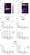
(A) Heat map of grooming time across body parts and time in controls and nf1P1 mutants. (B) Heat map of grooming time across body parts and time with pan-neuronal Nf1 knockdown (R57C10-Gal4>UAS-Nf1RNAi, UAS-dcr2). (C) Abdomen grooming across time in controls (wCS10) and nf1P1 mutants. Box plots: median = line, box = interquartile range; whiskers = min/max values, individual data points: circles. (D) Abdomen grooming with pan-neuronal Nf1 knockdown (R57C10-Gal4>UAS-Nf1RNAi,UAS-dcr2). (E) Head grooming in controls and nf1P1 mutants. (F) Head grooming with pan-neuronal Nf1 knockdown. (G) Wing grooming in controls and nf1P1 mutants. (H) Wing grooming with pan-neuronal Nf1 knockdown. Fly drawing modified from biorender.com.
Figure 3:
Figure 3:. Nf1 deficiency modulated a state-dependent behavioral switch from grooming to locomotion.
(A) Quantification of grooming (% time) in an open field arena when solid food was provided ad libitum (+Food), comparing control (wCS10) flies to nf1P1 mutants. (B) Still capture of fly locomotion tracking, with xy position tracks over 5 min for a representative control fly and nf1P1 mutant at 0 and 150 min (C) Total distance traveled for control flies and nf1P1 mutants. (D) Mean walking speed for control flies and nf1P1 mutants. (E) Diagram of the capillary feeding assay and protocol to test homeostatic feeding. Two cohorts each of controls and nf1P1 were tested: one kept on food continuously (“fed”) and one in which food was withheld at starting at t = 0 (“starved”). Food consumption was measured starting at t = 0 and t = 150 minutes. (F) Feeding in controls and nf1P1 mutants, comparing feeding in fed (fed +) and starved (fed −) conditions.
Figure 4:
Figure 4:. Knocking down Nf1 in sensory neurons and/or grooming command circuits shifted the pattern of grooming without affecting total grooming time.
Box plots: median = line, box = interquartile range; whiskers = min/max values, individual data points: circles. *p < 0.05, **p < 0.01, n.s. = not significant (Šidák; n = 16). (A) Simplified diagram of sensory neurons and antennal grooming command neurons, potential sites of modulation by Nf1 deficiency. RNAi was targeted to sensory neurons, command neurons, or both. (B) Effect of Nf1 knockdown in sensory neurons on head grooming (R81E10-Gal4>UAS- Nf1RNAi,UAS-dcr2). Experimental flies were compared to heterozygous Gal4/+ and UAS/+ controls. (C) Effect of Nf1 knockdown in eye/head grooming command neurons on head grooming (R23A07-Gal4>UAS-Nf1RNAi, UAS-dcr2). (D) Effect of Nf1 knockdown in wing grooming command neurons on wing grooming (R31H10-Gal4>UAS-Nf1RNAi, UAS-dcr2). (E) Effect of Nf1 knockdown in antennal grooming command neurons on head grooming (R18C11-Gal4>UAS-Nf1RNAi, UAS-dcr2). (F) Expression pattern of neurons labeled by R18C11-Gal4, focusing on the central brain. Box highlights the inset shown in panel G. GFP:green; brp:magenta. (G) Expanded view of the boxed region from panel F, including the somata of antennal descending neurons (white arrowheads). (H) Effect of Nf1 knockdown in antennal descending neurons (aDN) on head grooming using two different drivers (R71A06-Gal4 or R26B12-Gal4). (I) Effects of Nf1 knockdown in sensory neurons (R30B01-Gal4), wing grooming command neurons (R50B07-Gal4), and both, on wing grooming. (J) Effects of Nf1 knockdown in sensory neurons (R30B01-Gal4), eye/head command neurons (R23A07-Gal4), and both, on head grooming.
Figure 5:
Figure 5:. Loss of Nf1 altered the temporal evolution of stimulus-evoked grooming.
(A). Diagram of the experimental protocol. Flies were dusted, and amount of dust remining on each body part was imaged at t = 0, 8, 25, and 35 min. (B). Reference images of different body parts immediately after dusting, with images showing dust coverage after 0, 8, 25, and 35 min. (C). Dust removal in control (wCS10) flies. Dust coverage is the fraction of dust relative to those imaged at time 0. *p < 0.05; **p < 0.01, ***p < 0.001 re: time 0 (ANOVA/Šidák, n = 8). (D). Dust removal in nf1P1 mutants, plotted as in panel C. (E). Time course of dust removal (same data as in panels C,D) comparing controls and nf1P1 mutants at each time point. *p < 0.05; **p < 0.01, ***p < 0.001, comparing controls and nf1P1 mutants (ANOVA/Šidák, n = 8). Error bars = S.E.M.
Figure 6:
Figure 6:. Nf1 deficiency increased walking speed without altering gait or kinematics.
(A). Diagram of the experimental setup. Fly drawing modified from biorender.com. (B). Forward walking speed of controls (K33) vs. nf1P1 mutants (***p < 0.001 [Mann-Whitney], n = 70-90) and with pan-neuronal Nf1 knockdown (R57C10-Gal4>UAS-Nf1-RNAi) (*p < 0.05, ***p < 0.001 [Kruskal-Wallis/Dunn], n = 80-124). (C). Stance trajectory in control flies. 300 individual points plotted (randomly selected from >1000 steps). The dots indicate touch down locations, which are connected to the liftoff with a line. The black line is the mean of all trajectories. (D). Stance trajectory in nf1P1 mutants, plotted as in panel C. (E). Stance duration for the L1 leg, comparing K33 controls and nf1P1 mutants. Probability density is graphed as a heat map. (F). Step period for the L1 leg. (G). Swing duration for the L1 leg. (H). Diagram of leg coordination phase comparisons shown in panels I-L. (I). L1-R1 leg movement phase plot, comparing K33 controls and nf1P1 mutants. (J). L1-L2 leg movement phase plot. (K). L1-R2 leg movement phase plot. (L). L1-L3 leg movement phase plot.

References

    1. Anastasaki C, Mo J, Chen JK, Chatterjee J, Pan Y, Scheaffer SM, et al. Neuronal hyperexcitability drives central and peripheral nervous system tumor progression in models of neurofibromatosis-1. Nature communications. 2022;13(1):2785. Epub 20220519. doi: 10.1038/s41467-022-30466-6. - DOI - PMC - PubMed
    1. Costa RM, Federov NB, Kogan JH, Murphy GG, Stern J, Ohno M, et al. Mechanism for the learning deficits in a mouse model of neurofibromatosis type 1. Nature. 2002;415(6871):526–30. doi: 10.1038/nature711. - DOI - PubMed
    1. Wang Y, Nicol GD, Clapp DW, Hingtgen CM. Sensory neurons from Nf1 haploinsufficient mice exhibit increased excitability. Journal of neurophysiology. 2005;94(6):3670–6. doi: 10.1152/jn.00489.2005. - DOI - PubMed
    1. Contractor A, Klyachko VA, Portera-Cailliau C. Altered Neuronal and Circuit Excitability in Fragile X Syndrome. Neuron. 2015;87(4):699–715. doi: 10.1016/j.neuron.2015.06.017. - DOI - PMC - PubMed
    1. Ip JPK, Mellios N, Sur M. Rett syndrome: insights into genetic, molecular and circuit mechanisms. Nat Rev Neurosci. 2018;19(6):368–82. doi: 10.1038/s41583-018-0006-3. - DOI - PMC - PubMed

Publication types