[Preprint]. 2024 Aug 1:rs.3.rs-4792057.
doi: 10.21203/rs.3.rs-4792057/v1.

Bi-specific antibody engagers for cancer immunotherapy

Affiliations

Bi-specific antibody engagers for cancer immunotherapy

Hidde Ploegh et al. Res Sq. .

Update in

Abstract

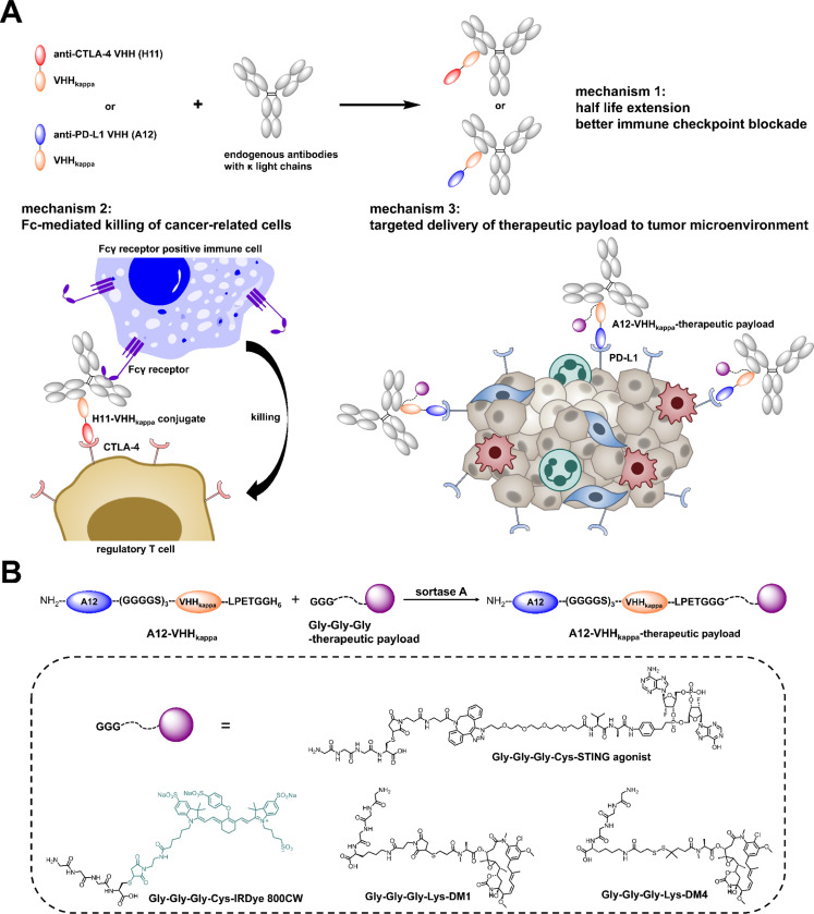
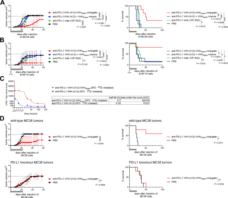
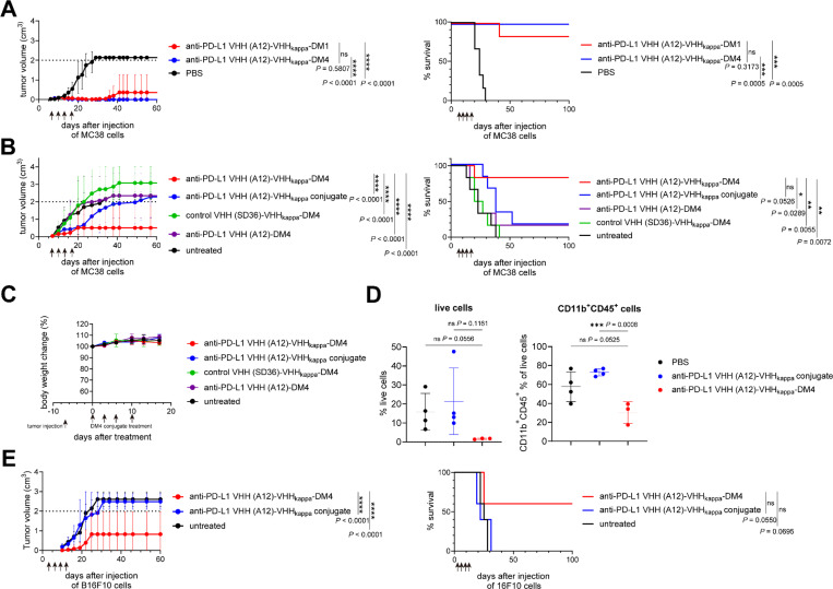
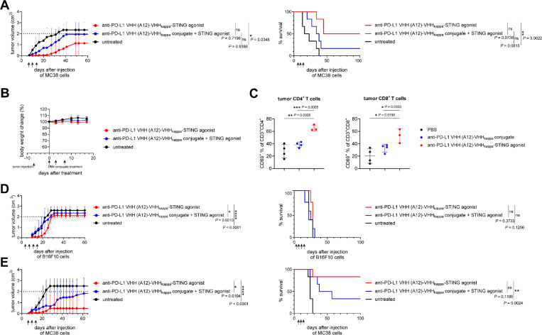
Bispecific antibody engagers are fusion proteins composed of a nanobody that recognizes immunoglobulin kappa light chains ( VHH kappa ) and a nanobody that recognizes either CTLA-4 or PD-L1. These fusions show strong antitumor activity in mice through recruitment of polyclonal immunoglobulins independently of specificity or isotype. In the MC38 mouse model of colorectal carcinoma, the anti-CTLA-4 VHH-VHH kappa conjugate eradicates tumors and reduces the number of intratumoral regulatory T cells. The anti-PD-L1 VHH-VHH kappa conjugate is less effective in the MC38 model, whilst still outperforming an antibody of similar specificity. The potency of the anti-PD-L1 VHH-VHH kappa conjugate was strongly enhanced by installation of the cytotoxic drug maytansine or a STING agonist. The ability of such fusions to engage the Fc-mediated functions of all immunoglobulin isotypes is an appealing strategy to further improve on the efficacy of immune checkpoint blockade, commonly delivered as a monoclonal immunoglobulin of a single defined isotype.


Conflict of interest statement

Declarations Competing interests X.L., and H.L.P. have filed a patent covering the technologies described in this publication (International Publication Number: WO2023141500A2). The other authors declare no competing interests. Additional Declarations: Yes there is potential Competing Interest. X.L., and H.L.P. have filed a patent covering the technologies described in this publication (International Publication Number: WO2023141500A2). The other authors declare no competing interests.

Figures

Figure 1
Figure 1. Recruitment of polyclonal immunoglobulins using nanobody-based immune checkpoint inhibitor-VHHkappa conjugates: proposed mechanism of action.
(A) Mechanism of action for VHHkappa-conjugated anti-CTLA-4 VHH (H11) and anti-PD-L1 VHH (A12) as anti-cancer immunotherapies: (1) the H11-VHHkappa or A12-VHHkappa conjugate binds to kappa light chains of polyclonal immunoglobulins, regardless of specificity or isotype, thereby prolonging the circulatory half-life of the conjugates; (2) the H11-VHHkappa conjugate recruits immunoglobulins to mediate Fc receptor-dependent depletion of CTLA-4-positive T regulatory cells; (3) the A12-VHHkappa conjugate can be further modified for delivery of drugs to the tumor microenvironment by targeting PD-L1, overexpressed by tumor and myeloid cells. (B) The triglycine-modified therapeutic payload is site-specifically attached to the C-terminus of A12-VHHkappa conjugate through a sortase reaction. These payloads include a near-infrared dye imaging agent, cytotoxic drugs, and immunostimulatory drugs.
Figure 2
Figure 2. Affinity, specificity and antibody-recruiting capability of the anti-CTLA-4 VHH (H11)-VHHkappa and the anti-PD-L1 VHH (A12)-VHHkappa conjugates.
(A) Saturation binding curves of biotinylated H11, A12, H11-VHHkappa conjugate, and A12-VHHkappa conjugate to murine CTLA-4 and PD-L1 (Data represent mean ± SD, n = 3). (B) Saturation binding curves of biotinylated H11-VHHkappa and A12-VHHkappa conjugates to murine polyclonal IgG (Data represent mean ± SD, n = 3). (C) Flow cytometry confirms that the A12-VHHkappa conjugate can recruit phycoerythrin (PE)-conjugated mouse IgG, IgM, and IgA, to PD-L1-positive B16-F10 cells. (D) Flow cytometry of MC38 tumor cells, isolated from tumor-bearing C57BL/6 mice and stained with H11-Cy5, reveals CTLA-4 expression on T regulatory cells within the tumor, with only marginal staining observed on splenic T cells. (E) Flow cytometry of cells recovered from PD-L1-positive and PD-L1 knockout MC38 tumors in C57BL/6 mice stained with A12-Cy5 confirms PD-L1 staining on wild-type MC38 cells (CD45-) and infiltrating CD11b+ immune cells (CD45+).
Figure 3
Figure 3. The anti-CTLA-4 VHH (H11)-VHHkappa conjugate shows better in vivo antitumor activity than the anti CTLA-4 antibody 9H10 or H11 VHH alone.
(A) Tumor growth and survival curves for MC38 tumor-bearing mice (n = 6) treated intraperitoneally with the indicated VHHs at 5 mg/kg on day 1 post-tumor injection, followed by 3 times a week for 3 weeks as indicated by the arrows. † Mouse died from an unknown cause. (B) Tumor growth and survival curves for B16-F10 tumor-bearing mice (n = 6) treated intraperitoneally with the indicated VHHs at 5 mg/kg on day 1 post-tumor injection, followed by 3 times a week for 3 weeks as indicated by the arrows. Mice were vaccinated with GM-CSF–secreting B16 cells (GVAX) on day 0. † Mice were sacrificed due to severe ulceration of their tumors. (C) Changes in T cell populations after treatment with the (H11)-VHHkappa conjugate. MC38 tumor-bearing mice were treated intraperitoneally with the indicated VHHs at 5 mg/kg on day 8, 10, 12, and 14 post-tumor injection. Single cell suspensions from tumors, spleens, and draining lymph nodes were analyzed by flow cytometry on day 15 post-tumor injection. The experiment was performed twice, and the data were combined (Data represent mean ± SD, n= 7, unpaired t test). (D) Changes in regulatory T cell populations after treatment with the H11-VHHkappa conjugate in wild-type, Fc receptor common gamma chain knockout mice, and complement-deficient mice. MC38 tumor-bearing mice were treated intraperitoneally with the indicated VHHs at 5 mg/kg on day 8, 10, 12, and 14 post-tumor injection. Single cell suspensions from tumors and spleens were analyzed by flow cytometry on day 15 post-tumor injection (Data represent mean ± SD, n= 4, unpaired t test). (E) Half-life measurements of 89Zr-labelled H11-VHHkappa conjugate and 89Zr-labelled H11 in mice (Data represent mean ± SD, n = 3). (F) Tumor growth and survival curves for MC38 tumor-bearing mice (n = 6) treated intravenously with the indicated VHHs at 5 mg/kg or anti-CTLA-4 antibody 9H10 at 12.5 mg/kg (equimolar) on day 7, 10, and 13 post-tumor injection as indicated by the arrows. † Mouse was sacrificed due to severe ulceration of its tumor.
Figure 4
Figure 4. Whole-body imaging and biodistribution of near-infrared dye-labelled anti-PD-L1 VHH (A12)-VHHkappaconjugate in MC38 tumor-bearing mice show its PD-L1-dependent accumulation in the tumor.
(A) Structure of near-infrared (NIR) dye (IRDye800CW)-labelled A12-VHHkappa conjugate. (B) Timeline of the imaging experiment. Wild-type and PD-L1 knockout MC38 tumor-bearing mice (n = 3) were treated intravenously with the indicated NIR dye-labelled VHHs (1 nmol) on day 14 post-tumor injection. Imaging was done 24 h later. (C) Whole-body imaging. Left: comparison of various NIR dye-labelled VHHs in tumor-bearing mice; right: individual images of mice from the same treatment group. SD36 is a VHH that recognizes influenza virus hemagglutinin (specificity control). Where indicated (30x), a 30-fold molar excess of unlabeled imaging agent was given as a competitor. (D) Left: Ex vivo NIR imaging of tumors from the mice treated with the indicated NIR dye-labelled VHHs; Right: quantification of the NIR dye-labelled VHHs accumulated in the tumors (Data represent mean ± SD, n = 3, unpaired t test). (E) Left: Ex vivo NIR imaging of organs from mice treated with the indicated NIR dye-labelled VHHs (from top to bottom: tumor, lung, heart, liver, stomach, intestine, spleen, and kidneys); Right: quantification of accumulation of the NIR dye-labelled VHHs (Data represent mean ± SD, n = 3, unpaired t test).
Figure 5
Figure 5. The anti-PD-L1 VHH (A12)-VHHkappa conjugate has superior in vivo antitumor activity compared to anti-PD-L1 antibody 10F.9G2 or A12 VHH alone.
(A) Tumor growth and survival curves for MC38 tumor-bearing mice (n = 6) treated intraperitoneally with the indicated VHHs at 5 mg/kg on day 1 post-tumor injection, followed by 3 times a week for 3 weeks as indicated by the arrows. (B) Tumor growth and survival curves for B16-F10 tumor-bearing mice (n = 6) treated intraperitoneally with the indicated VHHs at 5 mg/kg on day 1 post-tumor injection, followed by 3 times a week for 3 weeks, as indicated by the arrows. Mice were vaccinated with GM-CSF-secreting B16 cells (GVAX) on day 0. (C) Half-life measurements of 89Zr-labelled A12-VHHkappa conjugate and 89Zr-labelled A12 in mice (Data represent mean ± SD, n = 3). (D) Tumor growth and survival curves for wild-type and PD-L1 knockout MC38 tumor-bearing mice (n = 5) treated intraperitoneally with the indicated VHHs at 5 mg/kg on day 1 post-tumor injection, followed by three times a week for 3 weeks as indicated by the arrows.
Figure 6
Figure 6. Targeted delivery of the cytotoxicity drug maytansine enhances antitumor activity of the anti-PD-L1 VHH (A12)-VHHkappa conjugate.
(A) Tumor growth and survival curves for MC38 tumor-bearing mice (n = 6) treated intravenously with A12-VHHkappa-DM1 or A12-VHHkappa-DM4 at 5 mg/kg on day 7, 10,13, and 17 post-tumor injection as indicated by the arrows. (B) Tumor growth and survival curves for MC38 tumor-bearing mice (n = 6) treated intravenously with indicated VHH-DM4 conjugates at 5 mg/kg on day 7, 10,13, and 17 post-tumor injection as indicated by the arrows. (C) Body weight curves for the mice used in panel B (Data represent mean ± SD, n = 6). (D) Percentage of live cells and CD11b+ lymphocytes harvested from the tumors in mice treated with A12-VHHkappa-DM4 or A12-VHHkappa conjugate. MC38 tumor-bearing mice were treated intravenously with the indicated VHH conjugates at 5 mg/kg on day 11 and 13 post-tumor injection (Data represent mean ± SD, n = 3 or 4, unpaired t test). (E) Tumor growth and survival curves for B16-F10 tumor-bearing mice (n = 5) treated intravenously with indicated VHH-DM4 conjugates at 5 mg/kg on day 3, 6, 9, and 13 post-tumor injection as indicated by the arrows.
Figure 7
Figure 7. Targeted delivery of a STING agonist enhances antitumor activity of the anti-PD-L1 VHH (A12)-VHHkappaconjugate.
(A) Tumor growth and survival curves for MC38 tumor-bearing mice (n = 6) treated intravenously with A12-VHHkappa-STING agonist or A12-VHHkappa-conjugate plus free STING agonist (equimolar amount) at 5 mg/kg on day 7, 10, and 14 post-tumor injection as indicated by the arrows. (B) Body weight curves for the mice used in panel A (Data represent mean ± SD, n = 6). (C) Percentage of activated (CD69-positive) CD4+ and CD8+ T cells harvested from the tumors in mice treated with A12-VHHkappa-STING agonist or A12-VHHkappa conjugates. MC38 tumor-bearing mice were treated intravenously with the indicated VHH conjugates at 5 mg/kg on day 11 and 13 post-tumor injection. Single cell suspensions from tumors were analyzed by flow cytometry on day 15 post-tumor injection (Data represent mean ± SD, n = 3 or 4, unpaired t test). (D) Tumor growth and survival curves for B16-F10 tumor-bearing mice (n = 5) treated intravenously with A12-VHHkappa-STING agonist or A12-VHHkappa-conjugate plus free STING agonist (equimolar amount) on day 3, 7,11, and 15 post-tumor injection as indicated by the arrows. (E) Tumor growth and survival curves for MC38 tumor-bearing mice (n = 6) treated intravenously with A12-VHHkappa-STING agonist or A12-VHHkappa-conjugate plus free STING agonist (equimolar amount) at 10 mg/kg on day 7, 10, and 14 post-tumor injection as indicated by the arrows.

References

    1. Jiang X.-R. et al. Advances in the assessment and control of the effector functions of therapeutic antibodies. Nature reviews Drug discovery 10, 101–111 (2011). - PubMed
    1. Park H. I., Yoon H. W. & Jung S. T. The highly evolvable antibody Fc domain. Trends in biotechnology 34, 895–908 (2016). - PubMed
    1. Liu R., Oldham R. J., Teal E., Beers S. A. & Cragg M. S. Fc-engineering for modulated effector functions—improving antibodies for cancer treatment. Antibodies 9, 64 (2020). - PMC - PubMed
    1. Kretschmer A., Schwanbeck R., Valerius T. & Rösner T. Antibody isotypes for tumor immunotherapy. Transfusion Medicine and Hemotherapy 44, 320–326 (2017). - PMC - PubMed
    1. Mukai K., Tsai M., Starkl P., Marichal T. & Galli S. J. in Seminars in immunopathology. 581–603 (Springer; ). - PMC - PubMed

Publication types

LinkOut - more resources